. 2001 Nov;118(5):447-56.
doi: 10.1085/jgp.118.5.447.

Gap junctional coupling in lenses from alpha(8) connexin knockout mice

Affiliations

Gap junctional coupling in lenses from alpha(8) connexin knockout mice

G J Baldo et al. J Gen Physiol. 2001 Nov.

Abstract

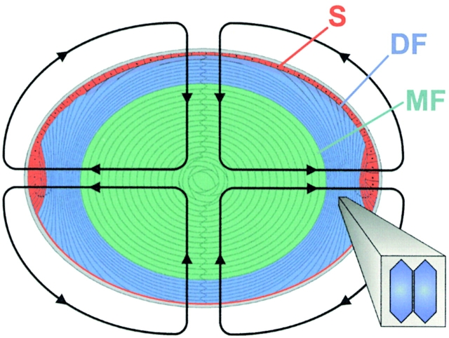
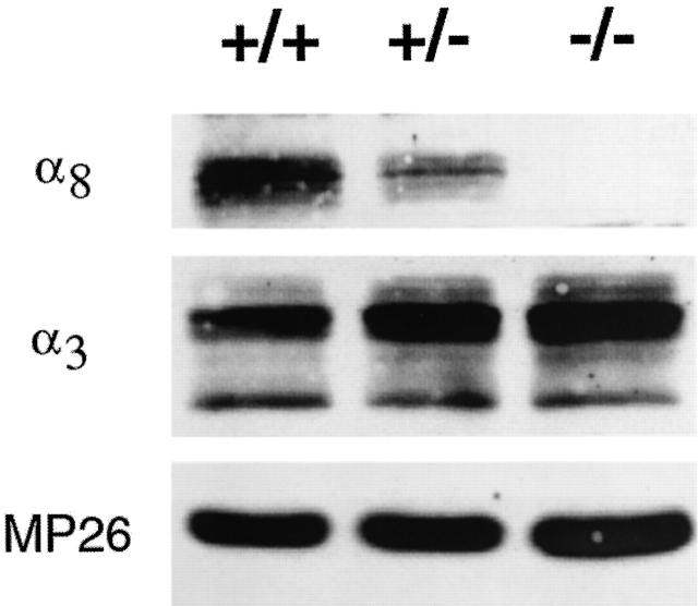
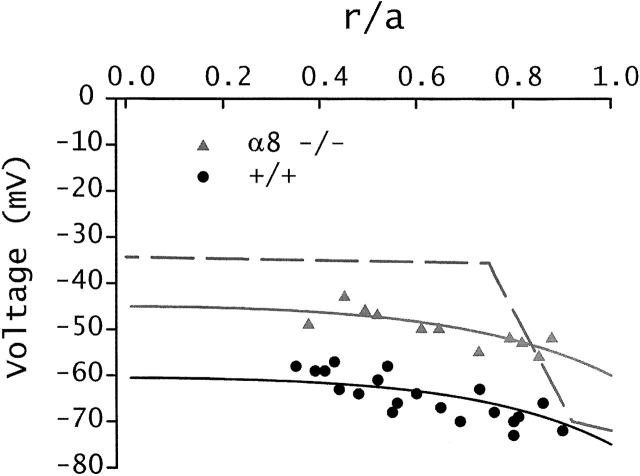
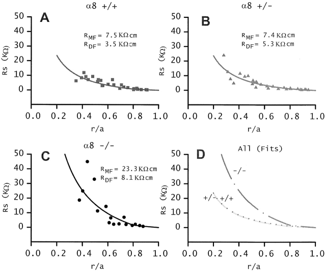
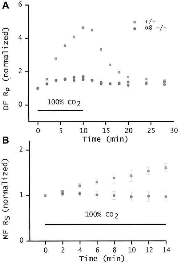
Lens fiber cell gap junctions contain alpha(3) (Cx46) and alpha(8) (Cx50) connexins. To examine the roles of the two different connexins in lens physiology, we have genetically engineered mice lacking either alpha(3) or alpha(8) connexin. Intracellular impedance studies of these lenses were used to measure junctional conductance and its sensitivity to intracellular pH. In Gong et al. 1998, we described results from alpha(3) connexin knockout lenses. Here, we present original data from alpha(8) connexin knockout lenses and a comparison with the previous results. The lens has two functionally distinct domains of fiber cell coupling. In wild-type mouse lenses, the outer shell of differentiating fibers (see 1, DF) has an average coupling conductance per area of cell-cell contact of approximately 1 S/cm(2), which falls to near zero when the cytoplasm is acidified. In the inner core of mature fibers (see 1, MF), the average coupling conductance is approximately 0.4 S/cm(2), and is insensitive to acidification of the cytoplasm. Both connexin isoforms appear to contribute about equally in the DF since the coupling conductance for either heterozygous knockout (+/-) was approximately 70% of normal and 30-40% of the normal for both -/- lenses. However, their contribution to the MF was different. About 50% of the normal coupling conductance was found in the MF of alpha(3) +/- lenses. In contrast, the coupling of MF in the alpha(8) +/- lenses was the same as normal. Moreover, no coupling was detected in the MF of alpha(3) -/- lenses. Together, these results suggest that alpha(3) connexin alone is responsible for coupling MF. The pH- sensitive gating of DF junctions was about the same in wild-type and alpha(3) connexin -/- lenses. However, in alpha(8) -/- lenses, the pure alpha(3) connexin junctions did not gate closed in the response to acidification. Since alpha(3) connexin contributes about half the coupling conductance in DF of wild-type lenses, and that conductance goes to zero when the cytoplasmic pH drops, it appears alpha(8) connexin regulates the gating of alpha(3) connexin. Both connexins are clearly important to lens physiology as lenses null for either connexin lose transparency. Gap junctions in the MF survive for the lifetime of the organism without protein turnover. It appears that alpha(3) connexin provides the long-term communication in MF. Gap junctions in DF may be physiologically regulated since they are capable of gating when the cytoplasm is acidified. It appears alpha(8) connexin is required for gating in DF.


Figures

Figure 1
Figure 1
The cellular structure of the lens. Based on transport properties, the lens can be divided into three radial zones. The surface (S) includes the basal lateral membrane of the anterior epithelium and the basal membrane of differentiating fibers (DF) cells in contact with the posterior surface. These membranes have a relatively high permeability for K+ and perform nearly all of the active transport in the lens. The DF and mature fibers (MF) are in communication with each other and with the surface membranes through an extensive network of gap junctions. However, the functional and structural properties of gap junctions coupling DF and MF differ, so these two domains are studied separately.
Figure 2
Figure 2
Immunoblot of the α8 connexin knockout mice. The NaOH-insoluble protein fractions were prepared from the lenses of +/+, α8 +/−, and α8 −/− mice at the age of 8 wk (Gong et al. 1997). Immunoblot analysis was performed using antibodies against α8 connexin (top), α3 connexin (middle), and MP26 (bottom), respectively. Equal portions of the samples (∼15 mg total proteins) were loaded in each lane.
Figure 3
Figure 3
Resting voltages. The value of the resting voltage is plotted as a function of normalized distance from the lens center (r/a), where r (in centimeters) is the distance from the center and a (in centimeters) is the lens radius. The points represent data from lenses of +/+ and α8 −/− mice. The solid smooth curves represent the expected distribution of resting voltages based on a model of the circulating current that flows into the lens along extracellular spaces, crosses the fiber cell membrane, and then flows toward the surface of the lens from cell to cell via gap junctions (for review see Mathias et al. 1997). The dashed line is an arbitrary fit to resting voltage data from lenses of α3 −/− mice (Gong et al. 1998). The α8 −/− lenses are depolarized at all locations, suggesting some indirect effects on surface cell membrane permeability. In contrast, the α3 −/− lenses have a normal voltage in DF, but the MF depolarize in a way that reflects loss of gap junctional coupling.
Figure 4
Figure 4
The cumulative series resistance (RS). The closed circles in A–C are Rs data from six lenses from +/+, +/−, and −/− α8 connexin knockout mice. The solid lines represent the best fit using and with a = 0.101 cm for +/+ and +/− lenses, a = 0.08 cm for −/− lenses, b = 0.8a, and with the values of RDF and RMF listed in each panel. The best fits are graphed together in D. The lenses from α8 −/− mice have many abnormalities that are secondary consequences of the knockout of α8 connexin. The lenses from α8 +/− mice are normal except for changes in coupling conductance. In these lenses, removal of ∼50% of α8 connexin significantly increases RDF, but has no effect on RMF, suggesting the α8 connexin may not be a functional component of gap junctions coupling MF.
Figure 5
Figure 5
pH-sensitive gating. A compares the coupling of DF from wild-type and α8 connexin −/− lenses when superfused with Tyrode bubbled with 100% CO2. The CO2-saturated solution was superfused during the time period indicated by the bar. The initial values of RP. RDF for the two −/− lenses was 5.9 and 10.1 KΩ-cm, whereas the initial value of RP in the +/+ lens was 3.4 KΩ-cm, and all of these values were stable before CO2 exposure. To facilitate comparison, each resistivity was normalized to its initial value, and then graphed as a function of time. In a 10-min exposure to 100% CO2, the RP of DF in the wild-type lens increased 4.7-fold. This was a typical response, similar to the responses we recorded from a number of lenses from wild-type mice of different litters. In contrast, RP of the DF in α8 −/− lenses increased only 1.6-fold. Thus, in the α8 −/− lens, homotypic channels made from α3 connexin have little or no sensitivity to pH. The surface cells express α1 (Cx43) connexin, so the limited pH gating seen in A could be due to α1. B compares the cumulative series resistance (RS in ) in lenses from +/+ and α8 −/− mice. Again, the CO2 exposure was during the period indicated by the bar and before t = 0, the values of RS were stable. As illustrated by , this resistance is dominated by the contribution of MF. Again, the resistances were normalized to their initial values, and then graphed as a function of time exposed to 100% CO2. The +/+ data were mean ± SD from four lenses. Although the MFs of +/+ lenses are not sensitive to pH, DFs are sensitive, hence, RS increases significantly. The α8 −/− data are mean ± SD from three lenses. In the α8 −/− lenses, all fiber cell gap junctions contain homotypic channels made up of α3 connexin. If the small increase in resistance of DF from α8 −/− mice shown in A is confined to DF or surface cells, it will be a very small fraction of the total RS shown in B. Indeed, the total RS of α8 −/− mice shows no significant increase on exposure of the lenses to 100% CO2.

References

    1. Baldo G.J., Mathias R.T. Spatial variations in membrane properties in the intact rat lens. Biophys. J. 1992;63:518–529. - PMC - PubMed
    1. Bassnett S., Croghan P.C., Duncan G. Diffusion of lactate and its role in determining intracellular pH in the lens of the eye. Exp. Eye Res. 1987;44:143–147. - PubMed
    1. Donaldson P., Kistler J., Mathias R.T. Molecular solutions to lens transparency. News Physiol. Sci. 2001;16:118–123. - PubMed
    1. Fischbarg J., Diecke F.P., Kuang K., Yu B., Kang F., Iserovich P., Li Y., Rosskothen H., Koniarek J.P. Transport of fluid by lens epithelium. Am. J. Physiol. 1999;276:C548–C557. - PubMed
    1. Gao J., Sun X., Yatsula V., Wymore R.S., Mathias R.T. Isoform-specific function and distribution of Na/K pumps in the frog lens epithelium. J. Membr. Biol. 2000;178:89–101. - PubMed

Publication types