

Membrane-associated quinoprotein formaldehyde dehydrogenase from Methylococcus capsulatus Bath
- PMID: 11698372
- PMCID: PMC95524
- DOI: 10.1128/JB.183.23.6832-6840.2001
Membrane-associated quinoprotein formaldehyde dehydrogenase from Methylococcus capsulatus Bath
Abstract
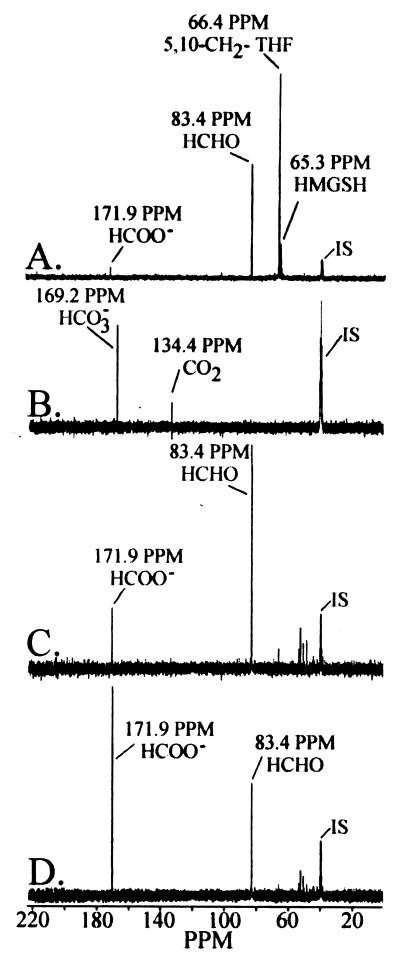
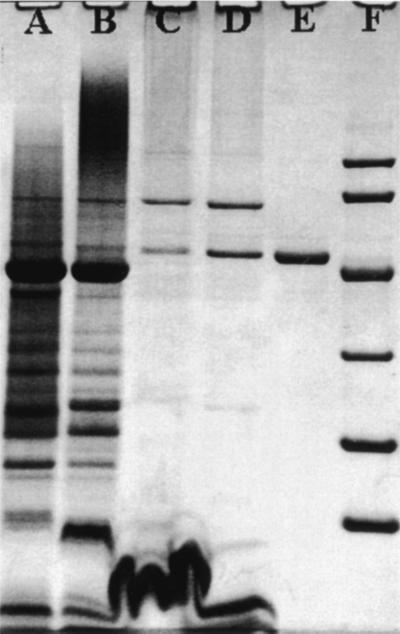
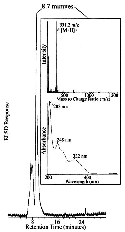
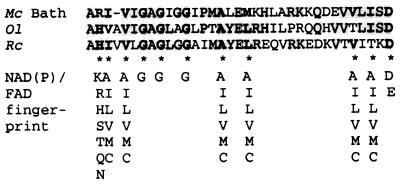
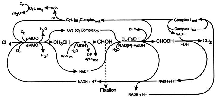
A membrane-associated, dye-linked formaldehyde dehydrogenase (DL-FalDH) was isolated from the obligate methylotroph Methylococcus capsulatus Bath. The enzyme was the major formaldehyde-oxidizing enzyme in cells cultured in high (above 1 micromol of Cu per mg of cell protein) copper medium and expressing the membrane-associated methane monooxygenase. Soluble NAD(P)(+)-linked formaldehyde oxidation was the major activity in cells cultured in low-copper medium and expressing the soluble methane monooxygenase (Tate and Dalton, Microbiology 145:159-167, 1999; Vorholt et al., J. Bacteriol. 180:5351-5356, 1998). The membrane-associated enzyme is a homotetramer with a subunit molecular mass of 49,500 Da. UV-visible absorption, electron paramagnetic resonance, and electrospray mass spectrometry suggest the redox cofactor of the DL-FalDH is pyrroloquinoline quinone (PQQ), with a PQQ-to-subunit stochiometry of approximately 1:1. The enzyme was specific for formaldehyde, oxidizing formaldehyde to formate, and utilized the cytochrome b(559/569) complex as the physiological electron acceptor.
Figures
Similar articles
-
Formaldehyde dehydrogenase preparations from Methylococcus capsulatus (Bath) comprise methanol dehydrogenase and methylene tetrahydromethanopterin dehydrogenase.Microbiology (Reading). 2004 Mar;150(Pt 3):707-713. doi: 10.1099/mic.0.26707-0. Microbiology (Reading). 2004. PMID: 14993320
-
Effect of methanobactin on the activity and electron paramagnetic resonance spectra of the membrane-associated methane monooxygenase in Methylococcus capsulatus Bath.Microbiology (Reading). 2005 Oct;151(Pt 10):3417-3426. doi: 10.1099/mic.0.28169-0. Microbiology (Reading). 2005. PMID: 16207923
-
The PmoB subunit of particulate methane monooxygenase (pMMO) in Methylococcus capsulatus (Bath): The CuI sponge and its function.J Inorg Biochem. 2019 Jul;196:110691. doi: 10.1016/j.jinorgbio.2019.04.005. Epub 2019 Apr 15. J Inorg Biochem. 2019. PMID: 31063931
-
Pyrroloquinoline quinone (PQQ) and quinoprotein enzymes.Antioxid Redox Signal. 2001 Oct;3(5):757-74. doi: 10.1089/15230860152664966. Antioxid Redox Signal. 2001. PMID: 11761326 Review.
-
Structure and mechanism of soluble glucose dehydrogenase and other PQQ-dependent enzymes.Biochim Biophys Acta. 2003 Apr 11;1647(1-2):143-51. doi: 10.1016/s1570-9639(03)00087-6. Biochim Biophys Acta. 2003. PMID: 12686124 Review.
Cited by
-
CorA is a copper repressible surface-associated copper(I)-binding protein produced in Methylomicrobium album BG8.PLoS One. 2014 Feb 3;9(2):e87750. doi: 10.1371/journal.pone.0087750. eCollection 2014. PLoS One. 2014. PMID: 24498370 Free PMC article.
-
Isolation, sequencing, and heterologous expression of the Paecilomyces variotii gene encoding S-hydroxymethylglutathione dehydrogenase (fldA).Appl Microbiol Biotechnol. 2015 Feb;99(4):1755-63. doi: 10.1007/s00253-014-6203-8. Epub 2014 Nov 16. Appl Microbiol Biotechnol. 2015. PMID: 25398285 Free PMC article.
-
Methanobactin and the Link between Copper and Bacterial Methane Oxidation.Microbiol Mol Biol Rev. 2016 Mar 16;80(2):387-409. doi: 10.1128/MMBR.00058-15. Print 2016 Jun. Microbiol Mol Biol Rev. 2016. PMID: 26984926 Free PMC article. Review.
-
Oxygen Generation via Water Splitting by a Novel Biogenic Metal Ion-Binding Compound.Appl Environ Microbiol. 2021 Jun 25;87(14):e0028621. doi: 10.1128/AEM.00286-21. Epub 2021 Jun 25. Appl Environ Microbiol. 2021. PMID: 33962982 Free PMC article.
-
Biological pincer complexes.ChemCatChem. 2020 Sep 4;12(17):4242-4254. doi: 10.1002/cctc.202000575. Epub 2020 May 8. ChemCatChem. 2020. PMID: 33072225 Free PMC article.
References
-
- Anthony C. The biochemistry of methylotrophs. London, England: Academic Press; 1982.
-
- Ariel B, Shahak Y, Taglicht D, Hauska G, Padan E. Purification and characterization of sulfide-quinone reductase, a novel enzyme driving anoxygenic photosynthesis in Oscillatoria limnetica. J Biol Chem. 1994;268:5705–5711. - PubMed
-
- Attwood M, Quayle J R. Formaldehyde as a central intermediary metabolite in methylotrophic metabolism. In: Crawford R L, Hanson R H, editors. Microbial growth on C1 compounds. Washington, D.C.: ASM Press; 1984. pp. 315–323.
Publication types
MeSH terms
Substances
LinkOut - more resources
Full Text Sources
