. 2001 Dec;183(23):6898-907.
doi: 10.1128/JB.183.23.6898-6907.2001.

Binding site of macrolide antibiotics on the ribosome: new resistance mutation identifies a specific interaction of ketolides with rRNA

Affiliations

Binding site of macrolide antibiotics on the ribosome: new resistance mutation identifies a specific interaction of ketolides with rRNA

G Garza-Ramos et al. J Bacteriol. 2001 Dec.

Abstract

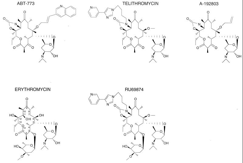
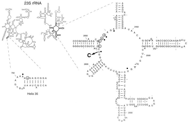
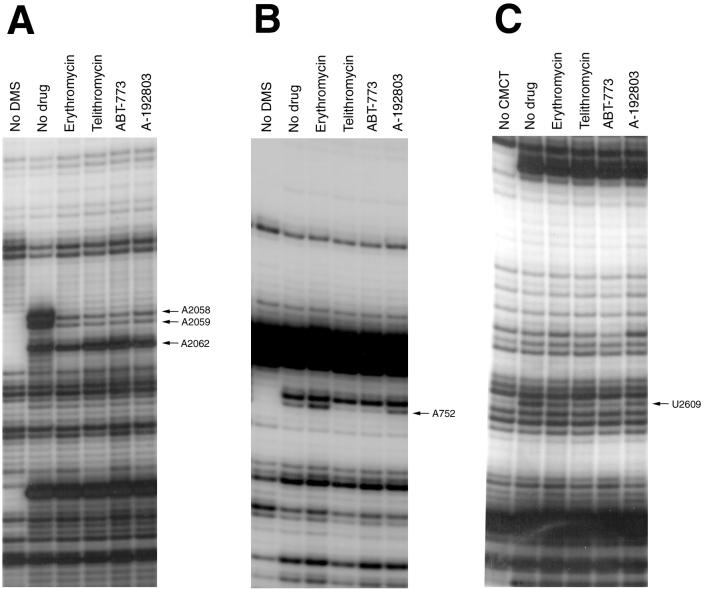
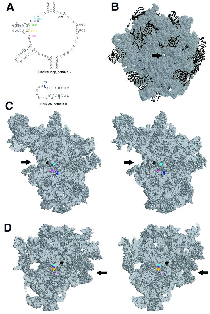
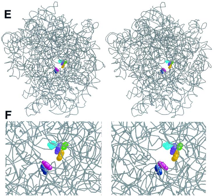
Macrolides represent a clinically important class of antibiotics that block protein synthesis by interacting with the large ribosomal subunit. The macrolide binding site is composed primarily of rRNA. However, the mode of interaction of macrolides with rRNA and the exact location of the drug binding site have yet to be described. A new class of macrolide antibiotics, known as ketolides, show improved activity against organisms that have developed resistance to previously used macrolides. The biochemical reasons for increased potency of ketolides remain unknown. Here we describe the first mutation that confers resistance to ketolide antibiotics while leaving cells sensitive to other types of macrolides. A transition of U to C at position 2609 of 23S rRNA rendered E. coli cells resistant to two different types of ketolides, telithromycin and ABT-773, but increased slightly the sensitivity to erythromycin, azithromycin, and a cladinose-containing derivative of telithromycin. Ribosomes isolated from the mutant cells had reduced affinity for ketolides, while their affinity for erythromycin was not diminished. Possible direct interaction of ketolides with position 2609 in 23S rRNA was further confirmed by RNA footprinting. The newly isolated ketolide-resistance mutation, as well as 23S rRNA positions shown previously to be involved in interaction with macrolide antibiotics, have been modeled in the crystallographic structure of the large ribosomal subunit. The location of the macrolide binding site in the nascent peptide exit tunnel at some distance from the peptidyl transferase center agrees with the proposed model of macrolide inhibitory action and explains the dominant nature of macrolide resistance mutations. Spatial separation of the rRNA residues involved in universal contacts with macrolides from those believed to participate in structure-specific interactions with ketolides provides the structural basis for the improved activity of the broader spectrum group of macrolide antibiotics.


Figures

FIG. 1
FIG. 1
Chemical structures of macrolide antibiotics used in this study. Ketolides, containing the 3-keto group, are represented by ABT-773, telithromycin, and A-192803. Erythromycin and RU69874 are examples of cladinosolides that contain a 3-cladinose residue.
FIG. 2
FIG. 2
E. coli 23S rRNA positions involved in interaction with 14-member macrolide antibiotics. A newly identified ketolide resistance mutation, U2609 to C, is shown by an arrow. Sites of the previously characterized macrolide-resistance mutations are circled. For references, see reference . Nucleotide residues whose accessibility to chemical modification is affected by macrolides are marked by dots. The secondary structure is presented following those in references and .
FIG. 3
FIG. 3
Effect of macrolides (cladinosolides and ketolides) on accessibility of 23S rRNA nucleotides to chemical modification. (A) Central loop of domain V, DMS modification. (B) Hairpin 35, DMS modification. (C) Central loop domain V, CMCT modification. “No DMS” represents the unmodified control. “No drug” represents ribosomes modified in the absence of antibiotics. All of the other samples contained respective antibiotics present at a 5 μM concentration (see Materials and Methods for experimental details). Nucleotides with modification affected by bound antibiotics are indicated by arrows.
FIG. 4
FIG. 4
Relative position of nucleotides involved in macrolide binding in the crystallographic structure of the H. marismortui 50S ribosomal subunit. (A) Secondary structure of the central loop of domain V and hairpin 35 of H. marismortui 23S rRNA. Nucleotide positions in domain V at which mutations (in E. coli ribosome) confer macrolide resistance are shown in color. The nucleotide position in helix 35 corresponding to the adenine base in the E. coli ribosome for which modification is differentially affected by ketolides and cladinosolides is shown in blue. A2451 located within the active site of the peptidyl transferase center is shown in black. The nucleotide numbering is that of E. coli 23S rRNA. (B) “Front” view of the 50S subunit from the interface side. The opening of the peptidyl transferase center leading into the exit tunnel is indicated by an arrow. To make the tunnel visible, the subunit was rotated by 35° versus the y axis and tilted by 35° versus the z axis. rRNA is shown in a space-fill representation (gray), and protein backbones are shown in black. Positions 2058 and 2059 important for macrolide binding are seen at the bottom of the peptidyl transferase cavity. (C) Stereo view of a vertical cross-cut of the 50S subunit through the exit tunnel. (D) Same as panel C, but rotated by 180° versus the vertical axis. Only the rRNA moiety of the 50S subunit is shown. The entrance into the peptidyl transferase cavity (located at the interface surface of the subunit), which leads to the nascent peptide exit tunnel, is shown by arrows. Nucleotides involved in interaction with macrolide antibiotics are shown in color (same as in panel A), and A2451 located in the peptidyl transferase active site is shown in black. (E) Spatial arrangement of the nucleotides involved in macrolide binding in the exit tunnel seen from the peptidyl transferase (subunit interface) side. rRNA is shown as a gray backbone, while nucleotides involved in interaction with macrolide antibiotics are colored in a space-fill representation. Proteins were removed for clarity. (F) Close-up view of the macrolide binding site (approximately fourfold enlargement compared to panel E).
FIG. 4
FIG. 4
Relative position of nucleotides involved in macrolide binding in the crystallographic structure of the H. marismortui 50S ribosomal subunit. (A) Secondary structure of the central loop of domain V and hairpin 35 of H. marismortui 23S rRNA. Nucleotide positions in domain V at which mutations (in E. coli ribosome) confer macrolide resistance are shown in color. The nucleotide position in helix 35 corresponding to the adenine base in the E. coli ribosome for which modification is differentially affected by ketolides and cladinosolides is shown in blue. A2451 located within the active site of the peptidyl transferase center is shown in black. The nucleotide numbering is that of E. coli 23S rRNA. (B) “Front” view of the 50S subunit from the interface side. The opening of the peptidyl transferase center leading into the exit tunnel is indicated by an arrow. To make the tunnel visible, the subunit was rotated by 35° versus the y axis and tilted by 35° versus the z axis. rRNA is shown in a space-fill representation (gray), and protein backbones are shown in black. Positions 2058 and 2059 important for macrolide binding are seen at the bottom of the peptidyl transferase cavity. (C) Stereo view of a vertical cross-cut of the 50S subunit through the exit tunnel. (D) Same as panel C, but rotated by 180° versus the vertical axis. Only the rRNA moiety of the 50S subunit is shown. The entrance into the peptidyl transferase cavity (located at the interface surface of the subunit), which leads to the nascent peptide exit tunnel, is shown by arrows. Nucleotides involved in interaction with macrolide antibiotics are shown in color (same as in panel A), and A2451 located in the peptidyl transferase active site is shown in black. (E) Spatial arrangement of the nucleotides involved in macrolide binding in the exit tunnel seen from the peptidyl transferase (subunit interface) side. rRNA is shown as a gray backbone, while nucleotides involved in interaction with macrolide antibiotics are colored in a space-fill representation. Proteins were removed for clarity. (F) Close-up view of the macrolide binding site (approximately fourfold enlargement compared to panel E).

References

    1. Arevalo M A, Tejedor F, Polo F, Ballesta J P. Protein components of the erythromycin binding site in bacterial ribosomes. J Biol Chem. 1988;263:58–63. - PubMed
    1. Asai T, Condon C, Voulgaris J, Zaporojets D, Shen B, Al-Omar M, Squires C, Squires C L. Construction and initial characterization of Escherichia coli strains with few or no intact chromosomal rRNA operons. J Bacteriol. 1999;181:3803–3809. - PMC - PubMed
    1. Asai T, Zaporojets D, Squires C, Squires C L. An Escherichia coli strain with all chromosomal rRNA operons inactivated: complete exchange of rRNA genes between bacteria. Proc Natl Acad Sci USA. 1999;96:1971–1976. - PMC - PubMed
    1. Ban N, Nissen P, Hansen J, Moore P B, Steitz T A. The complete atomic structure of the large ribosomal subunit at 2.4 Å resolution. Science. 2000;289:905–920. - PubMed
    1. Beauclerk A A, Cundliffe E. Sites of action of two ribosomal RNA methylases responsible for resistance to aminoglycosides. J Mol Biol. 1987;193:661–671. - PubMed

Publication types

LinkOut - more resources