. 2001 Nov 15;21(22):8746-57.
doi: 10.1523/JNEUROSCI.21-22-08746.2001.

Subcellular and subsynaptic localization of presynaptic and postsynaptic kainate receptor subunits in the monkey striatum

Affiliations

Subcellular and subsynaptic localization of presynaptic and postsynaptic kainate receptor subunits in the monkey striatum

J Z Kieval et al. J Neurosci. .

Abstract

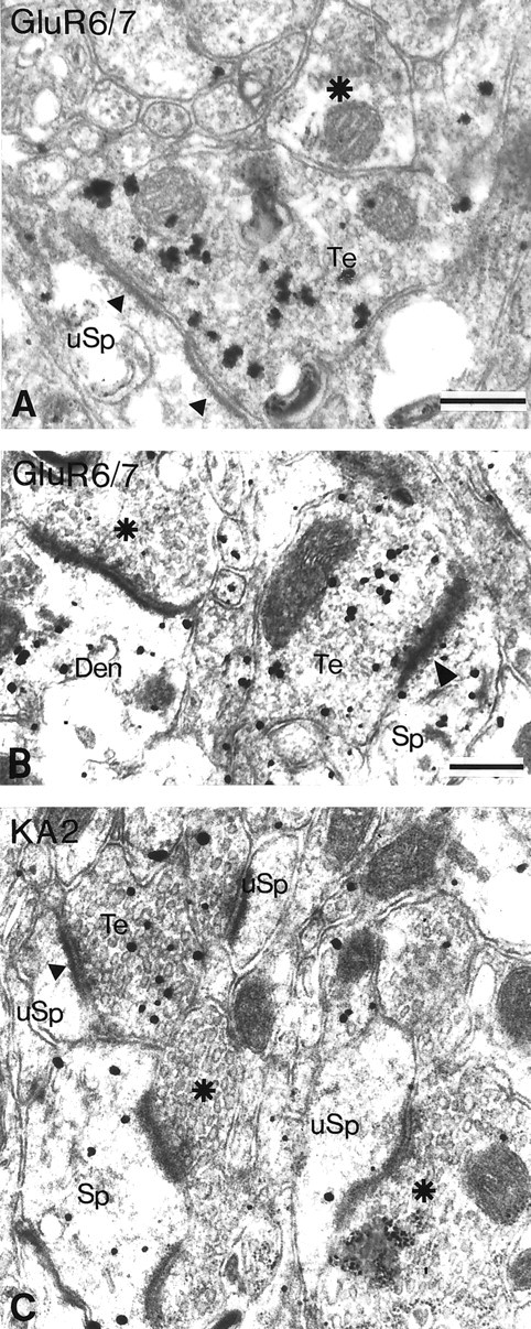
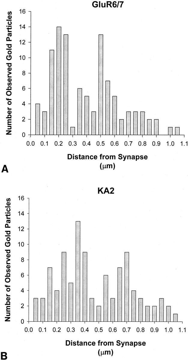
The localization and functions of kainate receptors (KARs) in the CNS are still poorly known. In the striatum, GluR6/7 and KA2 immunoreactivity is expressed presynaptically in a subpopulation of glutamatergic terminals and postsynaptically in dendrites and spines. The goal of this study was to further characterize the subcellular and subsynaptic localization of kainate receptor subunits in the monkey striatum. Immunoperoxidase data reveal that the relative abundance of GluR6/7- and KA2-immunoreactive terminals is homogeneous throughout the striatum irrespective of the differential degree of striatal degeneration in Huntington's disease. Pre-embedding and post-embedding immunogold data indicate that >70% of the presynaptic or postsynaptic GluR6/7 and KA2 labeling is expressed intracellularly. In material stained with the post-embedding immunogold method, approximately one-third of plasma membrane-bound gold particles labeling in axon terminals and spines is associated with asymmetric synapses, thereby representing synaptic kainate receptor subunits. On the other hand, >60% of the plasma-membrane bound labeling is extrasynaptic. Both GluR6/7 and KA2 labeling in glutamatergic terminals often occurs in clusters of gold particles along the membrane of large vesicular organelles located at various distances from the presynaptic grid. Anterograde labeling from the primary motor cortex or the centromedian thalamic nucleus indicate that both corticostriatal and thalamostriatal terminals express presynaptic GluR6/7 and KA2 immunoreactivity in the postcommissural putamen. In conclusion, these data demonstrate that kainate receptors in the striatum display a pattern of subcellular distribution different from other ionotropic glutamate receptor subtypes, but consistent with their metabotropic-like functions recently shown in the hippocampus.


Figures

Fig. 1.
Fig. 1.
Controls for the specificity of GluR6/7 (A, B) and KA2 (C,D) antisera on monkey striatal tissue. InB and D, the antisera were preadsorbed with 10 μg/ml of homologous peptides for 1 hr before incubation. Scale bars: A, 50 μm (valid for B);C, 50 μm (valid for D).
Fig. 2.
Fig. 2.
Western blot analysis demonstrating the specificity of the GluR6/7 antiserum. The antibodies detected a single band that corresponds to the approximate molecular weights predicted for GluR6 and GluR7 subunits (∼118 kDa). Immunoreactivity is completely abolished when antibodies are preadsorbed with the synthetic GluR6/7 peptide 1 hr before immunoblotting. Molecular weight standards are indicated on the left (103 molecular weight).
Fig. 3.
Fig. 3.
Relative distribution of GluR6/7 and KA2 immunoreactivity in different striatal regions. A, Histogram showing the relative proportion of neuronal elements immunoreactive for GluR6/7 in different striatal regions.B, GluR6/7-immunoreactive elements in the body of the caudate nucleus. Note the presence of labeled terminals (Te) forming asymmetric synapses (arrowheads) and immunoreactive dendrites (Den). The asterisks indicate unlabeled boutons. C, Histogram showing the relative distribution of neuronal elements immunoreactive for KA2 in different striatal regions. D, KA2-containing elements in the body of the caudate nucleus. Two immunoreactive terminals (Te), a labeled dendrite (Den), and an unmyelinated axon (Ax) are shown. The asterisks indicate unlabeled boutons. Statistical analysis revealed no significant difference in the relative abundance of GluR6/7 or KA2-containing elements among the different striatal regions (χ2;p < 0.001). Scale bars, 0.5 μm.
Fig. 4.
Fig. 4.
GluR6/7 and KA2 immunoreactivity in neuronal perikarya. A, A GluR6/7-containing neuronal perikaryon in the putamen. B, High magnification of gold particles labeling associated with the endoplasmic reticulum (ER) in this cell body. C, KA2 immunoreactivity in Golgi or ER-like tubular organelles in a neuronal perikaryon (arrows) of an interneuron (invaginated nuclear membrane; open arrow). Nu, Nucleus. Scale bars: A, 1.0 μm; B, 0.25 μm;C, 0.5 μm.
Fig. 5.
Fig. 5.
GluR6/7-immunoreactive (A, B) and KA2-immunoreactive (C) elements in the striatum as revealed with the silver-intensified pre-embedding immunogold method. A, B, GluR6/7-containing axon terminals (Te) forming asymmetric axospinous synapses (arrowheads) in the caudate nucleus. Nonimmunoreactive terminals (*) and unlabeled spines (uSp) are shown in the same region. Labeled dendrites (Den) and spines (Sp) are indicated.C, A KA2-immunoreactive terminal (Te) in the putamen. Nonimmunoreactive boutons (*) and spines (uSp) are shown in the same field. Note that most gold particles in axon terminals, dendrites (Den), and spines are located intracellularly. Scale bars: A, 0.25 μm;B, 0.25 μm (valid for C).
Fig. 6.
Fig. 6.
Post-embedding immunogold localization of GluR6/7 (A–D) and KA2 (E,F) immunoreactivity in the striatum.A, C, GluR6/7 labeling (double arrows) in the active zone of asymmetric axospinous synapses.B, Presynaptic and postsynaptic GluR6/7 labeling at an asymmetric axospinous synapse. D, Presynaptic GluR6/7 immunoreactivity along the surface of a large vesicular structures (arrows) in a terminal forming an asymmetric synapse with a spine. Note that gold particles in the axon terminal are aggregated at the presynaptic grid (arrows), whereas the spine labeling is associated with the postsynaptic density of the asymmetric postsynaptic specialization (arrowhead).E, Dense KA2 labeling (arrows) in the presynaptic grid of an asymmetric axospinous synapse. F,KA2 labeling (arrows) along the surface of a large vesicular organelle in an axon terminal apposed to a spine (Sp). Scale bars: A, 0.25 μm (valid for C, D,F); B, 0.25 μm (valid forE).
Fig. 7.
Fig. 7.
Histograms showing the distribution of gold particle labeling for GluR6/7 (A) and KA2 (B) in relation to the presynaptic grids of asymmetric synapses in the putamen. The mean (±SD) shortest distance of gold particles from the active zones is relatively similar for both KAR subunits antibodies (0.40 ± 0.20 μm for GluR6/7; 0.47 ± 0.25 μm for KA2). Fifteen terminals immunoreactive for GluR6/7 or KA2 were examined.
Fig. 8.
Fig. 8.
BDA injection sites in the primary motor cortex (A) and the centromedian nucleus (B) in squirrel monkeys. CM,Centromedian nucleus; CS, central sulcus;FR, fasciculus retroflexus; PF,parafascicular nucleus.
Fig. 9.
Fig. 9.
GluR6/7-immunoreactive (A,B) and KA2-immunoreactive (C) terminals (Te) labeled anterogradely from the centromedian thalamic nucleus (CM) or the primary motor cortex (M1) in the putamen. Nonimmunoreactive terminals are indicated by asterisks.Arrowheads point to asymmetric synapses. The open arrow indicates an unlabeled bouton that displays GluR6/7 immunoreactivity. Scale bars: A, 0.25 μm (valid forB); C, 0.5 μm.

Similar articles

Cited by

References

    1. Altschul SF, Madden TL, Schaffer AA, Zhang J, Zhang Z, Miller W, Lipman DJ. Gapped BLAST and PSI-BLAST: a new generation of protein database search programs. Nucleic Acids Res. 1997;25:3389–3402. - PMC - PubMed
    1. Anwyl R. Metabotropic glutamate receptors: electrophysiological properties and role in plasticity. Brain Res Rev. 1999;29:83–120. - PubMed
    1. Aronin N, Chase K, Young C, Sapp E, Schwarz C, Matta N, Kornreich R, Landwehrmeyer B, Bird E, Beal MF, Vonsattel JP, Smith T, Carraway R, Boyce FM, Young AB, Penney JB, DiFiglia M. CAG expansion affects the expression of mutant huntingtin in the Huntington's disease brain. Neuron. 1995;15:1193–1201. - PubMed
    1. Baude A, Nusser Z, Roberts JD, Mulvihill E, McIlhinney RAJ, Somogyi P. The metabotropic glutamate receptor (mGluR1a) is concentrated at perisynaptic membrane of neuronal subpopulations as detected by immunogold reaction. Neuron. 1993;11:771–787. - PubMed
    1. Bernard V, Somogyi P, Bolam JP. Cellular, subcellular, and subsynaptic distribution of AMPA-type glutamate receptor subunits in the neostriatum of the rat. J Neurosci. 1997;17:819–833. - PMC - PubMed

Publication types

MeSH terms

LinkOut - more resources