. 2001 Nov 15;21(22):8842-53.
doi: 10.1523/JNEUROSCI.21-22-08842.2001.

Stimulation of endothelin B receptors in astrocytes induces cAMP response element-binding protein phosphorylation and c-fos expression via multiple mitogen-activated protein kinase signaling pathways

Affiliations

Stimulation of endothelin B receptors in astrocytes induces cAMP response element-binding protein phosphorylation and c-fos expression via multiple mitogen-activated protein kinase signaling pathways

S Schinelli et al. J Neurosci. .

Abstract

The vasoconstrictor peptide endothelin (ET-1) exerts its physiological and pathological effects via activation of ET(A) and ET(B) receptor (ET-R) subtypes. In this study, we demonstrate that both ET-R subtypes are highly expressed in rat astrocytes in vivo, indicating that these cells are potential targets of the biological effects of ET-1 in the brain. In cultured cortical astrocytes, both ET-R subtypes are expressed, and selective stimulation of ET(B)-R with ET-1 induces phosphorylation of cAMP response element-binding protein (CREB). The signal transduction pathway activated by ET-1 includes the Rap1/B-Raf and the Ras/Raf-1 complexes, protein kinase C (PKC) together with extracellular signal-regulated kinases (ERK), and the ribosomal S6 kinase (RSK) isoforms RSK2 and RSK3, two kinases that lie immediately downstream of ERK and are able to phosphorylate CREB. Moreover, ET-1 activates the p38 mitogen-activated protein kinase (MAPK)-dependent, but not the c-jun N-terminal kinase (JNK)-dependent pathway. By using selective protein kinase inhibitors and expression of dominant-negative Rap1 protein, we also found that the Rap1/PKC/ERK-dependent pathway induces the phosphorylation of activating transcription factor-1, CREB, and Elk-1, whereas the p38MAPK-dependent pathway only causes CREB phosphorylation. ET-1-induced transcription of the immediate early gene c-fos requires the concomitant activation of both the PKC/ERK- and p38MAPK-dependent pathways, because inhibitors of either pathway block the ET-1-induced increase of c-fos mRNA. Our findings indicate that changes in the expression of cAMP response element-dependent immediate and delayed response genes could play a pivotal role in the physiological effects elicited by ET-1 in astrocytes.


Figures
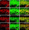
Fig. 1.
Fig. 1.
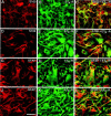
Rat astrocytes express ETA- and ETB-Rs in vivo. In the immature (P8) subcortical white matter (A–F), S100-β+ astrocytes express both ETA- and ETB-Rs. A similar staining pattern was also found in the adult (P30) white matter (G–L). Double immunostaining with antibodies against ETA-R (B, H; green) and ETB-R (E, K;green) and the astrocyte marker S100-β+(A, D, G,J; red) reveals coexpression in the white matter (yellow and orangestaining). C and I are overlays of S100-β+ and ETA-R, whereas Fand L are overlays of S100-β+ and ETB-R. Note that the staining pattern of ETA- and ETB-R in S100-β+ astrocytes of the developing and adult cerebellar white matter was very similar to that shown here for the subcortical white matter (data not shown). Scale bar: A–L, 100 μm; insets, 33 μm.
Fig. 2.
Fig. 2.
Astrocytes express ETA- and ETB-Rs in culture. S100-β+ astrocytes express both ETA- and ETB-Rs (A–F). A similar staining pattern was observed when astrocytes were stained with anti-GFAP antibodies (G–L). Double labeling of cultured astrocytes with antibodies against ETA-R (B,H; green) and ETB-R (E, K; green) and astrocyte markers (red). C andF are overlays of S100-β and ETA-R, and S100-β and ETB-R, respectively. I andL are overlays of GFAP and ETA-R, and GFAP and ETB-R, respectively. All astrocytes coexpress both ETA- and ETB-Rs. Scale bar, 50 μm.
Fig. 3.
Fig. 3.
ETA- and ETB-R proteins are expressed in astrocytes in culture and in cerebral cortex in vivo. Western blot analysis demonstrates that ETA- and ETB-R proteins with the same molecular weight are detected in cultured cells and in vivo. Lane 1, Cultured astrocytes (ASTR). Lane 2, Tissue extract from postnatal day 8 rat cerebral cortex (CX-P8). Lane 3, Brain tissue extracts from postnatal day 30 rat cerebral cortex (CX-P30). Eachlane contained 25 μg of total protein.Arrows indicate the bands corresponding to ETA-R (46 kDa) and ETB-R (39 kDa).
Fig. 4.
Fig. 4.
ET-1 induces CREB phosphorylation via stimulation of ETB-R in astrocytes. A, Time course of ET-1-induced CREB phosphorylation. Cortical astrocytes were stimulated with 50 nm ET-1 for the indicated times, and total cell lysates were analyzed by Western blot using an anti-P-CREB antibody (top panel) or an anti-CREB antibody (bottom panel) to normalize for the total (phosphorylated plus nonphosphorylated) CREB. B, Effects of specific ETA- and ETB-R antagonists on ET-1-induced CREB phosphorylation. Astrocytes were preincubated for 15 min with the ETA-R antagonist BQ123 (A; 500 nm) or with the ETB-R antagonist BQ788 (B; 50 nm) and then stimulated for 10 min with 50 nm ET-1. The samples were then processed for CREB phosphorylation analysis as in A. Data inA and B are representative of three independent experiments.
Fig. 5.
Fig. 5.
ET-1 induces CREB phosphorylation via a PKC/ERK-dependent pathway. A, PKC and MEK inhibitors prevented ET-1-induced CREB phosphorylation. Astrocytes were preincubated for 30 min with the PKC inhibitor Gö6976 (; 5 μm) or with the MEK inhibitor PD98059 (PD; 50 μm) and then stimulated for 10 min with 50 nm ET-1. Total cell lysates were then analyzed by Western blot using an anti-P-CREB antibody.B, PKC inhibition blocks ET-1-induced ERK activation. Astrocytes were preincubated for 30 min with the PKC inhibitor Gö6976 (; 5 μm) and then stimulated for 10 min with 50 nm ET-1. Total cell lysates were then analyzed by Western blot using an anti-P-ERK antibody (top panel) or an anti-ERK antibody (bottom panel) to normalize for the total (phosphorylated plus nonphosphorylated) ERK. C, ET-1 activates the ERK kinase MEK. Astrocytes were stimulated as inA, and total cell lysates were analyzed by Western blot using an anti-P-MEK antibody (top panel) or an anti-MEK antibody (bottom panel) to normalize for the total (phosphorylated plus nonphosphorylated) MEK.D, ET-1 activates the CREB kinases RSK2 and RSK3. Astrocytes were stimulated as in A, and RSK2 and RSK3 were immunoprecipitated from total cell lysates using specific anti-RSK2 and anti-RSK3 antibodies. The amount of P-RSK2 and P-RSK3 in the immunoprecipitates was then analyzed by Western blot using an anti-P-RSK antibody (top panel). The amount of total (phosphorylated plus nonphosphorylated) RSK2 and RSK3 in the immunoprecipitates (bottom panel) was analyzed with the same specific anti-RSK2 or anti-RSK3 antibodies used for the immunoprecipitation. Data in A–D were representative of three independent experiments.
Fig. 6.
Fig. 6.
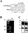
ET-1 activates the Rap1/B-Raf complex in astrocytes. A , ET-1 activates Rap1. Astrocytes were preincubated for 30 min with the PLC inhibitor U-73122 (10 μm) and then stimulated for 10 min with 50 nmET-1. Total cell lysates were processed for the pull-down assay of Rap1-GTP, as described in Materials and Methods. The amount of GTP-Rap1 (top panel) and total Rap1 (bottom panel) in the cell lysates was analyzed by Western blot with an anti-Rap1 antibody. Note that only 5% of the lysates that was used to determine GTP-Rap1 was used to determine the total amount of Rap1 (bottom panel). B, Expression of B-Raf isoforms in cortical astrocytes. RT-PCR of B-Raf transcripts. Total RNA extracted from cortical astrocytes was retrotranscribed to cDNA (RT+ lane). Untreated RNA was used as a negative control (RTlane). cDNAs were then amplified by PCR, as reported in Materials and Methods. PCR products were analyzed on a 3% agarose gel. Size markers (M lane) are indicated in base pairs. C, Expression of B-Raf proteins. Total cell lysates were analyzed by Western blot using an antibody raised against a synthetic peptide corresponding to residues 10–22 of human B-Raf (lane 1) or an antibody raised against a recombinant protein corresponding to amino acid 12–156, mapping at the N terminus of human B-Raf (lane 2). Data in A–C are representative of at least three independent experiments.
Fig. 7.
Fig. 7.
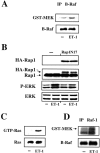
ET-1 activates the B-Raf and Raf-1 kinases.A, ET-1 activates the B-Raf kinase. Astrocytes were stimulated for 10 min with 50 nm ET-1. B-Raf activity was measured by an immunocomplex kinase assay, as described in Materials and Methods. The amount of the phosphorylated substrate GST-MEK was then analyzed by Western blot using an anti-P-MEK antibody (top panel). The amount of immunoprecipitated B-Raf (bottom panel) was analyzed with the same B-Raf antibody used for the immunoprecipitation. B, Rap1 is involved in ET-1-induced ERK activation. Astrocytes were transfected with a vector coding for HA-tagged dominant-negative Rap1 (Rap1N17), as described in Materials and Methods. Control cells (dash) were transfected with pMT2HA empty vector. Cells were then stimulated for 10 min with 50 nmET-1, and total cell lysates were analyzed by Western blot using an anti-HA antibody (top panel), an anti-Rap1 antibody (second panel), an anti P-ERK antibody (third panel), and an anti-ERK antibody (bottom panel) to normalize for the total (phosphorylated plus nonphosphorylated) ERK. C, ET-1 induces Ras activation. Astrocytes were stimulated for 10 min with 50 nm ET-1. Total cell lysates were processed for the pull-down assay of GTP-Ras, as described in Materials and Methods. The amount of GTP-Ras (top panel) and total Ras (bottom panel) in the cell lysates was analyzed by Western blot with an anti-Ras antibody. Note that only 5% of the lysates that was used to determine GTP-Ras was used to determine the total amount of Ras (bottom panel).D, ET-1 activates Raf-1 kinase. Astrocytes were stimulated for 10 min with 50 nm ET-1, and Raf-1 activity was measured by an immunocomplex kinase assay, as described in Materials and Methods. The substrate GST-MEK was separated by SDS-PAGE, and the radioactive phosphorylated band was measured by Phosphorimager (Molecular Dynamics) analysis (top panel). The amount of immunoprecipitated Raf-1 (bottom panel) was analyzed with the same Raf-1 antibody used for the immunoprecipitation (IP).
Fig. 8.
Fig. 8.
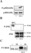
ET-1 induces CREB phosphorylation via the p38MAPK-dependent pathway. A, ET-1 activates p38MAPK. Astrocytes were stimulated for 10 min with 50 nm ET-1, and total cell lysates were analyzed by Western blot using an anti-P-p38MAPK antibody (top panel) or an anti-p38MAPK antibody (bottom panel) to normalize for the total (phosphorylated plus nonphosphorylated) p38MAPK.B, ET-1 does not activate the JNK-dependent pathway. Astrocytes were stimulated as in A, and total cell lysates were analyzed by Western blot using an anti-P-JNK antibody (top panel) or an anti-JNK antibody (bottom panel) to normalize for the total (phosphorylated plus nonphosphorylated) JNK. A whole-cell lysate of anisomycin-treated NIH/3T3 cells was analyzed as a positive control (pc) for the 54 kDa JNK isoform.C, The p38MAPK inhibitor SB203580 (SB) blocks ET-1-induced CREB phosphorylation. Astrocytes were pretreated for 30 min with the p38MAPK inhibitor SB203580 (10 μm) and stimulated as in A. Total cell lysates were analyzed by Western blot using an anti-P-CREB antibody. Data inA–C were representative of at least three independent experiments.
Fig. 9.
Fig. 9.
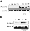
ET-1 induces ATF-1 and Elk-1 phosphorylation in astrocytes. A, ET-1 induces ATF-1 phosphorylation via a PKC/ERK-dependent but not a p38MAPK-dependent pathway. Astrocytes were pretreated for 30 min with the p38MAPK inhibitor SB203580 (SB; 10 μm), the PKC inhibitor Gö6976 (; 5 μm), or the MEK inhibitor PD98059 (PD; 50 μm) and then stimulated for 10 min with 50 nm ET-1. Total cell lysates were analyzed by Western blot using an anti-P-CREB antibody that recognizes also the phosphorylation consensus sequence of ATF-1 (top panel). P-ATF-1 was identified based on its molecular weight. Total ATF-1 (phosphorylated plus nonphosphorylated) was determined with an anti-ATF-1 antibody. B, ET-1 induces Elk-1 phosphorylation via an ERK-dependent pathway. Astrocytes were pretreated for 30 min with the MEK inhibitor PD98059 (PD; 50 μm) and then stimulated for 10 min with 50 nm ET-1. Elk-1 was immunoprecipitated from total cell lysate and then analyzed by Western blot using an anti-P-Elk-1 antibody (top panel) or the same anti-Elk-1 (phosphorylated plus nonphosphorylated) antibody (bottom panel) used for immunoprecipitation. Data inA and B are typical of at least three independent experiments.
Fig. 10.
Fig. 10.
ET-1 induces c-fos transcription via multiple MAPK-dependent pathways. Astrocytes were pretreated for 30 min with the p38MAPK inhibitor SB203580 (SB; 10 μm), the PKC inhibitor Gö6976 (; 5 μm), or the MEK inhibitor PD98059 (PD; 50 μm) and then stimulated for 30 min with 50 nm ET-1. Total RNA was analyzed by Northern blot using a radiolabeled probe for c-fos(top panel) or a radiolabeled probe for 18S ribosomal RNA (bottom panel), and the signals were detected by autoradiography. Data are representative of three independent experiments.
Fig. 11.
Fig. 11.
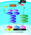
Representative scheme of signaling pathways involved in ET-1-induced/ETB-R-mediated CREB, ATF-1, and Elk-1 phosphorylation and c-fos expression in astrocytes. Question marks indicate unknown links between elements of signaling pathways. DAG, Diacylglycerol; MAPKAP2/3, MAP kinase-activated protein kinase 2/3.

Similar articles

Cited by

References

    1. Barnier JV, Papin C, Eychene A, Lecoq O, Calothy G. The mouse B-raf gene encodes multiple protein isoforms with tissue-specific expression. J Biol Chem. 1995;270:23381–23389. - PubMed
    1. Böhm M, Moellmann G, Cheng E, Alvarez-Franco M, Wagner S, Sassone-Corsi P, Halaban R. Identification of p90RSK as the probable CREB-Ser133 kinase in human melanocytes. Cell Growth Differ. 1995;6:291–302. - PubMed
    1. Bos JL. All in the family? New insight and questions regarding interconnectivity of Ras, Rap 1 and Ral. EMBO J. 1998;17:6776–6782. - PMC - PubMed
    1. Campbell SL, Khosravi-Far R, Rossman KL, Clark GJ, Der CJ. Increasing complexity of Ras signaling. Oncogene. 1998;17:1395–1413. - PubMed
    1. Cazaubon S, Chaverot N, Romero IA, Girault JA, Adamson P, Strosberg AD, Couraud PO. Growth factor activity of endothelin-1 in primary astrocytes mediated by adhesion-dependent and -independent pathway. J Neurosci. 1997;15:6203–6212. - PMC - PubMed

Publication types

MeSH terms