. 2001 Nov 15;21(22):8943-55.
doi: 10.1523/JNEUROSCI.21-22-08943.2001.

Anatomical distribution and cellular basis for high levels of aromatase activity in the brain of teleost fish: aromatase enzyme and mRNA expression identify glia as source

Affiliations

Anatomical distribution and cellular basis for high levels of aromatase activity in the brain of teleost fish: aromatase enzyme and mRNA expression identify glia as source

P M Forlano et al. J Neurosci. .

Abstract

Although teleost fish have higher levels of brain aromatase activity than any other vertebrate group, its function remains speculative, and no study has identified its cellular basis. A previous study determined aromatase activity in a vocal fish, the plainfin midshipman (Porichthys notatus), and found highest levels in the telencephalon and lower levels in the sonic hindbrain, which was dimorphic between and within (males) sexes. We have now localized aromatase-containing cells in the midshipman brain both by immunocytochemistry using teleost-specific aromatase antibodies and by in situ hybridization using midshipman-specific aromatase probes. Aromatase-immuno-reactivity and mRNA hybridization signal are consistent with relative levels of aromatase activity in different brain regions: concentrated in the dimorphic sonic motor nucleus, in a band just beneath the periaqueductal gray in the midbrain, in ventricular regions in the hypothalamus, and highest levels in the telencephalon especially in preoptic and ventricular areas. Surprisingly, double-label immunofluorescence does not show aromatase-immunoreactive colocalization in neurons, but instead in radial glia throughout the brain. This is the first study to identify aromatase expression mostly, if not entirely, in glial cells under normal rather than brain injury-dependent conditions. The abundance of aromatase in teleosts may represent an adaptation linked to continual neurogenesis that is known to occur throughout an individual's lifetime among fishes. The localization of aromatase within the intersexually and intrasexually dimorphic vocal-motor circuit further implies a function in the expression of alternative male reproductive phenotypes and, more generally, the development of natural, individual variation of specific brain nuclei.


Figures

Fig. 1.
Fig. 1.
Dorsal view of a midshipman brain indicating areas of transverse sections in Figure 2, posterior to anterior (A–K), which demonstrate anatomical locations of aromatase immunoreactivity. Cb, Cerebellum;M, midbrain; OB, olfactory bulb;OC, occipital nerve; OL, olfactory nerve;PLLN, posterior lateral line nerve; PN, pacemaker neurons; SMN, sonic motor nucleus;SP, spinal cord; T, telencephalon;VM, ventral medullary neurons; V, trigeminal nerve; VIII, eighth nerve; IX, glossopharyngeal nerve; X, vagus nerve.
Fig. 2.
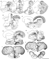
Fig. 2.
Distribution of aromatase immunoreactivity in the brain of P. notatus. Left half of each section is a camera lucida drawing that shows aromatase-IR cell and fiber distribution; right half is a photomicrograph of ICC processed tissue with Nissl counterstain. A,B, Aromatase-IR cells are concentrated dorsally and dorsolaterally within the sonic motor nucleus (SMN) and around the ventral half of the fourth ventricle (IV). C,D, Aromatase-IR cells remain prominent just lateral to the medial longitudinal fasciculus (MLF) in the rostral hindbrain. E, F, Large numbers of aromatase-IR cells line the periventricular areas of the fourth ventricle (IV) and cerebral aqueduct (CA) in the caudal (E) and rostral (F) midbrain, respectively; label is absent in the midbrain tectum (Te) and torus semicircularis (TS).G, H, Throughout the diencephalon, the third ventricle (III) is lined with aromatase-IR cells, which project ventrolaterally. I,J, Aromatase-IR cells (2–4 layers thick) line the entire periphery of the telencephalic hemispheres, as well as the medial ventricular surface; fiber projections course ventromedially. Aromatase-IR cells are prominent in preoptic areas (PPa, anterior parvocellular; PM, magnocellular).K, Label extends well into the olfactory bulbs (OB) in which numerous IR cells line the dorsomedial edge and fiber projections course ventrolaterally. CC, Cerebellar crest; Cg, granule cell layer of the corpus of the cerebellum; Cm, molecular layer of the cerebellum; CP c/d, compact/diffuse division of the central posterior nucleus; DD, dorsal division of the dorsal telencephalon; DL, dorsolateral telencephalon;DM, dorsomedial telencephalon; DP, dorsal posterior nucleus of the thalamus; Hv, ventral periventricular hypothalamus; iaf, internal arcuate fibers; ll, lateral lemniscus; OC, occipital nerve; ot, optic tract; PAG, periaqueductal gray; PCo, posterior commissure;Pe, periventricular cell layer of the torus semicircularis; PGl, lateral division of nucleus preglomerulosus; PGm, medial division of nucleus preglomerulosus; RF, reticular formation;SOv, ventral division of secondary octaval nucleus;Vd, dorsal nucleus of area ventralis;Vde, descending tract of the trigeminal nerve;Vl, vagal lobe; VP, posterior nucleus of area ventralis of the telencephalon; Vs, supracommissural nucleus of the ventral telencephalon;Vv, ventral nucleus of area ventralis;Xm, vagal motor nucleus. Scale bar, 1 mm.
Fig. 3.
Fig. 3.
Aromatase immunoreactivity in the hindbrain and midbrain. A, B, Low-magnification photomicrographs of aromatase immunoreactivity visualized by DAB chromogen corresponding to levels near Figure 2, A andB. C, Photomicrograph of the sonic motor nucleus fluorescently double-labeled with anti-aromatase (green) and neuronal specific anti-Hu (red). Note the prominent aromatase-IR fibers throughout the entire sonic motor nucleus. D, Low-magnification photomicrograph of aromatase immunoreactivity visualized by DAB chromogen at level near Figure 2F.E, Photomicrograph of the periventricular region in the midbrain (also near level 2F) fluorescently double-labeled with anti-aromatase (green) and neuronal-specific anti-Hu (red). Scale bars:A, B, D, 500 μm;C, 240 μm; E, 80 μm.
Fig. 4.
Fig. 4.
Aromatase immunoreactivity in the diencephalon and telencephalon. A, Low-magnification photomicrograph of aromatase immunoreactivity visualized by DAB chromogen at level near Figure 2G. Scale bar, 500 μm. B, Photomicrograph of the diencephalon (also near level G) fluorescently double-labeled with anti-aromatase (green) and neuronal-specific anti-Hu (red). Scale bar, 160 μm. C, Low-magnification photomicrograph of aromatase immunoreactivity visualized by DAB chromogen at level near Figure2I. Scale bar, 500 μm. D, Photomicrograph of dorsal telencephalon (also near level 2I) fluorescently double-labeled with anti-aromatase (green) and neuronal-specific anti-Hu (red). Scale bar, 80 μm.
Fig. 5.
Fig. 5.
Nucleotide and deduced amino acid sequence of aromatase cDNA clone isolated from midshipman brain.Bold amino acids indicate conserved regions of teleost brain aromatase (based on goldfish and Tilapia) to which degenerate primers were made for RT-PCR. Presumed functional domains are underlined and correspond to regions identified in goldfish brain aromatase by Gelinas et al. (1998). I, Helical region (partial); II, Ozols peptide;III, aromatic region; IV, heme binding region (partial).
Fig. 6.
Fig. 6.
Alignment of deduced amino acid sequences of midshipman, P. notatus (Pn), brain aromatase to known teleost brain (br) aromatases [T. mossambica (Tm); O. niloticus (On); C. auratus(Ca); D. rerio (Dr), as well as Tm and On ovarian (ov), zebra finch P. guttata(Pg) (ovarian), and human placental (Hum pl)]. Boxed areas, which includeP. notatus, indicate identical amino acids to midshipman brain aromatase. Bold amino acids indicate conserved regions of teleost brain aromatase (based on goldfish andTilapia) to which degenerate primers were made for RT-PCR. The antibody used in this study before the midshipman sequence was known (see Materials and Methods) corresponds to residues 50–68 and differs from midshipman at positions 50, 52, and 56.
Fig. 7.
Fig. 7.
Dark-field (A–J) and bright-field with Nissl counterstain (K–O) visualization of in situ hybridization throughout the brain of P. notatus using probes from partial cDNA cloning of midshipman brain aromatase. Pattern of signal in all brain regions is consistent with that found by ICC (Fig. 2-4).A, Aromatase mRNA signal clearly defines the sonic motor nucleus (SMN) boundary near level Figure2A; heaviest signal is around the dorsal and lateral periphery. B, Aromatase mRNA just below the fourth ventricle (IV) in the rostral medulla near Figure 2E. C, Hybridization signal in the caudal midbrain in between levels at Figure 2, Eand F. D, Aromatase mRNA expression in the diencephalon near Figure 2G. E, Aromatase mRNA expression in the diencephalon–telencephalon transition area near Figure2H. F, The caudal telencephalon between Figure 2, H and I.G, Strong hybridization in the anterior parvocellular division of the preoptic area (PPa), posterior nucleus of area ventralis of the telencephalon (VP), and ventrolateral area (near Fig. 2I).H, Section just caudal to Figure2J at the level of the anterior commissure (ac), which shows very strong punctate signal within thePPa. I, Anterior telencephalon (rostral to Fig.2J) has high aromatase mRNA signal, especially within ventrolateral (VL) and ventral nucleus of area ventralis (Vv), as well as within the optic tract (ot). J, Aromatase mRNA expression in olfactory bulb (OB, near Fig. 2K). Scale bars: A, B, 200 μm;C–J, 400 μm. K, Section through the hindbrain just caudal to the level at Figures 2Band 3B, which indicates high silver grain concentration in the dorsal rostral SMN; signal within the central SMN is much greater than in the MLF. Scale bar, 100 μm. L, Hybridization signal in the midbrain just below the periaqueductal gray (PAG) near the level at Figure 2F. Scale bar, 100 μm. M, ISH signal in the diencephalon at levels between Figure 2, G and H. Notice the clusters of strong signal along III, which expand into the dorsal posterior nucleus of the thalamus (DP). Scale bar, 100 μm. N, ISH signal in the lateral forebrain between levels at Figure 2, Iand J. The ventral and lateral boundaries between VL and DL telencephalon show a strong signal of hybridization. Scale bar, 250 μm. O, Intense ISH signal in Vv and inPPa between levels at Figure 2,I and J. P, ISH signal in a sagittal section through an ovary. Large, discrete clusters of aromatase mRNA hybridization appear between maturing oocytes (O). Scale bar, 500 μm. Hd, Dorsal periventricular hypothalamus; Hv, ventrolateral nucleus of the hypothalamus; tc, tela choroidea;Vm, molecular layer of the valvula; also see Figure2A–K.
Fig. 8.
Fig. 8.
Distribution of radial glia in the telencephalon.A, DAB visualization of radial glia using monoclonal anti-GFAP (MAB360) near the level of Figure 2J. Note the long fibers that extend throughout the lateral telencephalon and converge in the ventrolateral area, virtually identical to the pattern seen with anti-aromatase (Figs.2I,J, 4C).B, Higher magnification of the ventrolateral area fiber pattern in A (arrows indicate same position). C, High magnification of faintly labeled somata (arrows) and darkly labeled fibers in the dorsal telencephalon using MAB360. D, DAB visualization of radial glia in the lateral telencephalon (near the level of Fig.2I) using monoclonal anti-vimentin. Notice the characteristic pattern of labeled fibers similar to anti-GFAP inA. E, Polyclonal anti-GFAP (G-9269) labels glial cell bodies of the same shape and size as anti-aromatase (compare with Fig. 4D). Scale bar:A, 500 μm; B, 100 μm;C, 40 μm; D, 500 μm;E, 35 μm.
Fig. 9.
Fig. 9.
Colocalization of aromatase and GFAP in the telencephalon of P. notatus by double-label immunofluorescence using anti-aromatase and monoclonal anti-GFAP (MAB360) visualized by secondary fluorescein and Texas Red, respectively. A, Low-magnification photomicrograph that shows aromatase-IR cells (green) along the lateral telencephalic lobe whose projections are double-labeled with anti-GFAP (yellow-orange). Scale bar, 200 μm.B, Single cells with aromatase-IR labeled cell bodies (green) with yellow-orange fiber projections indicating that the same cell is labeled for aromatase and GFAP. Because GFAP immunofluorescence was often more robust in fibers than aromatase, fibers distal to the cell body often appeared morered-orange than yellow when viewed through a double-bandpass filter. Scale bar, 50 μm. C, Abundant colocalization (yellow) of aromatase and GFAP in the anterior parvocellular division of the preoptic area (bottom arrows). Again, fibers from aromatase-IR cells along the midline ventricle are also labeled by MAB360 (top arrow). Scale bar, 200 μm.

Similar articles

Cited by

References

    1. Alvarez-Buylla A, Kirn J. Birth, migration, incorporation, and death of vocal control neurons in adult songbirds. J Neurobiol. 1997;33:585–601. - PubMed
    1. Alvarez-Buylla A, Buskirk DR, Nottebohm F. Monoclonal antibody reveals radial glia in adult avian brain. J Comp Neurol. 1987;264:159–170. - PubMed
    1. Alvarez-Buylla A, Garcia-Verdugo JM, Tramontin AD. A unified hypothesis on the lineage of neural stem cells. Nat Rev Neurosci. 2001;2:287–293. - PubMed
    1. Balthazart J, Ball G. New insights into the regulation and function of brain estrogen synthase (aromatase). Trends Neurosci. 1998;21:243–248. - PubMed
    1. Bass AH. Dimorphic male brains and alternate reproductive tactics in a vocalizing fish. Trends Neurosci. 1992;15:139–145. - PubMed

Publication types

MeSH terms

LinkOut - more resources