. 2001 Nov 15;21(22):9027-35.
doi: 10.1523/JNEUROSCI.21-22-09027.2001.

Cloning, expression, and regulation of a glucocorticoid-induced receptor in rat brain: effect of repetitive amphetamine

Affiliations

Cloning, expression, and regulation of a glucocorticoid-induced receptor in rat brain: effect of repetitive amphetamine

D Wang et al. J Neurosci. .

Abstract

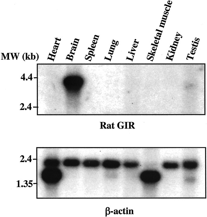
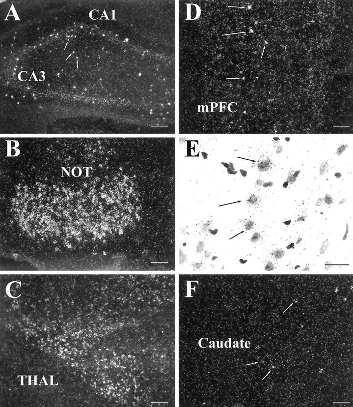
Behavioral sensitization to psychostimulants involves neuroadaptation of stress-responsive systems. We have identified and sequenced a glucocorticoid-induced receptor (GIR) cDNA from rat prefrontal cortex. The full-length GIR cDNA encodes a 422 amino acid protein belonging to G-protein-coupled receptor superfamily. Although the ligand for GIR is still unknown, the dendrogram construction indicates that GIR may belong to peptide receptor subfamily (e.g., substance P receptor), with more distant relationship to subfamilies of glycoprotein hormone receptors (e.g., thyrotropin receptor) and biogenic amine receptors (e.g., dopamine receptor). GIR shares 31-34% amino acid identity to the tachykinin receptors (substance P receptor, neurokinin A receptor, and neurokinin B receptor). GIR mRNA is expressed preferentially in brain, and its neuronal expression is relegated to limbic brain regions, particularly in forebrain. GIR transcript levels are increased significantly and persistently in prefrontal cortex for 7 d after discontinuation of chronic amphetamine exposure. The induction of GIR expression by amphetamine is associated with augmented behavioral activation. These findings suggest that modulation of GIR expression may be involved in behavioral sensitization, and GIR may play a role at the interface between stress and neuroadaptation to psychostimulants.


Figures

Fig. 1.
Fig. 1.
Hydropathy analysis. Plot of hydrophobicity and hydrophilicity of the rat GIR protein. Eight hydrophobic domains, including a putative signal sequence (S) and seven transmembrane spans (I–VII) are predicted.
Fig. 2.
Fig. 2.
Amino acid sequence of rat GIR and its comparison with other receptors. A, The amino acid sequences of rat GIR, mouse GIR, human GIR, rat substance P receptor (NK1R), rat neurokinin B receptor (NK3R), and rat neurokinin A receptor (NK2R) are aligned for best homology. Transmembrane domains (TM) of the rat GIR are predicted and labeled. Signal peptide is predicted andboxed. ▴ indicates potential glycosylation sites, ● indicates potential protein kinase C phosphorylation sites, and ○ indicates potential protein kinase A phosphorylation sites. ▪ indicates Cys residues, * indicates Asp residues, and ∧ indicates His residues. B, Relatedness of GIR to other members of the seven transmembrane receptor family. A dendrogram was constructed by Pileup (GCG program) of the amino acid sequences representing the cloned members of the seven transmembrane family receptors (TSHR, thyrotropin receptor;LHR, lutropin receptor; NY4R, neuropeptide Y4 receptor; NY1R, neuropeptide Y1 receptor; GIR, glucocorticoid-induced receptor;NK1R, substance P receptor; NK3R, neurokinin B receptor; NK2R, neurokinin A receptor;OPRK, Kapp-type opioid receptor; D3R, dopamine D3 receptor; D2R, dopamine D2 receptor;D5R, dopamine D5 receptor; D1R, dopamine D1 receptor; β2AR, adrenergic β2 receptor;β1AR, adrenergic β1 receptor; STE2, yeast α factor receptor).
Fig. 3.
Fig. 3.
Expression of GIR in adult rat tissues by Northern blot analysis. Poly(A)+ RNA (2 μg) from various rat tissues was hybridized with probes specific to rat GIR (top panel) and β-actin (bottom panel) on a nylon membrane. The origin of each RNA is shown at the top, and the molecular mass of standard markers (in kilobases) is shown on the left. GIR expression is detected in brain. No detectable hybridization signal is seen in heart, spleen, lung, liver, skeletal muscle, kidney, and testis. The blot was stripped and rehybridized with a β-actin probe.
Fig. 4.
Fig. 4.
Expression of GIR mRNA in the forebrain of the rat (A–F ). A, GIR mRNA is expressed in scattered neurons throughout the hippocampal formation.B, The highest levels of GIR mRNA are detected in the nucleus of the lateral nucleus of the olfactory tract (NOT). C, Signal is also observed in several diencephalic nuclei, including the anteromedial thalamic nucleus and nucleus reuniens. D, Dense GIR mRNA expression is observed in scattered cells of the cerebral cortex, including the medial prefrontal region. E, High-power photomicrograph of prefrontal cortex GIR hybridization, illustrating confinement of grains to neuronal nuclei (arrows).F, Localization of GIR mRNA in caudate putamen; weak labeling is observed in widely scattered cells throughout the structure. Scale bars: A, B, D, F, 100 μm;C, 200 μm; E, 25 μm.
Fig. 5.
Fig. 5.
Expression of GIR mRNA in caudate nucleus and nucleus accumbens of mouse (A) and rat (B). The same probe was used for panelsA and B. A, Note extensive GIR mRNA in the caudate putamen (Caudate) and nucleus accumbens (NAcc) of the mouse. B, Weak, but positive hybridization signal is observed in the shell of the nucleus accumbens (NAcc). No signal is observed in the caudate putamen (Caudate) at this magnification.
Fig. 6.
Fig. 6.
Effects of amphetamine on rat GIR expression in prefrontal cortex quantitatively determined by competitive PCR analysis and on locomotion activities measured by cumulative numbers of crossovers. A, Representative competitive PCR image.B, GIR mRNA levels calculated from linear regression plot of the ratio plotted logarithmically against initial amount of input competitor DNA. Values are expressed as mean ± SEM (n = 6). SEs are represented by bars. ▨ denotes SAL–AMPH rats. ▪ denotes AMPH–AMPH rats. C,Cumulative numbers of crossovers measured during the 12–60 min and the 60–150 min periods after challenge injection. Values are expressed as mean ± SEM (n = 6). SEs are represented by bars. ▨ denotes SAL–AMPH rats. ▪ denotes AMPH–AMPH rats.D, Bar graph represents the increased levels of GIR mRNA in prefrontal cortex after withdrawal from chronic amphetamine exposure in a separate experiment without amphetamine challenge. Values are expressed as mean ± SEM (n = 6). SEs are represented by bars. ■ denotes SAL rats. ▩ denotes AMPH rats.

References

    1. Antelman SM, Eichler AJ, Black CA, Kocan D. Interchangeability of stress and amphetamine in sensitization. Science. 1980;207:329–331. - PubMed
    1. Bouvier M, Collins S, O'Dowd BF, Campbell PT, de Blasi A, Kobilka BK, MacGregor C, Irons GP, Caron MG, Lefkowitz RJ. Two distinct pathways for cAMP-mediated down-regulation of the beta 2-adrenergic receptor. Phosphorylation of the receptor and regulation of its mRNA level. J Biol Chem. 1989;264:16786–16792. - PubMed
    1. Cha XY, Pierce RC, Kalivas PW, Mackler SA. NAC-1, a rat brain mRNA, is increased in the nucleus accumbens three weeks after chronic cocaine self-administration. J Neurosci. 1997;17:6864–6871. - PMC - PubMed
    1. Casada JH, Dafny N. Restraint and stimulation of the bed nucleus of the stria terminalis produce similar stress-like behaviors. Brain Res Bull. 1991;27:207–212. - PubMed
    1. Clark RB, Friedman J, Dixon RA, Strader CD. Identification of a specific site required for rapid heterologous desensitization of the beta-adrenergic receptor by cAMP-dependent protein kinase. Mol Pharmacol. 1989;36:343–348. - PubMed

Publication types

MeSH terms

Associated data