. 2001 Nov 20;98(24):13832-7.
doi: 10.1073/pnas.231418598. Epub 2001 Nov 6.

Extraordinary variation in a diversified family of immune-type receptor genes

Affiliations

Extraordinary variation in a diversified family of immune-type receptor genes

N A Hawke et al. Proc Natl Acad Sci U S A. .

Abstract

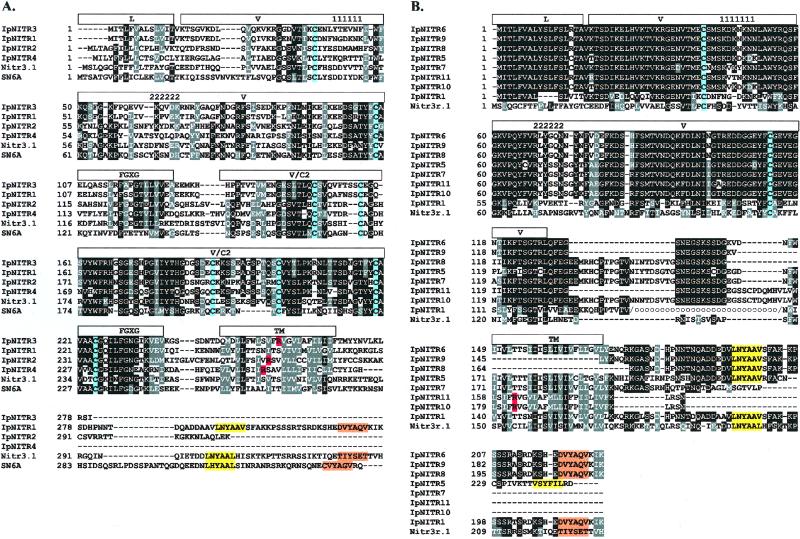
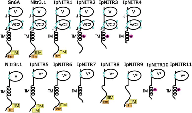
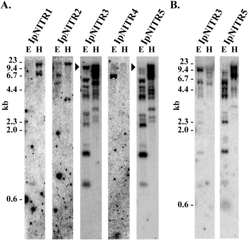
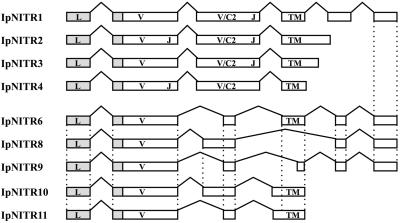
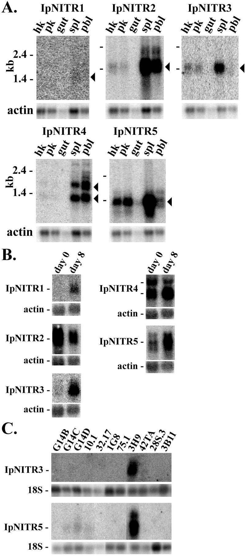
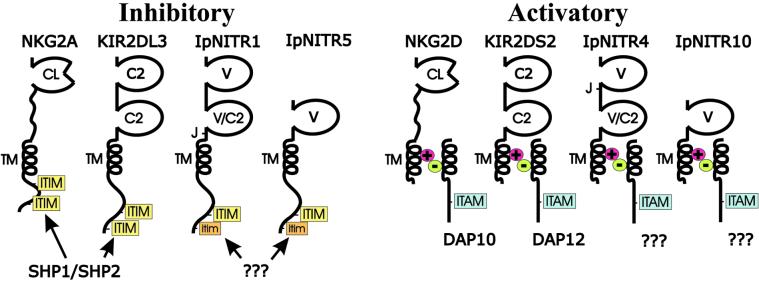
Immune inhibitory receptor genes that encode a variable (V) region, a unique V-like C2 (V/C2) domain, a transmembrane region, and a cytoplasmic tail containing immunoreceptor tyrosine-based inhibition motifs (ITIMs) have been described previously in two lineages of bony fish. In the present study, eleven related genes encoding distinct structural forms have been identified in Ictalurus punctatus (channel catfish), a well characterized immunological model system that represents a third independent bony fish lineage. Each of the different genes encodes an N-terminal V region but differs in the number of extracellular Ig domains, number and location of joining (J) region-like motifs, presence of transmembrane regions, presence of charged residues in transmembrane regions, presence of cytoplasmic tails, and/or distribution of ITIM(s) within the cytoplasmic tails. Variation in the numbers of genomic copies of the different gene types, their patterns of expression, and relative levels of expression in mixed leukocyte cultures (MLC) is reported. V region-containing immune-type genes constitute a far more complex family than recognized originally and include individual members that might function in inhibitory or, potentially activatory manners.


Figures

Figure 1
Figure 1
(A) clustalw alignment of the predicted translation products of IpNITRs 1–4. The prototypic pufferfish (SN6A) and zebrafish (Nitr3.1) are shown for comparison. Identical residues are reverse image (black), functionally equivalent residues are in reverse image (gray). Leader (L), V, V/C2, J (FGXG), transmembrane (TM), CDR1 (111111), and CDR2 (222222) are indicated. Conserved cysteines are shaded with blue. Positively charged residues within the TM are shaded with red. Consensus ITIMs are shaded with yellow and ITIM-related sequences are shaded with orange. (B) clustalw alignment of the predicted translation products of the single Ig domain-containing IpNITRs 5–11. The zebrafish Nitr3r.1 and portions of IpNITR1 in which the V/C2 region has been removed (circles) are shown for comparison; note the high level of identity of the C-terminal regions of IpNITRs 1, 6, 8, and 9. Labeling is as in A.
Figure 2
Figure 2
Diversity in IpNITR structure. V, V/C2, joining (J; FGXG), and transmembrane (TM) regions are indicated. Consensus ITIMs are highlighted in yellow, ITIM-like sequences are highlighted in orange. Conserved cysteines are indicated with a blue oval. Conserved positively charged residues (+) are highlighted in red. The sequences of the Ig domains of IpNITRs 5–11 are related closely and indicated by *. Pufferfish SN6A (2) and zebrafish Nitr3.1 and Nitr3r.1 (3) are shown for comparison.
Figure 3
Figure 3
Genomic complexity of IpNITRs. Southern blot transfers were hybridized in Expresshyb (A) or under conventional conditions (B) with probes complementing the V domains of IpNITRs 1–5 labeled to equivalent specific activity. Arrowheads are used to emphasize certain positions; size standards are indicated. Restriction sites for EcoRI and HindIII within V domains account for the differences in hybridization intensity for individual probes.
Figure 4
Figure 4
Exon organization of IpNITRs. Exons are boxed, intergenic sequence similarity is indicated by vertical dotted lines; leader (L), V, V/C2, joining (J), and transmembrane (TM) regions are indicated. Some components of the genomic organization of IpNITRs 1, 6, 8, and 9 are inferred from highly related genes.
Figure 5
Figure 5
Expression patterns of IpNITRs. (A) RNA blot [≈1 μg/track of poly(A)+ mRNA] of head kidney (hk), posterior kidney (pk), gut, spleen (spl), and peripheral blood leukocytes (pbl). Probes complementing the V region of IpNITRs 1–5 were labeled to equivalent specific activity. Predominant transcripts are indicated with arrowheads. β-actin was used as a loading control; size standards are indicated. (B) RNA blot comparing expression of IpNITRs 1–5 at time 0 and after day 8 of one-way MLC. Total RNA (10 μg) was loaded into each track. Probes, hybridization, and loading controls are as in A. (C) RNA blot comparing expression of IpNITRs 1–5 in Ictalurus cell lines derived from peripheral blood leukocytes. Cell lines used include TCRα/β+ T cells (G14B, G14C, G14D, 32.17, and 28S.3), IgM+IgD B cells (1G8), IgM+IgD+ B cells (3B11), TCRαβ, Ig cytotoxic cells (10.1, 75.1, and 3H9), and macrophages (42TA). Total RNA (2 μg) was loaded into each track. Probes and hybridization are as in A. 18S RNA served as a loading control.
Figure 6
Figure 6
Hypothetical signaling relationships of V domain-containing IpNITRs to LRC gene products. IpNITR1 and IpNITR5 are depicted as immune inhibitory receptors, which use two potential ITIMs and might signal through interactions with SHP1 and/or SHP2 as demonstrated previously for the mammalian receptors NKG2A and KIR2DL3. In contrast, IpNITR4 and IpNITR10 are depicted as immune activatory receptors lacking an ITIM and possessing an intramembranous positively charged residue and might functionally correspond to the mammalian receptors NKG2D and KIR2DS2 (13), which are associated with the negative charge-containing adaptors DAP10 and DAP12, respectively. Abbreviations are as in Fig. 2. C-type lectin domains (CL) and immunoreceptor tyrosine-based activation motifs (ITAM) also are shown.

References

    1. Litman G W, Anderson M K, Rast J P. Annu Rev Immunol. 1999;17:109–147. - PubMed
    1. Strong S J, Mueller M G, Litman R T, Hawke N A, Haire R N, Miracle A L, Rast J P, Amemiya C T, Litman G W. Proc Natl Acad Sci USA. 1999;96:15080–15085. - PMC - PubMed
    1. Yoder J A, Mueller M G, Wei S, Corliss B C, Prather D M, Willis T, Litman R T, Djeu J Y, Litman G W. Proc Natl Acad Sci USA. 2001;98:6771–6776. . (First Published May 29, 2001; 10.1073/pnas.121101598) - PMC - PubMed
    1. Litman G W, Hawke N A, Yoder J A. Immunol Rev. 2001;181:250–259. - PubMed
    1. Barten R, Torkar M, Haude A, Trowsdale J, Wilson M J. Trends Immunol. 2001;22:52–57. - PubMed

Publication types

Substances

LinkOut - more resources