. 2001 Nov 20;98(24):13577-82.
doi: 10.1073/pnas.241397198. Epub 2001 Nov 6.

Bacillus subtilis arsenate reductase is structurally and functionally similar to low molecular weight protein tyrosine phosphatases

Affiliations

Bacillus subtilis arsenate reductase is structurally and functionally similar to low molecular weight protein tyrosine phosphatases

M S Bennett et al. Proc Natl Acad Sci U S A. .

Abstract

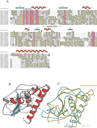
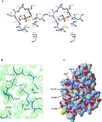
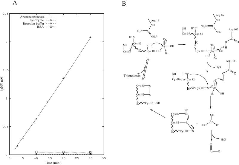
Arsenate is an abundant oxyanion that, because of its ability to mimic the phosphate group, is toxic to cells. Arsenate reductase (EC; encoded by the arsC gene in bacteria) participates to achieve arsenate resistance in both prokaryotes and yeast by reducing arsenate to arsenite; the arsenite is then exported by a specific transporter. The crystal structure of Bacillus subtilis arsenate reductase in the reduced form with a bound sulfate ion in its active site is solved at 1.6-A resolution. Significant structural similarity is seen between arsenate reductase and bovine low molecular weight protein tyrosine phosphatase, despite very low sequence identity. The similarity is especially high between their active sites. It is further confirmed that this structural homology is relevant functionally by showing the phosphatase activity of the arsenate reductase in vitro. Thus, we can understand the arsenate reduction in the light of low molecular weight protein tyrosine phosphatase mechanism and also explain the catalytic roles of essential residues such as Cys-10, Cys-82, Cys-89, Arg-16, and Asp-105. A "triple cysteine redox relay" is proposed for the arsenate reduction mechanism.


Figures

Figure 1
Figure 1
(A) Structure-based multiple sequence alignment by CLUSTAL X (36) with Gram-positive bacterial arsenate reductases, bacterial PTPase homologues, and mammalian LMW PTPases. The protein sequences are obtained from the SWISS-PROT database, and the alignment is drawn by the program ALSCRIPT (37). AB loop refers to the CX5R motif with Cys-10 and Arg-16 as filled and open circles; flexible region is indicated. Filled arrowheads refer to the Cys-82, Cys-89 pair and the open arrowhead to Asp-105. The PTPase homologues are widespread in most bacteria with known genome sequences. However, the functions of these bacterial PTPase homologues are largely unknown. (B) The overall structure of B. subtilis arsenate reductase in the reduced form with the secondary structure elements and N and C termini labeled. Some key residues are also shown. (C) The Cα trace of B. subtilis arsenate reductase (yellow) superimposed with bovine LMW PTPase (BPTP) (blue) with N and C termini labeled. Note the different positions of the sulfate ions (yellow sulfate for B. subtilis arsenate reductase and blue sulfate for BPTP) in these two structures.
Figure 2
Figure 2
(A) Stereoview of the active site with the residues labeled. (B) The AB loop, sulfate ion, and surroundings are superimposed with 3Fobs − 2Fcalc density map calculated from the refined model contoured at 1.0 σ. (C) The space-filling model to show the half-buried active site and the triple cysteine residues, Cys-10, Cys-82, and Cys-89. The structure figures in Figs. 1 and 2 are made by the programs RIBBONS (38) and MOLSCRIPT (39).
Figure 3
Figure 3
(A) The PNPP hydrolysis by B. subtilis arsenate reductase. See Materials and Methods for detailed description of the experiments; cysteine-containing proteins lysozyme and BSA are used as controls. pNP, p-nitrophenolate. (B) The proposed catalytic mechanism for Gram-positive bacterial arsenate reductase. The notation H+S refers to the sulfhydryl group of a cysteine residue that is prone to be deprotonated when placed close to a NHformula image group. The first half of arsenate reduction is analogous to the first step in the PTPase mechanism. Namely, the arsenic atom in H2AsOformula image is subjected to a nucleophilic attack by Cys-10 thiolate formed because of the lowered pKa and helped by Asp-105 in an in-line associative mechanism to form an arsenylated enzyme–substrate (ES) intermediate; a water molecule is the leaving group. Then, this ES intermediate is attacked by the adjacent Cys-82 thiolate ion stabilized by Arg-16. The bound arsenate ion gets reduced to arsenite (first H2AsOformula image, then most likely loses one water molecule quickly to become AsOformula image) by obtaining two electrons from cysteines 10 and 82. Cys-10 and Cys-82 form a transient mixed disulfide bond similar to the mechanism of disulfide reduction involving Cys-32 in E. coli thioredoxin (40). On the other hand, the reduced Cys-89 can come close to the active site because of the flexible region. By the same token, the positive charge of Arg-16 nearby can lower the pKa value of the Cys-89 as well and make it prone to become an activated thiolate. Thus, upon the activation of Cys-89, Cys-82 and Cys-89 can be oxidized to form a disulfide bridge and leave the Cys-10 reduced for the next cycle. The disulfide bond Cys-82–Cys-89 will be reduced by the thioredoxin and thioredoxin reductase system to regenerate the whole system. In E. coli and yeast, arsenate reductases lack the essential cysteine pair; the role of the cysteine pair is proposed to be carried out by glutathione molecules.

References

    1. Silver S, Keach D. Proc Natl Acad Sci USA. 1982;79:6114–6118. - PMC - PubMed
    1. Rensing C, Ghosh M, Rosen B P. J Bacteriol. 1999;181:5891–5897. - PMC - PubMed
    1. Gladysheva T B, Oden K L, Rosen B P. Biochemistry. 1994;33:7288–7293. - PubMed
    1. Ji G, Garber E A, Armes L G, Chen C M, Fuchs J A, Silver S. Biochemistry. 1994;33:7294–7299. - PubMed
    1. Liu J, Gladysheva T B, Lee L, Rosen B P. Biochemistry. 1995;34:13472–13476. - PubMed

Publication types

MeSH terms

Associated data

LinkOut - more resources