. 2002 Feb 1;277(5):3530-6.
doi: 10.1074/jbc.M107031200. Epub 2001 Nov 7.

Expression of Galpha 13 (Q226L) induces P19 stem cells to primitive endoderm via MEKK1, 2, or 4

Affiliations

Expression of Galpha 13 (Q226L) induces P19 stem cells to primitive endoderm via MEKK1, 2, or 4

Hsien-yu Wang et al. J Biol Chem. .

Abstract

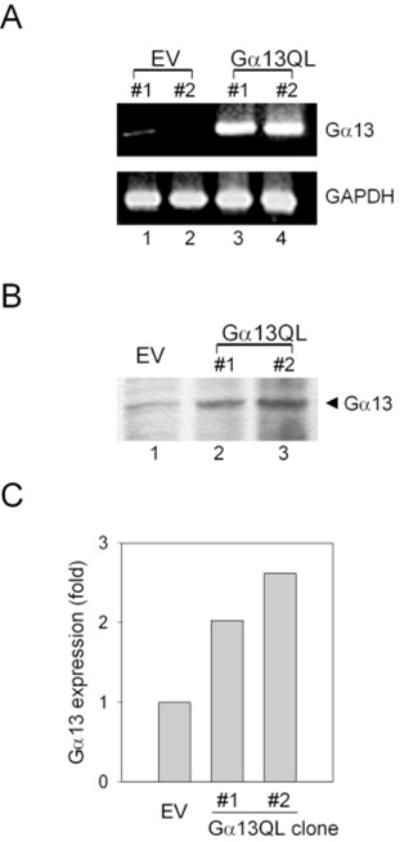
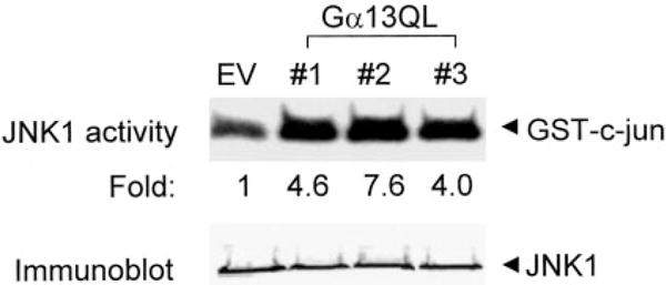
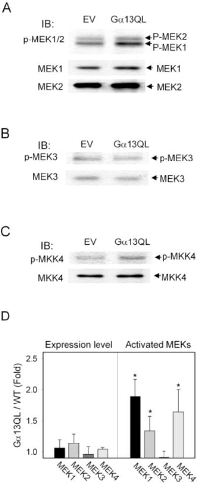
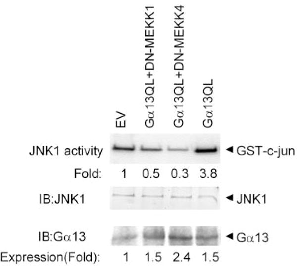
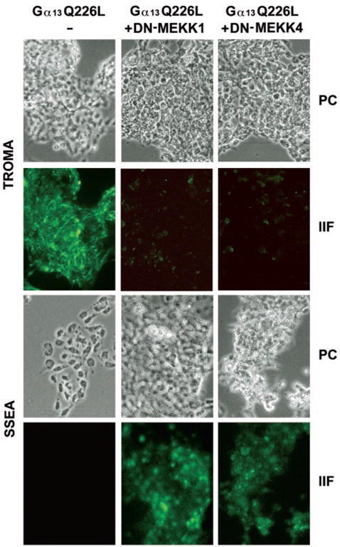
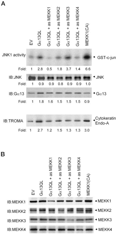
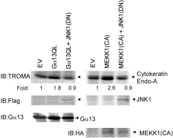
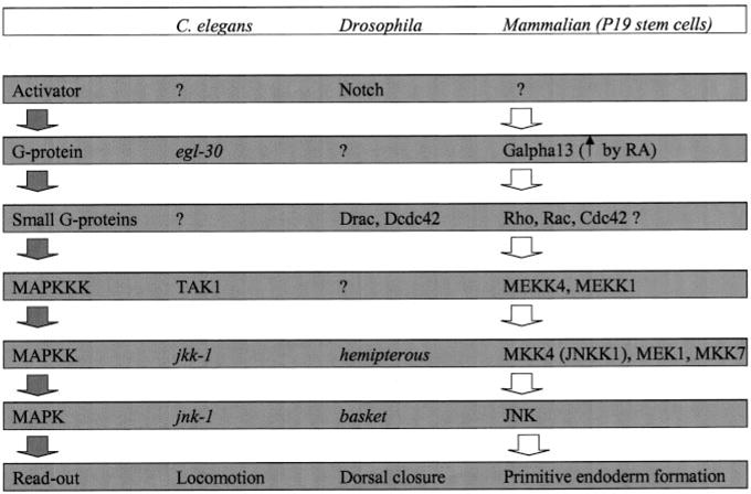
Galpha13 mediates the ability of the morphogen retinoic acid to promote primitive endoderm formation from mouse P19 embryonal carcinoma stem cells, a process that includes the obligate activation of Jun N-terminal kinase. Expression of the constitutively activated (Q226L) GTPase-deficient form of Galpha13 mimics retinoic acid and was used to investigate the signaling upstream of primitive endoderm formation. Jun N-terminal kinase 1 activity, MEK1,2, MKK4, and MEKK1 were constitutively activated in clones stably transfected to express Q226L Galpha13. Dominant negative forms of MEKK1 and MEKK4 were expressed stably in the clones harboring Q226L Galpha13. Expression of dominant negative versions of either MEKK1 or MEKK4 effectively blocks both the activation of Jun N-terminal kinase as well as the formation of primitive endoderm. Depletion of MEKK1, -2, or -4 by antisense oligodeoxynucleotides suppressed signaling from Q226L Galpha13 to JNK1 and primitive endoderm formation. We demonstrate that the signal linkage map from Galpha13 activation to primitive endoderm formation in these stem cells requires activation at three levels of the mitogen-activated protein kinase cascade: MEKK1, -2, or -4 for MAP kinase kinase kinase; MKK4 and/or MEK1 for MAP kinase kinase; and JNK1 for MAP kinase.


Figures

FIG. 1
FIG. 1. Expression of Gα13 mRNAs and immunoreactive Gα13 is increased in P19 embryonal carcinoma clones transfected with the cDNA of Q226L Gα13
A, reverse transcriptase-PCR amplification of total cellular RNA isolated from stable transfectant clones harboring empty vector (pCDNA3, lanes 1 and 2) or pCDNA3 vector harboring the cDNA for the constitutively active mutant form of Gα13Q226L (lanes 3 and 4). Simultaneous amplification was performed with primers specific for GAPDH, demonstrating equivalent loading for lanes 1–4. B, crude membranes were prepared from P19 clones harboring either the empty vector (pCDNA3) or pCDNA3 expression vector harboring the cDNA encoding the constitutively active mutant form of Gα13 (Q226L). An aliquot (200 μg of protein/lane) of crude membranes was separated on a 10% SDS-polyacrylamide gel, transferred to nitrocellulose membranes, and probed with a primary antibody against Gα13. Immunoblots stained for Gα13 were made visible by use of the chemiluminescence reagent. The data shown are representative of at least three independent experiments performed on separate occasions. C, quantification of the amount of expression of Gα13 (wild-type plus Q226L) immunoreactivity in crude membranes of P19 clones.
FIG. 2
FIG. 2. Activation of JNK1 in P19 embryonal carcinoma clones expressing the Q226L mutant version of Gα13
Cell lysates were prepared from cells harboring the empty expression vector or the expression vector harboring the cDNA to Q226L Gα13. JNK1 activity was measured using the solid-phase kinase assay that employs immunoprecipitation of JNK1 with a polyclonal antibody to JNK 1. Bacterially expressed GST-cJun was used as substrate. The phosphorylation reaction products were analyzed on 10% SDS-PAGE. The gel was stained, dried, and subjected to autoradiography. Replicates of the immunoprecipitants used for the solid-phase kinase assay were subjected to immunoblotting and stained with an antibody to JNK1. JNK1 protein was equivalent in all of the clones studied. The data shown are representative of at least three separate experiments.
FIG. 3
FIG. 3. Activation of MEK1,2 and MKK4 in P19 embryonal carcinoma clones expressing the Q226L version of Gα13
Crude whole-cell lysates were prepared from P19 clones harboring the empty vector (pCDNA3) or expression vector containing the cDNA for Q226L Gα13. Crude whole-cell lysates were subjected to electrophoresis and then immunoblotting. The immunoblots were probed with antibodies specific for MEK1 and MEK2 (A), MEK3 (B), and MKK4 (C) to measure protein expression as well as with antibodies specific for phospho-MEK1/2 (phospho-Ser-217 and -221), phospho-MEK3 (phospho-Ser-189 and -207), and MKK4 (phospho-Ser-219 and phospho-Thr-261). The combined results (mean ± S.E.) from three experiments for MEK expression and activation are shown (D). Asterisks denote values statistically significant from the control with p ≤ 0.05.
FIG. 4
FIG. 4. Activation of MEKK1 in P19 embryonal carcinoma clones expressing the constitutively active Q226L mutant form of Gα13QL
Crude cell lysates were prepared from P19 clones harboring empty vector (pCDNA3) or the expression vector containing the cDNA for Q226L Gα13. MEKK1 was immunoprecipitated from the crude whole-cell lysates using a polyclonal antibody to MEKK1. A solid-phase kinase assay was performed using the recombinant MEK 1 protein as substrate. The kinase assay products were separated on a10% SDS-PAGE, stained, dried and exposed to autoradiography. Immunoblots of samples from the whole-cell lysates were prepared and stained with a rabbit polyclonal antibody to MEKK1. The data displayed are representative of three replicate experiments.
FIG. 5
FIG. 5. Expression of dominant negative versions of either DN-MEKK1 or DN-MEKK4 blocks the constitutive activation of JNK1 in clones expressing Q226L Gα13
P19 cells were transfected stably with Q226L Gα13 alone as well as in combination with DN-MEKK1 or with DN-MEKK4. Clones were selected based on the expression of both Q226L Gα13 and dominant negative forms of the MEKKs. Cell lysates were prepared from the three types of clones and subjected to immunoprecipitation using an antibody specific for JNK1. The solid-phase kinase assay was performed using the JNK1 immunoprecipitates in combination with bacterially expressed GST-cJun as the substrate. The phosphorylated products were resolved on a 10% SDS-PAGE gel and subjected to autoradiography. Replicates of the immunoprecipitates were subjected to immunoblotting and stained with an antibody specific for JNK1. The level of JNK1 protein expression was equivalent among the various clones. The results displayed are representative of at least three separate experiments.
FIG. 6
FIG. 6. Expression of dominant negative versions of either dominant negative MEKK1 or MEKK4 blocks the ability of Q226L Gα13 to induce formation of primitive endoderm
Cells were transfected with empty vector (pCDNA3), expression vector harboring Q226L Gα13 alone, or Q226L Gα13 in combination with the dominant negative form of either DN-MEKK1 or DN-MEKK4. Selected clones were grown in chamber slides, and the cells were processed for immunocytochemistry using either a monoclonal antibody to the embryonic marker SSEA-1 antigen or a monoclonal antibody to the cytokeratin endo A (TROMA 1) marker protein for primitive endoderm. Fluorescein isothiocyanate-conjugated secondary antibodies were employed in tandem with indirect epifluorescence to detect the immune complexes. The phase-contrast images (PC) and the indirect immunofluorescence images (IIF) are shown from an experiment representative of four independent experiments.
FIG. 7
FIG. 7. Treatment with oligodeoxynucleotides antisense to MEKK1, 2, or 4 blocks the ability of Q226L Gα13 to activate JNK1 activity
Cells were transiently transfected with empty vector (EV), the expression vector harboring Q226L Gα13 alone, or the empty vector and the Q226L Gα13 vector in combination with the oligodeoxynucleotides antisense to MEKK1–4. Cells were treated for 12 h prior with the antisense ODNs and transiently transfected with the empty vector (pCDNA3) or expression vector harboring Q226L Gα13 for the next 48 h in the presence of the antisense ODNs. The ODNs were replenished every 24 h for the entire 60-h period. At the end of the transient transfection, JNK1 activity, JNK expression, Gα13 immunoreactivity, and PE formation were assayed in the cells (A). The results shown are representative of at least three separate experiments. The amounts of MEKK1, MEKK2, MEKK3, and MEKK4 were determined by immunoblotting for all of the clones treated with antisense ODNs specifically targeting one of the MEKK family (B). A representative experiment is displayed. The suppression of MEKK forms in response to treatment with antisense ODNs under these conditions ranged from 70–85%.
FIG. 8
FIG. 8. Co-expression of dominant negative JNK1 (JNK1(DN)) blocks PE formation stimulated by the expression of either Q226L Gα13 or constitutively active MEKK1 (MEKK1(CA))
Cells were transiently transfected with empty vector (EV), the expression vector harboring Q226L Gα13 alone, or the empty vector and the Q226L Gα13 vector in combination with expression vector harboring FLAG-tagged JNK1(DN). Cells were transiently transfected for 48 h. At the end of the transient transfection, PE formation was assayed by immunoblotting of the TROMA antigen, which is a hallmark of PE. The expression of JNK1(DN) was assayed by immunoblotting with antibodies to the FLAG epitope. MEKK1 and MEKK1(CA) were hemagglutinin antigen-tagged, and expression was determined by immunoblotting. The expression of Gα13 Q226L was determined by immunoblotting with antibodies that stain endogenous and mutant Gα13 proteins alike. The results shown are representative of at least three separate experiments.
FIG. 9
FIG. 9. Q226L Gα13 regulates the MAP kinase regulatory network in promoting the formation of primitive endoderm
A comparison of emerging signaling patterns in C. elegans, Drosophila, and mammalian stem cells is shown. See the “Discussion” section.

Similar articles

Cited by

References

    1. Ip YT, Davis RJ. Curr Opin Cell Biol. 1998;10:205–219. - PubMed
    1. Morris AJ, Malbon CC. Physiol Rev. 1999;79:1373–1430. - PubMed
    1. Gao P, Malbon CC. J Biol Chem. 1996;271:9002–9008. - PubMed
    1. Crespo P, Xu N, Simonds WF, Gutkind JS. Nature. 1994;369:418–420. - PubMed
    1. Noselli S. Trends Gen. 1998;14:33–38. - PubMed

Publication types

MeSH terms

Substances