

Review
doi: 10.1136/bmj.323.7321.1115.
ABC of the upper gastrointestinal tract: Upper gastrointestinal haemorrhage
- PMID: 11701581
- PMCID: PMC1121602
- DOI: 10.1136/bmj.323.7321.1115
Item in Clipboard
Review
ABC of the upper gastrointestinal tract: Upper gastrointestinal haemorrhage
BMJ.
.
No abstract available
Figures

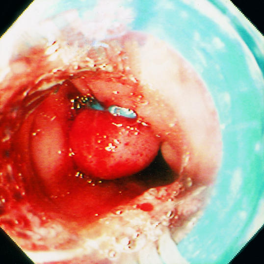
Endoscopic stigmata associated with high risk of further gastrointestinal bleeding. Top left: an active, spurting haemorrhage from a peptic ulcer is associated with an 80% risk of continuing bleeding or rebleeding in shocked patients. Top right: a non-bleeding, visible vessel represents either a pseudoaneurysm of an eroded artery or a closely adherent clot, and 50% of such patients rebleed in hospital. Left: large varices with red spots are also strongly associated with bleeding

Endoscopic stigmata associated with high risk of further gastrointestinal bleeding. Top left: an active, spurting haemorrhage from a peptic ulcer is associated with an 80% risk of continuing bleeding or rebleeding in shocked patients. Top right: a non-bleeding, visible vessel represents either a pseudoaneurysm of an eroded artery or a closely adherent clot, and 50% of such patients rebleed in hospital. Left: large varices with red spots are also strongly associated with bleeding

Endoscopic stigmata associated with high risk of further gastrointestinal bleeding. Top left: an active, spurting haemorrhage from a peptic ulcer is associated with an 80% risk of continuing bleeding or rebleeding in shocked patients. Top right: a non-bleeding, visible vessel represents either a pseudoaneurysm of an eroded artery or a closely adherent clot, and 50% of such patients rebleed in hospital. Left: large varices with red spots are also strongly associated with bleeding

Causes of acute upper gastrointestinal haemorrhage

Gross ascites and distended abdominal veins in advanced cirrhosis

Algorithm for diagnosis and management of upper gastrointestinal bleeding (SRH=stigmata of recent haemorrhage, TIPPS=transjugular intrahepatic portosystemic shunt)

Minnesota tube

Endoscopic treatment of varices. Intravariceal injection of sclerosant (left) and band ligation of oesophageal varices (right)
Endoscopic treatment of varices. Intravariceal injection of sclerosant (left) and band ligation of oesophageal varices (right)
Publication types
MeSH terms
LinkOut - more resources
Full Text Sources
Medical