. 2001 Nov;126(2):250-8.
doi: 10.1046/j.1365-2249.2001.01544.x.

Quantification of mucosal leucocyte endothelial cell interaction by in vivo fluorescence microscopy in experimental colitis in mice

Affiliations

Quantification of mucosal leucocyte endothelial cell interaction by in vivo fluorescence microscopy in experimental colitis in mice

S Farkas et al. Clin Exp Immunol. 2001 Nov.

Abstract

Leucocyte recruitment to sites of intestinal inflammation is a crucial, multi-step process that leads ultimately to the accumulation of cells in the inflamed tissue. We established a new in vivo model system of experimental colitis to quantify leucocyte-endothelial cell interaction and leucocyte extravasation in the inflamed mucosa of the colon. Furthermore, we investigated the pathophysiological role of ICAM-1 in the intestinal microcirculation in vivo. Using the model of dextran sodium sulphate (DSS)-induced acute and chronic colitis in mice, in vivo microscopy was performed in the colonic submucosal postcapillary venules and the submucosal collecting venules in normal or inflamed murine colonic segments. ICAM-1 expression was blocked by an anti-ICAM-1 monoclonal antibody or by suppressing NF-kappaB activation by gliotoxin. Significant increases in leucocyte adhesiveness (51-fold in postcapillary venules, 30-fold in collecting venules, P < 0.01) and extravasation (6.5-fold) could be demonstrated as early as day 2 of DSS-application in acute colitis (P < 0.01). This was paralleled by increases in both the histological damage scores and myeloperoxidase activities. In chronic dextran sodium sulphate-induced colitis significant increases in leucocyte-endothelium interactions and leucocyte extravasation were observed. Blocking ICAM-1 expression with a monoclonal antibody or gliotoxin, leucocyte sticking and extravasation were significantly down-regulated in vivo compared to controls (> 70%; P < 0.01). This new model system offers the possibility to specifically assess the role of adhesion molecules in the colonic mucosa in vivo as well as to investigate and quantify the effectiveness of experimental therapeutic approaches in acute or chronic intestinal inflammation.


Figures

Fig. 1
Fig. 1
In vivo microscopy set-up. The mouse is placed on a heating pad and an intra-arterial catheter for continuous recording of blood pressure (MAP) and an intravenous catheter (i.v.) for injection of fluorescent dye is inserted. All images are recorded by a videocamera (VC) attached to the microscope (M) and connected to a monitor and a video recorder (VCR) for off-line analysis.
Fig. 2
Fig. 2
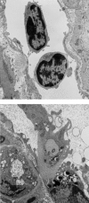
Micrograph of mononuclear cells adhering (a) and penetrating (b) through endothelial cells in a colonic postcapillary venule 4 days after starting DSS application.
Fig. 3
Fig. 3
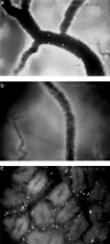
In vivo microscopy of mucosal microcirculation in acute DSS-induced colitis. (a) Sticking leucocytes in the submucosal CV of a control, non-inflamed colonic segment. (b) Sticking leucocytes in the submucosal CV 7 days after first DSS application. (c) Colonic mucosa with visible crypts and extravasated leucocytes 7 days after starting DSS application.
Fig. 4
Fig. 4
Rolling, sticking and leucocyte extravasation in acute DSS-induced colitis. Colitis was induced as described in Table 1. Leucocyte rolling (a) as well as leucocyte sticking (b) is significantly increased on day 2 of DSS-induced colitis in submucosal PV and CV compared to control animals. Stickers are defined as cells adhering to the endothelium longer than 30 s. (c) Leucocyte extravasation is significantly increased as early as day 2 after start of DSS-application and persisted until day 14. *P < 0·01, #P < 0·05 (n = 5/group or n = 5/time-point after start of DSS application). Box plots represent median in solid lines, mean with a broken line. (a and b) □, Collecting venules; ░, postcapillary venules.
Fig. 5
Fig. 5
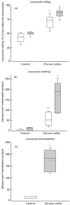
Rolling, sticking and leucocyte extravasation in chronic DSS-induced colitis 4 weeks after the last application of DSS. Leucocyte rolling (a) as well as leucocyte sticking (b) is significantly increased in chronic DSS-induced colitis in submucosal PV and CV compared to control animals. (c) Leucocyte extravasation is still significantly increased 4 weeks after the last cycle of DSS in chronic colitis compared to control animals. *P < 0·01, #P < 0·05 (n = 5/group). Box plots represent median in solid lines, mean with a broken line. (a and b) □, Collecting venules; ░, postcapillary venules; (c) □, control; ░, chronic colitis.
Fig. 6
Fig. 6
Expression of ICAM-1 in colonic mucosa of control animals, animals with DSS-induced colitis treated with vehicle or gliotoxin as described in the Methods section (magnification × 40).
Fig. 7
Fig. 7
Leucocyte sticking and extravasation but not leucocyte rolling is significantly down-regulated after application of MoAb against ICAM-1 compared to animals which were treated with an isotype-matched antibody. Treatment with intraperitoneal administration of anti-ICAM-1 (40 µg/mouse) (Pharmingen) or an isotype-control antibody was performed on day 3 after the start of DSS administration (n = 5/group). Four hours after the antibody application in vivo microscopy was performed. *P < 0·01, #P < 0·05 (n = 5/group). Box plots represent median in solid lines, mean with a broken line. □, Collecting venules; ░, postcapillary venules; (c) isotype control antibody, ░, anti-ICAM antibody.

References

    1. Panes J, Granger DN. Leukocyte–endothelial cell interactions. Molecular mechanisms and implications in gastrointestinal disease. Gastroenterology. 1998;114:1066–90. - PubMed
    1. Vainer B. Role of cell adhesion molecules in inflammatory bowel diseases. Scand J Gastroenterol. 1997;32:401–10. - PubMed
    1. Binion DG, West GA, Volk EE, et al. Acquired increase in leucocyte binding by intestinal microvascular endothelium in inflammatory bowel disease. Lancet. 1998;352:1742–6. - PubMed
    1. Kojouharoff G, Hans W, Obermeier F, et al. Neutralization of tumour necrosis factor (TNF) but not of IL-1 reduces inflammation in chronic dextran sulphate sodium-induced colitis in mice. Clin Exp Immunol. 1997;107:353–8. - PMC - PubMed
    1. Mahler M, Bristol IJ, Leiter EH, et al. Differential susceptibility of inbred mouse strains to dextran sulfate sodium-induced colitis. Am J Physiol. 1998;274:G544–G551. - PubMed

Publication types

MeSH terms