Case Reports
. 2001 Dec;69(6):1395-400.
doi: 10.1086/324648. Epub 2001 Oct 18.

Spinal neurofibromatosis without café-au-lait macules in two families with null mutations of the NF1 gene

Affiliations
Case Reports

Spinal neurofibromatosis without café-au-lait macules in two families with null mutations of the NF1 gene

D Kaufmann et al. Am J Hum Genet. 2001 Dec.

Abstract

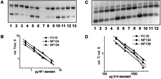
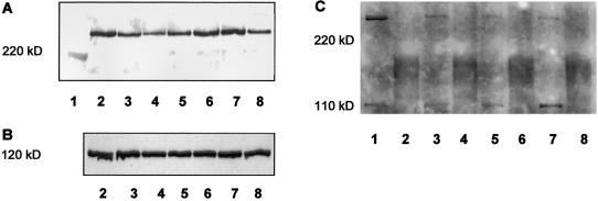
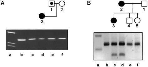
Spinal neurofibromatosis (SNF) is considered to be an alternative form of neurofibromatosis, showing multiple spinal tumors and café-au-lait macules. Involvement of the neurofibromatosis type 1 (NF1) locus has been demonstrated, by linkage analysis, for three families with SNF. In one of them, a cosegregating frameshift mutation in exon 46 of the NF1 gene was identified. In the present study, we report four individuals from two families who carry NF1 null mutations that would be expected to cause NF1. Three patients have multiple spinal tumors and no café-au-lait macules, and the fourth has no clinical signs of NF1. In the first family, a missense mutation (Leu2067Pro) in NF1 exon 33 was found, and, in the second, a splice-site mutation (IVS31-5A-->G) enlarging exon 32 by 4 bp at the 5' end was found. The latter mutation has also been observed in an unrelated patient with classical NF1. Both NF1 mutations cause a reduction in neurofibromin of approximately 50%, with no truncated protein present in the cells. This demonstrates that typical NF1 null mutations can result in a phenotype that is distinct from classical NF1, showing only a small spectrum of the NF1 symptoms, such as multiple spinal tumors, but not completely fitting the current clinical criteria for SNF. We speculate that this phenotype is caused by an unknown modifying gene that compensates for some, but not all, of the effects caused by neurofibromin deficiency.


Figures

Figure  1
Figure 1
Mutations of NF134-3 and of NF138-3, causing no alteration in the amount of NF1 mRNA (A and B) in relation to the expression of S14 (C and D). Fibroblasts from NF134-3, NF138-3, and a healthy donor (FC16) were seeded at a density of 4,000 cells/cm2, were cultured for 2 d, and were total-RNA isolated. For each cell culture, 4× 1 μg RNA was reverse transcribed with random hexamers and different amounts of standard RNAs for NF1 and S14 and then were PCR amplified with primer pair NF1-H/NF1-R and S14-H/S14-R. Measurement of the band intensities of the PCR products was performed as described by Kaufmann et al. (1999), with the following modification: the volume of the heteroduplex band was additionally measured, with half of its amount being added to both the volume of the transcript and the standard band. A, RT-PCR products for measurement of NF1 mRNA amounts, 1 μg of total RNA each: FC16 (lanes 1–4), NF134-3 (lanes 5–8), and NF138-3 (lanes 9–12). The products were separated on a 1.5% agarose gel stained with ethidium bromide. Lower band (345 bp), NF1 standard; middle band, heteroduplexes; upper band (395 bp), NF1 transcript. Amounts of NF1 standard RNA added: lanes 1, 5, and 9, 5 pg; lanes 2, 6, and 10, 2.5 pg; lanes 3, 7, and 11, 1.25 pg; lanes 4, 8, and 12, 0.625 pg. B, Graphical representation with linear regression for each of the NF1 mRNA measurements (vol. = integration of the volume of the band areas; T = transcript; S = standard). C, RT-PCR products for measurement of S14 mRNA amounts, 1 μg of total RNA each: FC16 (lanes 1–4), NF134-3 (lanes 5–8), and NF138-3 (lanes 9–12). The products were separated on a 1.5% agarose gel stained with SYBR-Gold. Lower band (148 bp), S14 standard; middle band (197 bp), S14 transcript; upper band, heteroduplexes. Amounts of S14 standard RNA added: lanes 1, 5, and 9, 2,000 pg; lanes 2, 6, and 10, 1,000 pg; lanes 3, 7, and 11, 500 pg; lanes 4, 8, and 12, 250 pg. D, Graphical representation with linear regression for each of the S14 mRNA measurements. Abbreviations are the same as those listed for figure 1B.
Figure  2
Figure 2
Reduction in the amount of neurofibromin in fibroblasts of NF134-3 and NF138-3 (A and B). No additional shortened neurofibromin band is detectable in either cell culture (C). A, Detection of neurofibromin in lysates, by western blotting of immunoprecipitated neurofibromin. Cells were cultured in parallel, were seeded at a density of 4,000 cells/cm2, were cultured for 3 d, and were lysed as described elsewhere (Griesser et al. 1995). Neurofibromin was immunoprecipitated from equal amounts of total protein (lanes 2–7, 600 μg; lane 8, 300 μg) by incubation with an antibody against the amino-terminal domain of neurofibromin (sc-68; Santa Cruz Biotechnology) for 1.5 h, followed by incubation with protein-A-Sepharose (Amersham Pharmacia Biotech). The samples were boiled in SDS-sample buffer and were separated with SDS-PAGE. After blotting on polyvinylidene fluoride (PVDF) membrane, immunodetection was performed using an antibody against the carboxy-terminal domain of neurofibromin (sc-67; Santa Cruz Biotechnology) and the ECL system. The resulting bands were evaluated using a video-densitometric system that allows neurofibromin quantification (Griesser et al. ; Klose et al. 1998). Lane 1, molecular weight marker, myosin (220 kD); lanes 2 and 6, fibroblasts from a healthy donor (FC7); lane 3, fibroblasts of NF71 carrying a stop mutation (Stark et al. 1992); lane 4, fibroblasts of NF138-3; lane 5, fibroblasts of NF134-3; lane 7, fibroblasts from a healthy donor (FC4); lane 8, fibroblasts from a peripheral nerve of a healthy donor (N21). The densitometric units (100% = median of the values of the four controls) are as follows—FC7: lane 2, 240 (108%), and lane 6, 218 (98%); FC4: 223 (100%); NF71: 126 (57%); NF138-3: 86 (37%); NF134-3: 126 (57%); peripheral nerve cells (300 μg protein): 104 (47%, corresponding to 94% for 600 μg protein). B, Detection of p120GAP by western blotting. Equal amounts of total protein (20 μg) were separated with SDS-PAGE and were blotted on PVDF membrane. Immunodetection was performed by incubation with anti-p120GAP antibody (G12920; Transduction Laboratories) for 2 h, followed by detection with the ECL system. The densitometric units are as follows—FC7: lane 2, 263 and lane 6, 212; FC4: 234; NF71: 268; NF138-3: 204; NF134-3: 202; peripheral nerve cells: 220. C, Investigation of shortened neurofibromin by western blotting of immunoprecipitated neurofibromin, using sc-68. Lane 1, FC7; lane 3, NF134-3; lane 5, NF138-3; lane 7, NF71. To evaluate unspecific signals, sc-68 was preincubated with the corresponding peptide (lanes 2, 4, 6, and 8), and the same detection was performed. 240 kD: neurofibromin; 110 kD: unspecific product. The expected neurofibromin should be detected at ∼225 kD.
Figure  3
Figure 3
NF1 mutations in family NF134 (A) and family NF138 (B). In the pedigrees, blackened symbols denote affected individuals, unblackened symbols denote unaffected indivuals, and unblackened symbols with a blackened circle denote carriers. A, Detection of the T→C mutation in NF1 exon 33 in family NF134. gDNA was amplified using PCR primers ex33mut (5′-TTT TAG CAC GCT ACA TGC TGG TGC) and ex33r (5′-TTT TGG TAA TAT TTC ATG TCA TTA CTG). Primer ex33mut was designed such that an additional NlaIV restriction site is created if the T→C mutation is present. Lanes b and c show the PCR product derived from gDNA of a healthy donor, both uncut (335 bp) and restricted with NlaIV, respectively. Lane d shows PCR products derived from the index patient (NF134-3), lane e from her father (NF134-1), and lane f from her mother (NF134-2), cut with NlaIV. The lower band (313 bp) in lanes d and e demonstrates the presence of the T→C mutation in the index patient and in her father. Lane a shows a size marker. B, Detection of A→G exchange in NF1 intron 31 in family NF138. gDNA PCR products including NF1 exon 32, using the primers ex32h (5′-TGA CAG GCC TGT AAA TAA AAT CTA G) and ex32r (5′-TTT CCA GAA GCC AAA ACT ACA G) are shown, from a healthy control (lane b), the index patient (NF138-3, lane c), the mother (NF138-2, lane d), her brother (NF138-4, lane e), and her sister (NF138-5, lane f), all restricted with AluI; lane a shows a 100-bp ladder (200-bp and 300-bp bands). The upper band corresponds to the wild-type PCR product of 225 bp. The lower band (144 bp) seen in lanes c and d results from the restriction at an additional AluI site created by the mutation in intron 31. The index patient and her mother carry this NF1 mutation.

References

Electronic-Database Information

    1. Online Mendelian Inheritance in Man (OMIM), http://www.ncbi.nlm.nih.gov/Omim/ (for SNF [MIM 162210], NF1 [MIM 162200], and NF2 [MIM 101100])

References

    1. Ars E, Kruyer H, Gaona A, Casquero P, Rosell J, Volpini V, Serra E, Lázaro C, Estivill X (1998) A clinical variant of neurofibromatosis type 1: familial spinal neurofibromatosis with a frameshift mutation in the NF1 gene. Am J Hum Genet 62:834–841 - PMC - PubMed
    1. Eisenbarth I, Beyer K, Krone W, Assum G (2000) Toward a survey of somatic mutation of the NF1 gene in benign neurofibromas of patients with neurofibromatosis type 1. Am J Hum Genet 66:393–401 - PMC - PubMed
    1. Fahsold R, Hoffmeyer S, Mischung C, Gille C, Ehlers C, Kücükceylan N, Abdel-Nour M, Gewies A, Peters H, Kaufmann D, Buske A, Tinschert S, Nürnberg P (2000) Minor lesion mutational spectrum of the entire NF1 gene does not explain its high mutability but points to a functional domain upstream of the GAP-related domain. Am J Hum Genet 66:790–818 - PMC - PubMed
    1. Griesser J, Kaufmann D, Eisenbarth I, Bauerle C, Krone W (1995) Ras-GTP regulation is not altered in cultured melanocytes with reduced levels of neurofibromin derived from patients with neurofibromatosis 1 (NF1). Biol Chem Hoppe Seyler 376:91–101 - PubMed
    1. Griesser J, Kaufmann D, Maier B, Mailhammer R, Kuehl P, Krone W (1997) Post-transcriptional regulation of neurofibromin level in cultured human melanocytes in response to growth factors. J Invest Dermatol 108:275–280 - PubMed

Publication types