. 2001 Dec;69(12):7326-33.
doi: 10.1128/IAI.69.12.7326-7333.2001.

Evidence that mycobacterial PE_PGRS proteins are cell surface constituents that influence interactions with other cells

Affiliations

Evidence that mycobacterial PE_PGRS proteins are cell surface constituents that influence interactions with other cells

M J Brennan et al. Infect Immun. 2001 Dec.

Abstract

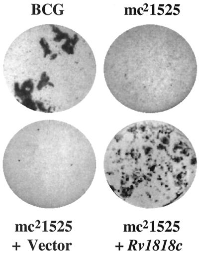
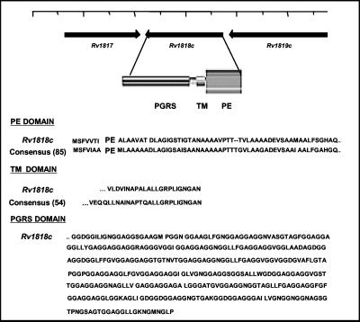
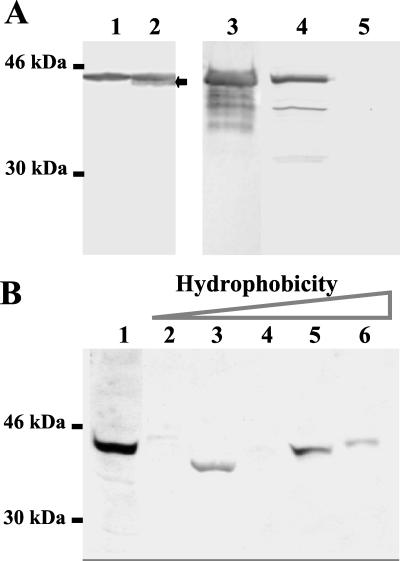
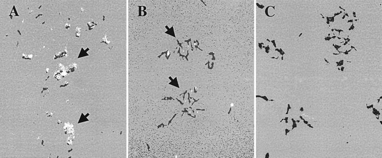
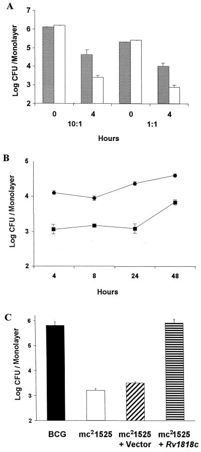
The elucidation of the genomic sequence of Mycobacterium tuberculosis revealed the presence of a novel multigene family designated PE/PE_PGRS that encodes numerous, highly related proteins of unknown function. In this study, we demonstrate that a transposon insertion in a PE_PGRS gene (1818(PE_PGRS)) found in Mycobacterium bovis BCG Pasteur, which is the BCG homologue of the M. tuberculosis H37Rv gene Rv1818c, introduces new phenotypic properties to this BCG strain. These properties include dispersed growth in liquid medium and reduced infection of macrophages. Complementation of the 1818(PE_PGRS)::Tn5367 mutant with the wild-type gene restores both aggregative growth (clumping) in liquid medium and reestablishes infectivity of macrophages to levels equivalent to those for the parent BCG strain. Western blot analysis using antisera raised against the 1818(PE_PGRS) protein shows that PE_PGRS proteins are found in cell lysates of BCG and M. tuberculosis H37Ra and in the cell wall fraction of M. tuberculosis H37Rv. Moreover, immunofluorescent labeling of mycobacteria indicates that certain PE_PGRS proteins are localized at the cell surface of BCG and M. tuberculosis. Together these results suggest that certain PE_PGRS proteins may be found at the surface of mycobacteria and influence both cell surface interactions among mycobacteria as well as the interactions of mycobacteria with macrophages.


Figures

FIG. 1
FIG. 1
Comparison of the phenotypic growth patterns of the BCG Pasteur parent line, the transposon variant mc21525, and mc21525 transformed with the pMV1-18 vector only and with the pMV1-23 vector carrying the Rv1818c gene. Cultures were seeded at a similar cell number into 7H9 broth and incubated at 36°C for 12 days.
FIG. 2
FIG. 2
Properties of the PE_PGRS protein encoded by the Rv1818c gene. The N-terminal domain of the 1818PE_PGRS protein has significant homology with the PE domain consensus sequence derived from 85 members of the PE multigene family; it contains a putative transmembrane domain (TM) found in 54 members of the family, and the PGRS domain extending to the C terminus is composed of 21 GGAGGX repeats. The position of the Rv1818c gene in the genome of the M. tuberculosis strain H37Rv (12) indicating the putative direction of transcription is shown.
FIG. 3
FIG. 3
Immunoblot detecting the presence of PE_PGRS proteins in mycobacterial preparations. (A) A 4 to 20% gradient SDS-polyacrylamide gel containing a cell lysate of mc21525 transformed with the vector only (lane 1), a cell lysate of mc21525 transformed with Rv1818c (lane 2), 3 μg of purified His-tagged 1818PE_PGRS protein (lane 3), 10 μg (protein) of a M. tuberculosis H37Rv cell wall fraction (lane 4), and 10 μg (protein) of a M. tuberculosis H37Rv culture filtrate preparation (lane 5) were transferred to nitrocellulose and probed with antisera directed against 1818PE_PGRS protein. The arrow shows the additional reactive band present in the complemented mc21525 strain. (B) Western blot containing proteins from cell lysates of M. tuberculosis H37Ra and partially purified by DEAE chromatography (lane 1) followed by phenyl-Sepharose chromatography and subsequent elution of hydrophobic material (lanes 2 to 6). Aliquots of the eluted fractions were separated by SDS-PAGE and transferred to nitrocellulose membranes, and blots were incubated with sera obtained from mice immunized with a DNA vaccine encoding 1818PE_PGRS. The position of the molecular mass standards is shown.
FIG. 4
FIG. 4
Localization of PE_PGRS antigens at the cell surface of BCG by immunofluorescence. The BCG parent strain (A and C) and mc21525 (B) were incubated with anti-1818PE_PGRS sera (A and B) or with preimmune mouse sera (C) followed by FITC-conjugated anti-mouse sera. Arrows designate the pronounced surface staining of clusters of BCG in panel A and the reduced punctate staining observed on mc21525 cells in panel B. Fluorescence was visualized using a Nikon Optiphot-2 microscope with a 100× fluorescent objective. Images were taken using a SPOT RT digital camera and were posterized using Adobe Photoshop software. (×1,000).
FIG. 5
FIG. 5
Comparison of the infection of J774 cells or mouse BMMφ by BCG Pasteur, the mc21525 mutant, and mc21525 complemented with the Rv1818c gene. (A) The number of parent BCG (solid bars) and mc21525 (clear bars) in log CFU per monolayer of J774 cells is shown at the time added (0) and following a 4-h incubation period. For the experiments shown, approximately 2.5 × 10⁶ mycobacteria per well were added for an MOI of 10:1 and approximately 2 × 10⁵ mycobacteria per well for an MOI of 1:1. Cell counts showed that there were 2.2 × 10⁵ J774 cells per monolayer for the 10:1 experiment and 2.5 × 10⁵ cells per monolayer for the 1:1 experiment. The results are an average of triplicate wells and show that the number of mc21525 associated with J774 cells is significantly less after 4 h than the parent BCG at P < 0.01, as measured by the t test. Three independent experiments gave results like those shown here. (B) Approximately 2.2 × 10⁵ BMMφ cells per monolayer were incubated for 4 h with about 3 × 10⁵ mycobacteria (BCG = 3.2 × 10⁵ per well and mc21525 = 3.7 × 10⁵ per well), and log CFU per monolayer were determined at 4, 8, 24, and 48 h. The results shown are an average of triplicate wells and show that the number of mc21525 organisms (solid squares) associated with BMMφ cells is significantly less after 4 h and throughout the test period than the number of parent BCG organisms (solid circles) as measured by the t test. (C) Approximately 4 × 10⁵ organisms of BCG Pasteur, the mc21525 mutant, mc21525 transformed with the vector only, and mc21525 transformed with the Rv1818c gene were incubated with 2 × 10⁵ J774 cells per monolayer for 4 h, and the numbers of CFU were determined from triplicate wells. The infectivity of J774 cells by mc21525 alone and mc21525 containing only the vector is similar and significantly reduced, as determined by t test measurements, compared to their infectivity by both BCG and mc21525 complemented with Rv1818c. Similar results were obtained in three separate experiments.

References

    1. Abou-Zeid C, Garbe T, Lathigra R, Wiker H G, Harboe M, Rook G A, Young D B. Genetic and immunological analysis of Mycobacterium tuberculosis fibronectin-binding proteins. Infect Immun. 1991;59:2712–2718. - PMC - PubMed
    1. Abraham S N, Jonsson A B, Normark S. Fimbriae-mediated host-pathogen cross-talk. Curr Opin Microbiol. 1998;1:75–81. - PubMed
    1. Arruda S, Bomfim G, Knights R, Huima-Byron T, Riley L W. Cloning of an M. tuberculosis DNA fragment associated with entry and survival inside cells. Science. 1993;261:1454–1457. - PubMed
    1. Bardarov S, Kriakov J, Carriere C, Yu A, Vaamonde C, McAdam R A, Bloom B R, Hatfull G F, Jacobs W R., Jr Conditionally replicating mycobacteriophages: a system for transposon delivery to Mycobacterium tuberculosis. Proc Natl Acad Sci USA. 1997;94:10961–10966. - PMC - PubMed
    1. Behr M A, Wilson M A, Gill W P, Salamon H, Schoolnik G K, Rane S, Small P M. Comparative genomics of BCG vaccines by whole-genome DNA microarray. Science. 1999;284:1520–1523. - PubMed

LinkOut - more resources