. 2001 Dec;69(12):7341-8.
doi: 10.1128/IAI.69.12.7341-7348.2001.

CD8(+)-T-cell depletion ameliorates circulatory shock in Plasmodium berghei-infected mice

Affiliations

CD8(+)-T-cell depletion ameliorates circulatory shock in Plasmodium berghei-infected mice

W L Chang et al. Infect Immun. 2001 Dec.

Abstract

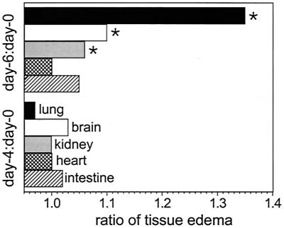
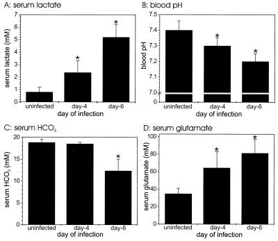
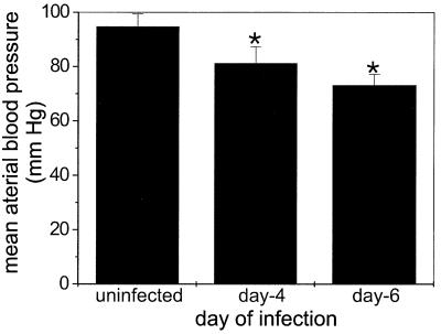
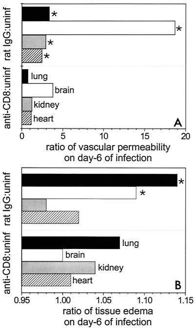
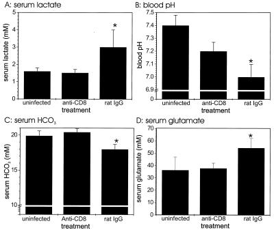
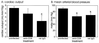
The Plasmodium berghei-infected mouse model is a well-recognized model for human cerebral malaria. Mice infected with P. berghei exhibit (i) metabolic acidosis (pH < 7.3) associated with elevated plasma lactate concentrations, (ii) significant (P < 0.05) vascular leakage in their lungs, hearts, kidneys, and brains, (ii) significantly (P < 0.05) higher cell and serum glutamate concentrations, and (iv) significantly (P < 0.05) lower mean arterial blood pressures. Because these complications are similar to those of septic shock, the simplest interpretation of these findings is that the mice develop shock brought on by the P. berghei infection. To determine whether the immune system and specifically CD8(+) T cells mediate the key features of shock during P. berghei malaria, we depleted CD8(+) T cells by monoclonal antibody (mAb) treatment and assessed the complications of malarial shock. P. berghei-infected mice depleted of CD8(+) T cells by mAb treatment had significantly reduced vascular leakage in their hearts, brains, lungs, and kidneys compared with infected controls treated with rat immunoglobulin G. CD8-depleted mice were significantly (P < 0.05) protected from lactic acidosis, glutamate buildup, and diminished HCO(3)(-) levels. Although the blood pressure decreased in anti-CD8 mAb-treated mice infected with P. berghei, the cardiac output, as assessed by echocardiography, was similar to that of uninfected control mice. Collectively, our results indicate that (i) pathogenesis similar to septic shock occurs during experimental P. berghei malaria, (ii) respiratory distress with lactic acidosis occurs during P. berghei malaria, and (iii) most components of circulatory shock are ameliorated by depletion of CD8(+) T cells.


Figures

FIG. 1
FIG. 1
Tissue edema during P. berghei malaria. The ratios of wet weight to dry weight for selected tissues were determined at zero time and on days 4 and 6 of infection for groups of five mice. The ratios for days 4 and 6 were divided by ratios for zero time. An asterisk indicates statistical significance (P < 0.05) for a comparison of a group of infected mice and uninfected controls.
FIG. 2
FIG. 2
Acidosis of blood during P. berghei malaria. Serum lactate (A), HCO3 (C), and glutamate (D) levels were measured for groups of five mice at zero time and on days 4 and 6 of P. berghei malaria. The blood pH (B) was measured in a separate set of experimental animals. Similar results were obtained in duplicate experiments. In addition, the results for controls used in the experiments whose results are shown in Fig. 5 were similar. An asterisk indicates statistical significance (P < 0.05) for a comparison of a group of infected mice and uninfected controls.
FIG. 3
FIG. 3
Mean arterial blood pressure during P. berghei malaria. Mean arterial blood pressure was measured for groups of five mice at zero time and on days 4 and 6 of P. berghei malaria. Similar results were obtained a duplicate experiment. An asterisk indicates statistical significance (P < 0.05) for a comparison of a group of infected mice and uninfected controls.
FIG. 4
FIG. 4
Ratios of vascular permeability (A) and tissue edema (B) in CD8+ T-cell-depleted mice (anti-CD8) infected with P. berghei malaria to vascular permeability and tissue edema in uninfected controls (uninf). These ratios are compared with the ratios of vascular permeability (A) and tissue edema (B) in rat IgG-treated and infected mice (rat IgG) to vascular permeability and tissue edema in uninfected controls. Vascular permeability was determined by the Evans Blue technique for selected tissues from groups of eight mice. The ratios of wet weight to dry weight for selected tissues were determined on day 6 of infection for groups of five mice. The vascular permeability or ratio of wet weight to dry weight for infected groups of animals (anti-CD8 mAb and rat IgG mAb treated) were divided by values for uninfected animals. Similar results were obtained in replicate experiments for vascular permeability and in duplicate experiments for tissue edema. An asterisk indicates statistical significance (P < 0.05) for a comparison of a group of infected CD8-depleted mice and infected rat IgG-treated controls.
FIG. 5
FIG. 5
Acidosis of blood in CD8+ T-cell-depleted mice infected with P. berghei malaria. Serum lactate (A), HCO3 (C), and glutamate (D) levels were measured in groups of four mice at zero time and on day 6 of P. berghei malaria. The groups of infected mice were treated with either anti-CD8 mAb or control rat IgG. The blood pH (B) was measured with a separate set of experimental animals. Two of five mice in the control rat IgG-injected group died on day 6 of infection before blood was obtained for pH measurement, so the pH values are averages based on the data for three mice. Similar results were obtained in a duplicate experiment. An asterisk indicates statistical significance (P < 0.05) for a comparison of a group of infected mice and uninfected controls.
FIG. 6
FIG. 6
Cardiac output (A) and mean arterial blood pressure (B) in CD8+ T-cell-depleted mice infected with P. berghei. The mean arterial blood pressure was measured for groups of five mice at zero time and on day 6 of P. berghei malaria. The groups of infected mice were treated with either anti-CD8 mAb or control rat IgG. This experiment was performed once. An asterisk indicates statistical significance (P < 0.05) for a comparison of a group of infected mice and uninfected controls.

References

    1. Aikawa M, Iseki M, Barnwell J W, Taylor D, Oo M M, Howard R J. The pathology of human cerebral malaria. Am J Trop Med Hyg. 1990;43:30–37. - PubMed
    1. Brown H C, Chau T T, Mai N T, Day N P, Sinh D X, White N J, Hien T T, Farrar J, Turner G D. Blood-brain barrier function in cerebral malaria and CNS infections in Vietnam. Neurology. 2000;55:104–111. - PubMed
    1. Bruneel F, Gachot B, Timsit J F, Wolff M, Bedos J P, Regnier B, Vachon F. Shock complicating severe falciparum malaria in European adults. Intensive Care Med. 1997;23:698–701. - PubMed
    1. Carter P, Welbourne T. Glutamate transport regulation of renal glutaminase flux in vivo. Am J Physiol. 1997;273:E521–E527. - PubMed
    1. Chang-Ling T, Neill A L, Hunt N H. Early microvascular changes in murine cerebral malaria detected in retinal whole mounts. Am J Pathol. 1992;140:1121–1130. - PMC - PubMed

Publication types