. 2001 Dec;69(12):7387-95.
doi: 10.1128/IAI.69.12.7387-7395.2001.

Bacterial fimbriae and their peptides activate human gingival epithelial cells through Toll-like receptor 2

Affiliations

Bacterial fimbriae and their peptides activate human gingival epithelial cells through Toll-like receptor 2

Y Asai et al. Infect Immun. 2001 Dec.

Abstract

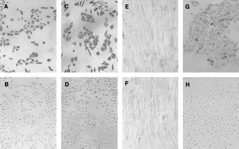
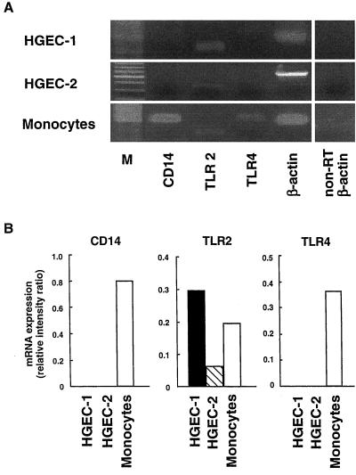
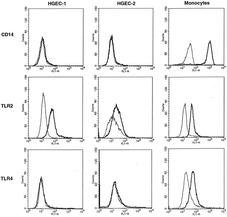
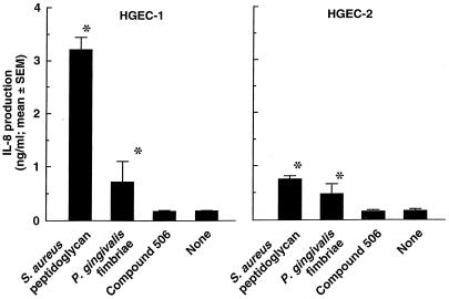
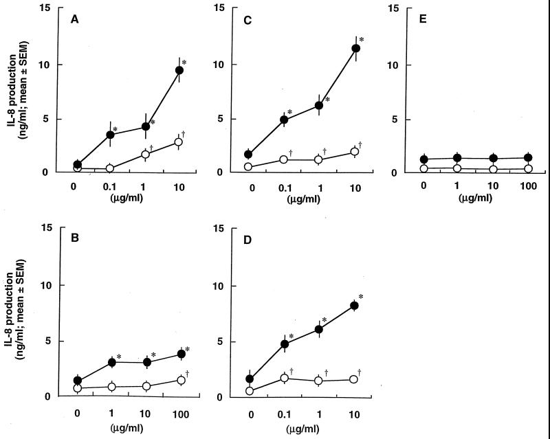
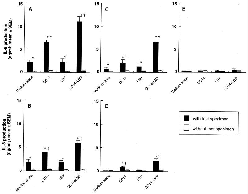
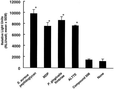
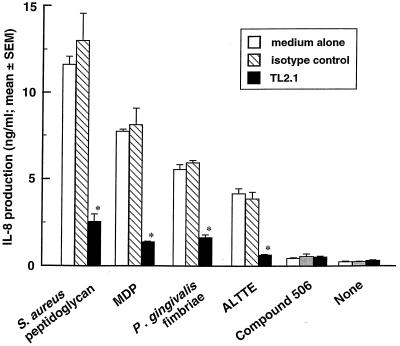
Gingival epithelial cells are a central component of the barrier between oral microflora and internal tissues. Host responses to periodontopathic bacteria and surface components containing fimbriae are thought to be important in the development and progression of periodontal diseases. To elucidate this mechanism, we established immortalized human gingival epithelial cells (HGEC) that were transfected with human papillomavirus. HGEC predominantly expressed Toll-like receptor (TLR) 2, but not TLR4 or CD14. They also induced interleukin-8 (IL-8) production when stimulated with Porphyromonas gingivalis fimbriae and Staphylococcus aureus peptidoglycan, but not Escherichia coli-type synthetic lipid A. Furthermore, an active synthetic peptide composed of residues 69 to 73 (ALTTE) of the fimbrial subunit protein, derived from P. gingivalis and similar to a common component of cell wall peptidoglycans in parasitic bacteria, N-acetylmuramyl-L-alanyl-D-isoglutamine (MDP), significantly induced IL-8 production and NF-kappaB activation in HGEC, and these cytokine-producing activities were augmented by a complex of soluble CD14 and lipopolysaccharide-binding protein (LBP). IL-8 production in HGEC stimulated with these bacterial components was clearly inhibited by mouse monoclonal antibody to human TLR2. These findings suggest that P. gingivalis fimbrial protein and its active peptide are capable of activating HGEC through TLR2.


Figures

FIG. 1
FIG. 1
Immunohistochemical staining with anti-total keratin and anti-involucrin antibodies. The stains were used to determine total keratin-positive and -negative cells, respectively, in HGEC-1 (A, B, G, and H), HeLa (C and D), and Gin-1 (E and F) cells. Cells were stained with mouse monoclonal antibody to human total keratin (AE1/AE3) (A, C, and E) and an isotype control, IgG1κ (B, D, and F). HGEC-1 were incubated with medium containing 2.0 mM (G) or 0.15 mM (H) calcium for 24 h or with mouse monoclonal antibody to human involucrin for involucrin detection.
FIG. 2
FIG. 2
CD14, TLR2, and TLR4 mRNA expression in HGEC. (A) Expression of human CD14, TLR2, and TLR4 mRNA was analyzed by RT-PCR as detailed in Materials and Methods. Human monocytes were used as positive sources of CD14, TLR2, and TLR4 mRNA expression to confirm the specificity of the primers and PCR. The β-actin gene was assayed as a positive control. PCR products of non-RT samples were examined as a negative control. Lane M, Size markers. (B) Ethidium bromide-stained PCR products were photographed, and then the images were digitized and analyzed. Results are expressed as the ratio of each PCR product to β-actin band density. Data represent three independent experiments. PCR was performed in duplicate for each assay.
FIG. 3
FIG. 3
CD14, TLR2, and TLR4 expression in HGEC. Expression of CD14, TLR2, and TLR4 in HGEC was determined by staining with specific antibodies (bold line) or their isotype as a control (thin line) as detailed in Materials and Methods. Human monocytes were used as positive sources of CD14, TLR2, and TLR4.
FIG. 4
FIG. 4
IL-8-producing activities in HGEC by various bacterial components. Cells were stimulated at 37°C for 24 h with 10 μg/ml of each test specimen in HuMedia-KG2 in the absence of FBS. After incubation, the supernatants were collected, and IL-8 production was determined by ELISA. Experiments were done at least three times, and representative results are presented. Each assay was done in triplicate wells, and the data are expressed as the mean ± SEM. *, significant difference between the groups with and without the test specimens (P < 0.01).
FIG. 5
FIG. 5
Effect of FBS on IL-8-producing activities in HGEC stimulated with S. aureus peptidoglycan (A), MDP (B), P. gingivalis fimbriae (C), ALTTE (D), or compound 506 (E). HGEC-1 were incubated at 37°C for 24 h with the indicated doses of the test specimens with (solid circles) or without (open circles) FBS. The experimental protocols were the same as described in the legend to Fig. 4. Significant differences were seen between the groups with and without the test specimens and with (*, P < 0.01) or without (†, P < 0.01) FBS.
FIG. 6
FIG. 6
Effect of soluble CD14 and LBP on IL-8-producing activities in HGEC stimulated with (solid bars) or without (open bars) S. aureus peptidoglycan (A), MDP (B), P. gingivalis fimbriae (C), ALTTE (D), or compound 506 (E). HGEC-1 were incubated at 37°C for 24 h with 1 μg/ml of each test specimen supplemented with CD14 (0.5 μg/ml) and/or LBP (0.05 μg/ml). The experimental protocols are the same as described in the legend to Fig. 4. Significant differences were seen between the groups with and without the bacterial specimens (*, P < 0.01), and between the groups with bacterial specimens with and without CD14 and/or LBP (†, P < 0.01).
FIG. 7
FIG. 7
NF-κB activation in HGEC in response to various bacterial components. HGEC-1 were transfected with 1 μg of pNK-κB-Luc plasmid and then incubated with or without 1 μg/ml of each test specimen with CD14 and LBP in HuMedia-KG2 at 37°C for 7 h. After incubation, the cells were lysed, and then luciferase activity was estimated with a luminometer. The experimental protocols were the same as described in the legend to Fig. 4. Significant differences were seen between the groups with and without the test specimens (*, P < 0.01).
FIG. 8
FIG. 8
Effect of mouse monoclonal antibody to human TLR2 on IL-8-producing activities in HGEC stimulated with various bacterial components. HGEC-1 were incubated with 1 μg/ml of mouse monoclonal antibody to human TLR2, TL2.1, or mouse IgG2a for 30 min at room temperature before the addition of various test specimens (each 1 μg/ml) in the presence of CD14 and LBP. After 24 h of incubation, the supernatants were collected, and IL-8 production was estimated by ELISA. The experimental protocols were the same as described in the legend to Fig. 4. A significant difference was seen from the medium alone (*, P < 0.01).

Similar articles

Cited by

References

    1. Akashi S, Ogata H, Kirikae F, Kirikae T, Kawasaki K, Nishijima M, Shimazu R, Nagai Y, Fukudome K, Kimoto M, Miyake K. Regulatory roles for CD14 and phosphatidylinositol in the signaling via Toll-like receptor 4-MD-2. Biochem Biophys Res Commun. 2000;168:172–177. - PubMed
    1. Aliprantis A O, Yang R B, Mark M R, Suggett S, Devaux B, Radolf J D, Klimpel G R, Godowski P, Zychlinsky A. Cell activation and apoptosis by bacterial lipoproteins through toll-like receptor-2. Science. 1999;285:736–739. - PubMed
    1. Böyum A. Isolation of mononuclear cells and granulocytes from human blood. Scand J Clin Lab Investig. 1968;97:77–89. - PubMed
    1. Brightbill H D, Libraty D H, Krutzik S R, Yang R B, Belisle J T, Bleharski J R, Maitland M, Norgard M V, Plevy S E, Smale S T, Brennan P J, Bloom B R, Godowski P J, Modlin R L. Host defense mechanisms triggered by microbial lipoproteins through toll-like receptors. Science. 1999;285:732–736. - PubMed
    1. Cario E, Rosenberg I M, Brandwein S L, Beck P L, Reinecker H-C, Podolsky D K. Lipopolysaccharide activates distinct signaling pathways in intestinal epithelial cell lines expressing Toll-like receptors. J Immunol. 2000;164:966–972. - PubMed

Publication types

MeSH terms