. 2001 Dec;69(12):7729-35.
doi: 10.1128/IAI.69.12.7729-7735.2001.

Role of complement in Mycobacterium avium pathogenesis: in vivo and in vitro analyses of the host response to infection in the absence of complement component C3

Affiliations

Role of complement in Mycobacterium avium pathogenesis: in vivo and in vitro analyses of the host response to infection in the absence of complement component C3

S S Bohlson et al. Infect Immun. 2001 Dec.

Abstract

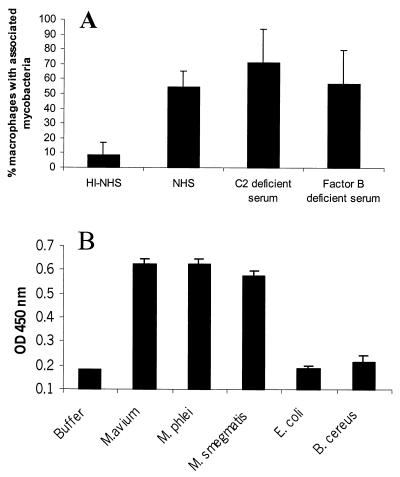
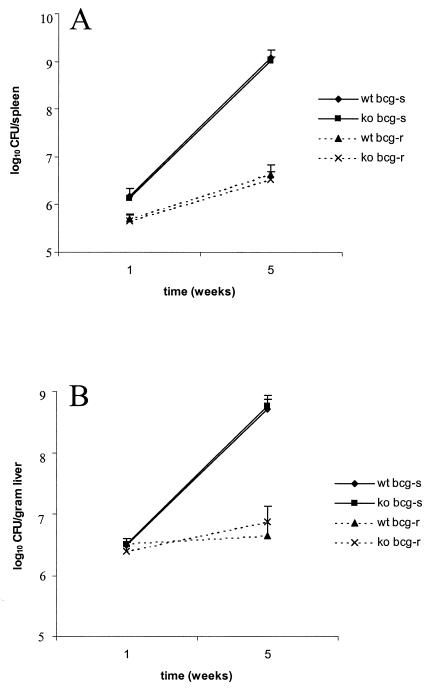
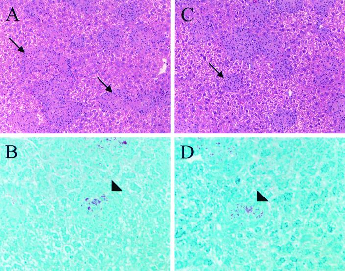
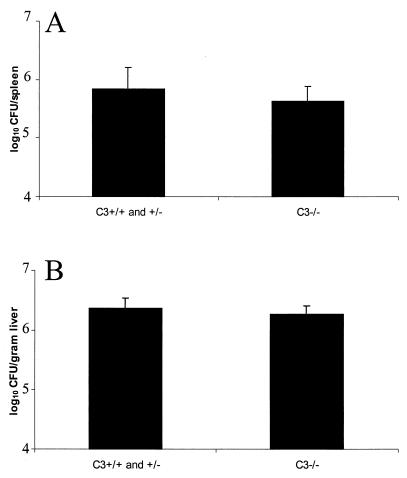
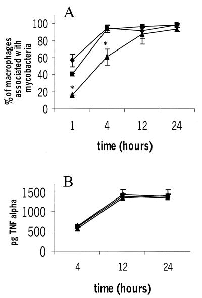
We investigated the importance of the host complement system in the pathogenesis of disease mediated by the intramacrophage pathogen Mycobacterium avium. Mycobacteria opsonized with complement are efficiently ingested by macrophages through various complement receptors. Furthermore, unlike other bacteria, mycobacteria can activate both the alternative and classical complement pathways in the absence of specific antibodies. Therefore, to examine the role of complement in the mycobacterial infection process in vivo, mice deficient in complement component C3 were infected with M. avium. Surprisingly, C3-deficient mice infected intravenously with M. avium displayed no difference in bacterial burden or granulomatous response compared to wild-type control mice. C3-sufficient mice and C3-deficient mice were equally susceptible to infection by M. avium regardless of the genotype at the bcg locus, a locus known to confer susceptibility to infection with intracellular pathogens. In vitro studies using mouse bone marrow-derived macrophages resulted in significant M. avium invasion of macrophages in the absence of C3; however, the kinetics of infection were delayed compared to complement-mediated invasion. The data indicate that complement does not play an essential role in mediating M. avium infections in the mouse and suggest either that other invasion mechanisms can compensate for the absence of complement-mediated entry or that complement is not a major mycobacterial opsonin in vivo.


Figures

FIG. 1
FIG. 1
Mycobacteria activate the classical complement pathway in the absence of specific antibody. M. avium 724 was opsonized with 10% heat-inactivated normal human serum (HI-NHS), normal human serum (NHS), human C2-deficient serum, or human factor B-deficient serum for 2 h at 37°C and then incubated for an additional 2 h at 37°C with J774 cells at a ratio of bacteria to cells of 10:1. The mycobacteria associated with macrophages were visualized by fluorescence microscopy. At least 100 cells were counted for each sample. (A) Representative results of three experiments in which107 bacteria were incubated with purified C1, C2, C3, and C4 for 2 h at 37°C and C3 cleavage was assessed by ELISA for C3a. The solid bars indicate the means and the error bars indicate the standard deviations based on triplicate experimental samples. (B) Optical densities at 450 nm (OD 450 nm) of different preparations.
FIG. 2
FIG. 2
Bacterial loads in spleens (A) and livers (B) of C3-sufficient and C3-deficient mice were similar after 1 or 5 weeks of infection. C3-sufficient and C3-deficient mice with both resistant (bcg-r) and susceptible (bcg-s) backgrounds were retroorbitally infected with 106 M. avium 724 cells, and the mice were maintained for 1 or 5 weeks. The mice were sacrificed, spleens and livers were homogenized, and serial dilutions were plated onto Middlebrook 7H10 agar to determine the numbers of CFU per organ. The data are representative of at least two experiments, and each data point represents the data from four to seven mice; the error bars indicate standard deviations. wt, wild type; ko, C3 knockout.
FIG. 3
FIG. 3
C3 deficiency does not alter granuloma formation in response to M. avium 724 infection. Liver sections from infected mice were processed and stained with hematoxylin and eosin (A and C) and Ziehl-Neelsen (B and D). C3-deficient mice (C and D) contained numbers of granulomas and acid-fast organisms within granulomas similar to the numbers in wild-type controls (A and B). Liver sections from bcg ss mice following 5 weeks of infection with M. avium 724 are shown. Hematoxylin- and eosin-stained sections were visualized by using a magnification of ×200, and Ziehl-Neelsen-stained sections were visualized by using a magnification of ×400. The arrows indicate hematoxylin- and eosin-stained granulomas, and the arrowheads indicate granulomas containing acid-fast bacilli.
FIG. 4
FIG. 4
Presence of C3 does not alter the efficiency of mycobacterial localization to spleens and livers 24 h after infection. C3-sufficient and C3-deficient bcg rr mice were infected with 107 CFU M. avium 724 cells. At 24 h after challenge the mice were sacrificed, and the bacterial loads in the organs were assessed as described in the legend to Fig. 2. The data are representative of three experiments. The solid bars indicate the means and the error bars indicate the standard deviations based on at least three mice.
FIG. 5
FIG. 5
Bone marrow-derived macrophages ingest mycobacteria and produce TNF-α in response to mycobacteria in the absence of C3. Bone marrow-derived macrophages and serum were recovered from C3-sufficient and C3-deficient mice, and macrophages were exposed to serum-opsonized mycobacteria for 1, 4, 12, and 24 h; this was followed by extensive washing and fixation. Mycobacteria associated with macrophages were visualized by fluorescence staining and subsequent microscopy. The data are representative of two experiments. Each data point represents the mean based on triplicate samples, and the error bars indicate standard deviations. Statistical differences were determined for macrophages in the absence of C3 compared to macrophages in the presence of C3. Symbols: ⧫, C3+/+ macrophages and serum; ▪, C3−/− macrophages and C3+/+ serum; ▴, C3−/− macrophages and serum. (A) An asterisk indicates that the P value was <0.05, as determined by an unpaired t test. (B) Supernatants from infected macrophages assayed for the presence of TNF-α by ELISA.

References

    1. Bellamy R. The natural resistance-associated macrophage protein and susceptibility to intracellular pathogens. Microbes Infect. 1999;1:23–27. - PubMed
    1. Bermudez L E, Goodman J, Petrofsky M. Role of complement receptors in uptake of Mycobacterium avium by macrophages in vivo: evidence from studies using CD18-deficient mice. Infect Immun. 1999;67:4912–4916. - PMC - PubMed
    1. Collins F M, Stokes R W. Mycobacterium avium-complex infections in normal and immunodeficient mice. Tubercle. 1987;68:127–136. - PubMed
    1. Cywes C, Hoppe H C, Daffe M, Ehlers M R. Nonopsonic binding of Mycobacterium tuberculosis to complement receptor type 3 is mediated by capsular polysaccharides and is strain dependent. Infect Immun. 1997;65:4258–4266. - PMC - PubMed
    1. Ehlers S, Benini J, Kutsch S, Endres R, Rietschel E T, Pfeffer K. Fatal granuloma necrosis without exacerbated mycobacterial growth in tumor necrosis factor receptor p55 gene-deficient mice intravenously infected with Mycobacterium avium. Infect Immun. 1999;67:3571–3579. - PMC - PubMed

Publication types

MeSH terms