. 2001 Dec;69(12):7820-31.
doi: 10.1128/IAI.69.12.7820-7831.2001.

Dynamic nature of host-pathogen interactions in Mycobacterium marinum granulomas

Affiliations

Dynamic nature of host-pathogen interactions in Mycobacterium marinum granulomas

D M Bouley et al. Infect Immun. 2001 Dec.

Abstract

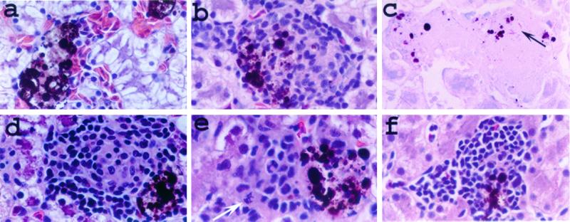
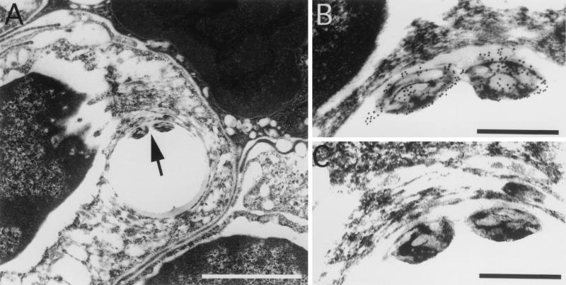
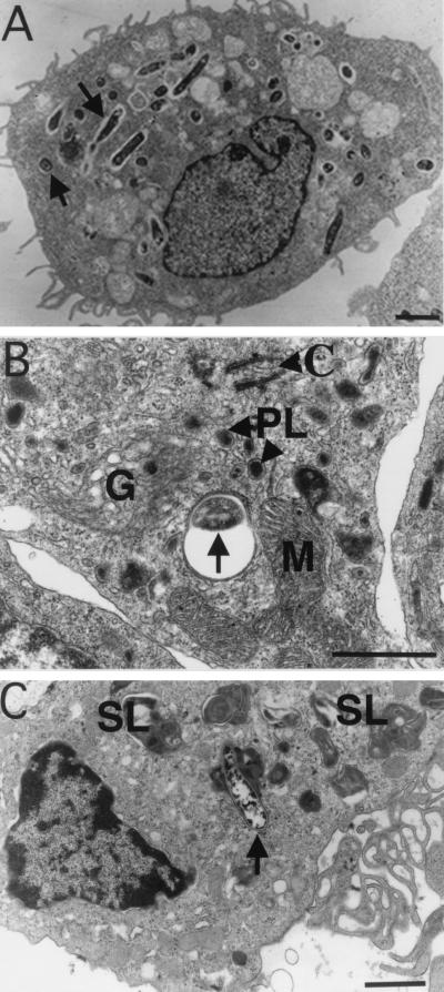
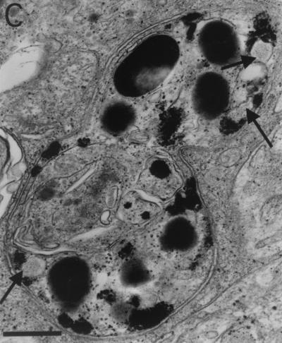
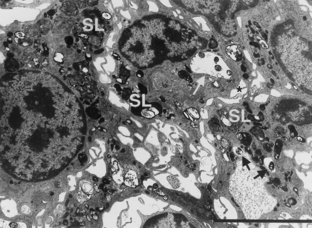
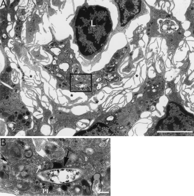
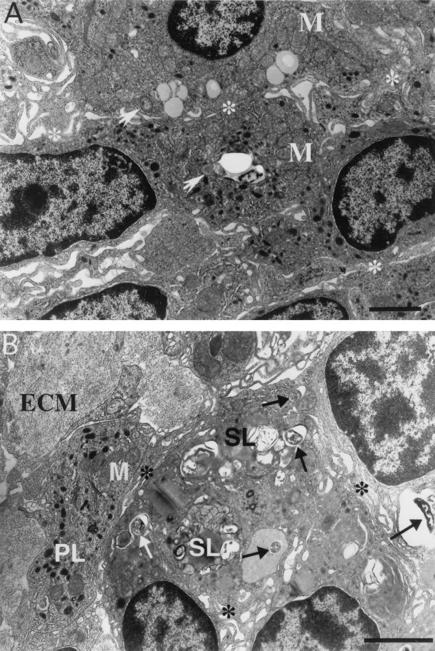
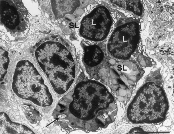
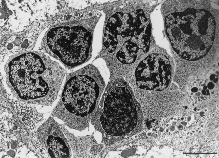
Mycobacterium marinum causes long-term subclinical granulomatous infection in immunocompetent leopard frogs (Rana pipiens). These granulomas, organized collections of activated macrophages, share many morphological features with persistent human tuberculous infection. We examined organs of frogs with chronic M. marinum infection using transmission electron microscopy in conjunction with immunohistochemistry and acid phosphatase cytochemistry to better define the bacterium-host interplay during persistent infection. Bacteria were always found within macrophage phagosomes. These phagosomes were often fused to lysosomes, in sharp contrast to those formed during in vitro infection of J774 macrophage-like cells by M. marinum. The infected macrophages in frog granulomas showed various levels of activation, as evidenced by morphological changes, including epithelioid transformation, recent phagocytic events, phagolysosomal fusion, and disintegration of bacteria. Our results demonstrate that even long-term granulomas are dynamic environments with regard to the level of host cell activation and bacterial turnover and suggest a continuum between constantly replicating bacteria and phagocytic killing that maintains relatively constant bacterial numbers despite an established immune response. Infection with a mutant bacterial strain with a reduced capacity for intracellular replication shifted the balance, leading to a greatly reduced bacterial burden and inflammatory foci that differed from typical granulomas.


Figures

FIG. 1
FIG. 1
Lesions produced in M. marinum-infected frogs. Sections from livers 8 weeks (a to d and f) and 2 weeks (e) postinfection were examined by light microscopy. (a) Uninfected liver section showing normal melanophage aggregates. Such aggregates of brown-black pigment-laden cells were distributed throughout the livers of uninfected frogs and associated with the granulomas of infected frogs (b and d to f). Such aggregates of pigmented cells are normal in the livers of R. pipiens and other species of frogs and are composed of melanin-containing macrophages (melanophages) (7). (b to e) Livers infected with wild-type M. marinum. (b and c) Typical granulomas during infection with M. marinum (arrow). (d) Less typical but not rare granuloma variation in wild-type infected livers. Note the mitotic figure in panel e (white arrow), suggestive of cell proliferation. (f) Typical mononuclear cell aggregate found in frogs infected with mutant strain L1D. All sections were stained with H&E except that in panel c, which was stained with an acid-fast (modified Ziehl-Neelsen) stain. Magnification, ×400.
FIG. 2
FIG. 2
M. marinum is located intracellularly within phagosomes in granulomas. (A) Immunohistochemistry of a 10-month spleen granuloma demonstrates bacteria within a phagosome (arrow). (B) Higher magnification of the M. marinum organisms in panel A showing specific labeling of the bacteria with antibody-conjugated gold particles. (C) Serial section stained without the primary antibody. No particles are seen. The phagosomes were often distorted due to artifactual tearing while exposed to the high beam of the electron microscope. The tearing was specific to regions that contained the bacteria and occurred over a range of infiltration and fixation procedures tested. Bars, 5 μm (A) and 0.5 μm (B and C).
FIG. 3
FIG. 3
M. marinum resides in either phagosomes or phagolysosomes in macrophages comprising granulomas. (A) TEM of M. marinum phagosomes (arrows) in J774 cells 7 days postinfection. The paucity of organelles is characteristic of quiescent or relatively inactive cells. (B and C) Macrophages in granulomas in spleens of frogs infected for 10 and 12 months, respectively. (B) Several organelles, indicative of cellular activation, are present in this macrophage, which contains a phagocytosed bacterium (arrow). (C) Cell exhibiting a high level of activation, including phagolysosomes (arrow), abundant secondary lysosomes, and prominent pseudopodia (ruffled edge). Bars, 2 μm (A and C) and 0.5 μm (B). SL, secondary lysosomes; PL, primary lysosomes; M, mitochondria; G, Golgi; C, centriole.
FIG. 4
FIG. 4
Analysis of phagolysosomal fusion by acid phosphatase cytochemistry. (A) Macrophage in a granuloma from liver 17 weeks postinfection containing bacterial phagosomes not fused to lysosomes (arrow and inset B) and those fused to lysosomes, forming phagolysosomes (inset C). (B) Bacteria (arrows) in phagosomes lacking acid phosphatase activity characterized by black deposits. (C) Granules within phagosomes indicative of phagolysosomal fusion. Arrows, partially degraded bacteria in the phagolysosome. Bars, 2 μm (A) and 0.5 μm (B and C).
FIG. 4
FIG. 4
Analysis of phagolysosomal fusion by acid phosphatase cytochemistry. (A) Macrophage in a granuloma from liver 17 weeks postinfection containing bacterial phagosomes not fused to lysosomes (arrow and inset B) and those fused to lysosomes, forming phagolysosomes (inset C). (B) Bacteria (arrows) in phagosomes lacking acid phosphatase activity characterized by black deposits. (C) Granules within phagosomes indicative of phagolysosomal fusion. Arrows, partially degraded bacteria in the phagolysosome. Bars, 2 μm (A) and 0.5 μm (B and C).
FIG. 5
FIG. 5
Recent phagocytic events ongoing in highly activated macrophages of 17-week liver granulomas. White arrow, four bacteria in a phagosome close to the plasma membrane (asterisk) of the macrophage (considered recently formed). The phagosome has not yet undergone fusion to lysosomes, but primary lysosomes surrounding it suggest that fusion is imminent. Similarly, two bacteria (black arrows) in an adjacent macrophage are within nonfused phagosomes and are surrounded by primary lysosomes. The presence of numerous secondary lysosomes (SL), many fused to bacterial phagosomes, suggests a high level of activation in these resident macrophages found within this granuloma. Bar, 5 μm.
FIG. 6
FIG. 6
Highly activated mature macrophages with loose intercellular interdigitations are present in mature granulomas. (A) One-year granulomas in liver in which the macrophages have loose interdigitations (asterisks), many phagocytosed bacteria, and secondary lysosomes. Lymphocytes (L) can occasionally be seen within granulomas of any duration. (B) Bacterial phagosome (black arrow) that is in the process of fusing with primary lysosomes (PL). Other markers of cellular activation, including secondary lysosomes, rough endoplasmic reticulum (asterisk), smooth endoplasmic reticulum (white arrow), free ribosomes (arrowhead), and intermediate filaments (F), are seen. Bars, 5 μm (A) and 0.5 μm (B).
FIG. 7
FIG. 7
Two types of epithelioid cell can be found in epithelioid granulomas. (A) Ten-month spleen granuloma made up of cells with closely apposed and tightly interdigitated pseudopodia (asterisks). These epithelioid cells are moderately active, contain primary but not secondary lysosomes, and demonstrate a relatively low level of phagocytosis. Two phagosomes (arrows) in the field are juxtaposed to primary lysosomes, indicating fusion initiation. (B) Ten-month liver granuloma also comprised of confluent cells with tightly interdigitated pseudopodia (asterisks) but containing numerous bacterial phagosomes (arrows) and secondary lysosomes (SL). Extracellular matrix (ECM) proteins, often associated with chronic granulomas, are present. PL, primary lysosomes; M, mitochondria; Bars, 5 μm.
FIG. 8
FIG. 8
Both viable and nonviable intracellular bacteria are found in granulomas. This 10-month liver granuloma is made up of a mixture of cell types, including lymphocytes (L), moderately activated macrophages, and highly activated macrophages containing numerous secondary lysosomes (SL). Black arrow, intact, presumably viable bacterium; white arrow, partially disintegrated bacterium in a large secondary lysosome. Bar, 5 μm.
FIG. 9
FIG. 9
Loose aggregates of monocytes/immature macrophages in an 8-week granuloma from a frog infected with the L1D mutant strain. Note the absence of ruffled cell membranes and interdigitations and a greater nuclear-to-cytoplasmic ratio in these cells compared to the activated macrophages in previous figures. Bar, 5 μm.

References

    1. Adams D O. The granulomatous inflammatory response. A review. Am J Pathol. 1976;84:164–191. - PMC - PubMed
    1. Adams D O. The structure of mononuclear phagocytes differentiating in vivo. I. Sequential fine and histologic studies of the effect of Bacillus Calmette-Guerin (BCG) Am J Pathol. 1974;76:17–48. - PMC - PubMed
    1. Adams D O. The structure of mononuclear phagocytes differentiating in vivo. II. The effect of Mycobacterium tuberculosis. Am J Pathol. 1975;80:101–116. - PMC - PubMed
    1. Ando M, Dannenberg A M., Jr Macrophage accumulation, division, maturation, and digestive and microbicidal capacities in tuberculous lesions. IV. Macrophage turnover, lysosomal enzymes, and division in healing lesions. Lab Investig. 1972;27:466–472. - PubMed
    1. Ando M, Dannenberg A M, Jr, Shima K. Macrophage accumulation, division, maturation and digestive and microbicidal capacities in tuberculous lesions. II. Rate at which mononuclear cells enter and divide in primary BCG lesions and those of reinfection. J Immunol. 1972;109:8–19. - PubMed

Publication types

MeSH terms