. 2001 Dec;69(12):7851-7.
doi: 10.1128/IAI.69.12.7851-7857.2001.

Evaluation of a tetracycline-inducible promoter in Staphylococcus aureus in vitro and in vivo and its application in demonstrating the role of sigB in microcolony formation

Affiliations

Evaluation of a tetracycline-inducible promoter in Staphylococcus aureus in vitro and in vivo and its application in demonstrating the role of sigB in microcolony formation

B T Bateman et al. Infect Immun. 2001 Dec.

Abstract

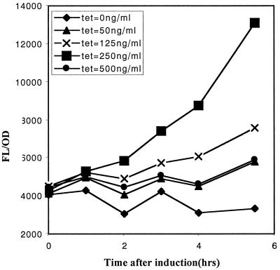
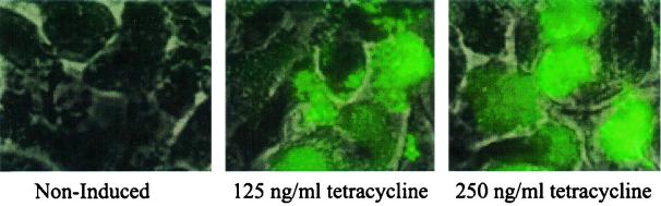
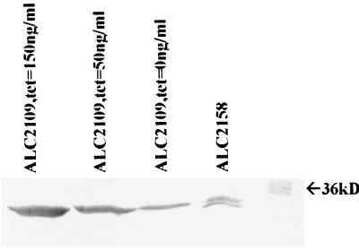
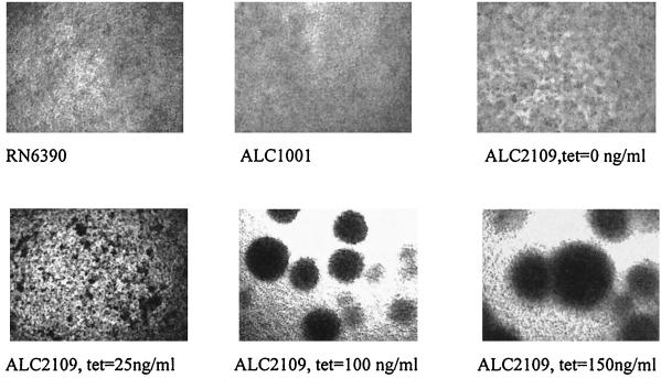
An inducible promoter system provides a powerful tool for studying the genetic basis for virulence. A variety of inducible systems have been used in other organisms, including pXyl-xylR-inducible promoter, the pSpac-lacI system, and the arabinose-inducible P(BAD) promoter, but each of these systems has limitations in its application to Staphylococcus aureus. In this study, we demonstrated the efficacy of a tetracycline-inducible promoter system in inducing gene expression in S. aureus in vitro and inside epithelial cells as well as in an animal model of infection. Using the xyl/tetO promoter::gfp(uvr) fusion carried on a shuttle plasmid, we demonstrated that dose-dependent tetracycline induction, as measured by bacterial fluorescence, occurred in each of the above environments while basal activation under noninduced conditions remained low. To ascertain how the system can be used to elucidate the genetic basis of a pathogenic phenotype, we cloned the sigB gene downstream of the inducible promoter. Induction of SigB expression led to dose-dependent attachment of the tested strain to polystyrene microtiter wells. Additionally, bacterial microcolony formation, an event preceding mature biofilm formation, also increased with tetracycline induction of SigB.


Figures

FIG. 1
FIG. 1
Induction of the xyl/tetO promoter linked to the gfpuvr reporter with increasing concentrations of tetracycline in strain ALC2085. Following overnight culture, the cells were diluted 1:100 in fresh TSB and then grown to an OD650 of 0.5. Tetracycline was then added to the final concentrations shown above. The cells were sampled hourly following induction (100 μl each, in triplicate). Fluorescence and OD650 measurements were obtained using a multipurpose spectrophotometer (FL600; Biotek Instrument). Results are presented as reported fluorescence (FL) units/OD650 to account for variations in fluorescence due to cell density. Data are given as the mean of the triplicate measurements. Error bars are too small to be shown. The experiment was repeated twice, with one representative experiment shown.
FIG. 2
FIG. 2
Tetracycline-induced GFP expression of ALC2085 inside CFT-1 epithelial cells. ALC2085, grown to an OD650 of 0.6, was added to CFT-1 monolayers at an MOI of 10:1 for 2 h. Extracellular bacteria were removed by washing followed by the addition of gentamicin (200 μg/ml) for 4 h to kill any remaining extracellular microorganisms. Tetracycline was added to some wells to facilitate induction. Cells were then examined by fluorescence microscopy. Most of the bacteria were intracellular, as evidenced by the failure of these bacteria to stain with anti-protein A antibody conjugated to Texas Red (data not shown). The experiment was repeated twice, with one representative experiment shown. Upon tetracycline induction of the xyl/tetO promoter of intracellular bacteria driving the expression of GFP, green fluorescence was observed only in tissue culture wells containing tetracycline.
FIG. 3
FIG. 3
Flow cytometry of lavaged bacteria obtained from the murine airpouch. An airpouch was created on the posterior side of the mouse. ALC2085 (RN6390 with a plasmid carrying the inducible promoter driving gfpuvr) was injected into the airpouch at 108 CFU. Tetracycline (100 μg) or a PBS injection was given every 12 h for 48 h. At 12 h after the final injection, the airpouch was lavaged with 1 ml of PBS. The aspirant was then diluted 1:1,000 in PBS (pH 7.4) and analyzed on a FACScan (Becton Dickinson). Ten thousand events were detected by forward scatter and a histogram was generated, showing the number of counts as a function of fluorescence. The experiment was repeated with two mice, with one representative experiment shown.
FIG. 4
FIG. 4
Immunoblot of cell extracts of ALC2109 (RN6390 with a plasmid containing the inducible promoter driving sigB) and ALC2158 (RN6390 with the vector control) as detected by anti-SigB monoclonal antibody 1D1. The strain was grown overnight in TSB with the amount of tetracycline indicated. Cell extracts were prepared, and 50 μg of total protein each was resolved by SDS-PAGE and blotted onto nitrocellulose. SigB protein was detected with anti-SigB monoclonal antibody 1D1 (1:1,000 dilution). The dose-dependent induction of SigB was confirmed by densitometry of the reactive bands (see text). The experiment was repeated twice, with one representative experiment shown.
FIG. 5
FIG. 5
Bacterial attachment to polystyrene microtiter wells. Cells were grown in TSB for 24 h in a 96-well polystyrene tissue culture plate. The plate was then washed three times gently with distilled water. The wells were stained with safranin O for 10 min, washed, and air dried. Then 100 μl of 30% acetic acid was added to each well to solubilize the stain. The OD405 for each well was then determined. Each of the conditions was replicated in eight wells. The mean OD405 values for each condition are displayed, with the error bar showing the standard deviation from the mean.
FIG. 6
FIG. 6
Microcolony formation with various levels of ςB induction in response to increasing tetracycline concentrations. Strains were grown in a 96-well polystyrene tissue culture plate under the same conditions used in the attachment assay (see Materials and Methods). Microscopy was done at 10× magnification. Strain RN6390 disclosed a very low level of intercellular aggregation, while the isogenic sigB mutant ALC1001 did not exhibit any tendency toward cellular aggregation. Upon induction of ςB expression by increasing concentrations of tetracycline, intercellular aggregation, as reflected by the size of the microcolony, was found to increase.

References

    1. Blake M S, Johnston K H, Russell-Jones G J, Gotschlich E C. A rapid sensitive method for detection of alkaline phosphatase-conjugated anti-antibody on Western blots. Anal Biochem. 1984;136:175–179. - PubMed
    1. Boyce J M. Epidemiology and prevention of nosocomial infections. In: Crossley K B, Archer G L, editors. The staphylococci in human disease. New York, N.Y: Churchill Livingstone; 1997. pp. 309–329.
    1. Cheung A L, Chien Y T, Bayer A S. Hyperproduction of alpha-hemolysin in a sigB mutant is associated with elevated SarA expression in Staphylococcus aureus. Infect Immun. 1999;67:1331–1337. - PMC - PubMed
    1. Chien Y, Cheung A L. Molecular interactions between two global regulators. sar and agr, in Staphylococcus aureus. J Biol Chem. 1998;274:2645–2652. - PubMed
    1. Costerton J W, Stewart P S, Greenberg E P. Bacterial biofilms: a common cause of persistent infections. Science. 1999;284:1318–1322. - PubMed

Publication types