

doi: 10.1128/IAI.69.12.7922-7926.2001.
Limited mycobacterial infection of the liver as a consequence of its microanatomical structure causing restriction of mycobacterial growth to professional phagocytes
Affiliations
- PMID: 11705978
- PMCID: PMC98892
- DOI: 10.1128/IAI.69.12.7922-7926.2001
Item in Clipboard
Limited mycobacterial infection of the liver as a consequence of its microanatomical structure causing restriction of mycobacterial growth to professional phagocytes
Infect Immun.
2001 Dec.
Abstract
Among sites of extrapulmonary growth of Mycobacterium tuberculosis, the liver is the least infected. Our data suggest that this is due to the complete restriction of mycobacterial growth to liver macrophages. Unlike in organs more persistently seeded by M. tuberculosis, in the liver the bacteria do not infect cell types other than professional phagocytes.
Figures
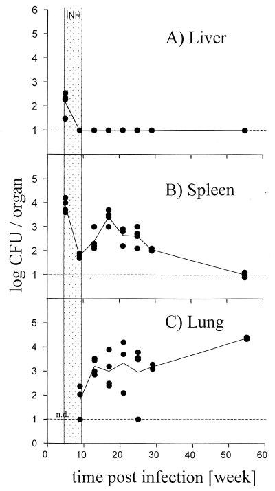
M. tuberculosis titers in vivo. C57BL/6 mice were infected intravenously with 104 CFU of M. tuberculosis strain Erdman. Five weeks postinfection, mice were treated with isoniazide (INH; 0.5 g/liter) in the drinking water for 4 weeks. Log CFU were determined in liver (A), spleen (B), and lung (C) at the indicated time points. Shown are values for four individual mice per group (solid circles) and the means for four mice (line) per time point and group for all three organs. The detection limit of the assay is indicated by the broken line. Hatched area, time interval of isoniazide treatment. n.d., first time point CFU not determined for lung. Shown is one representative experiment of two similar ones.

Confocal analysis of liver cell infection in vitro. TIB-75 mouse hepatocyte and J774A.1 mouse macrophage cell lines were infected with rBCG-GFP at an MOI of 10 for 1 h, resulting in an infection rate of 5 to 10% of the cells and analyzed after 1 h (A and C) and 24 h (B and D), respectively. For 24 h of infection, cultures were supplemented after 1 h of incubation with 25 mg of gentamicin sulfate per liter. Prior to analysis, cells were fixed with 4% PFA and stained with phalloidin-TRITC in order to visualize actin. The rBCG-GFP were localized within the cells by confocal laser microscopy. Shown are projections of eight confocal laser scans. Original magnification, ×1,000. Shown is one representative experiment of two similar ones.
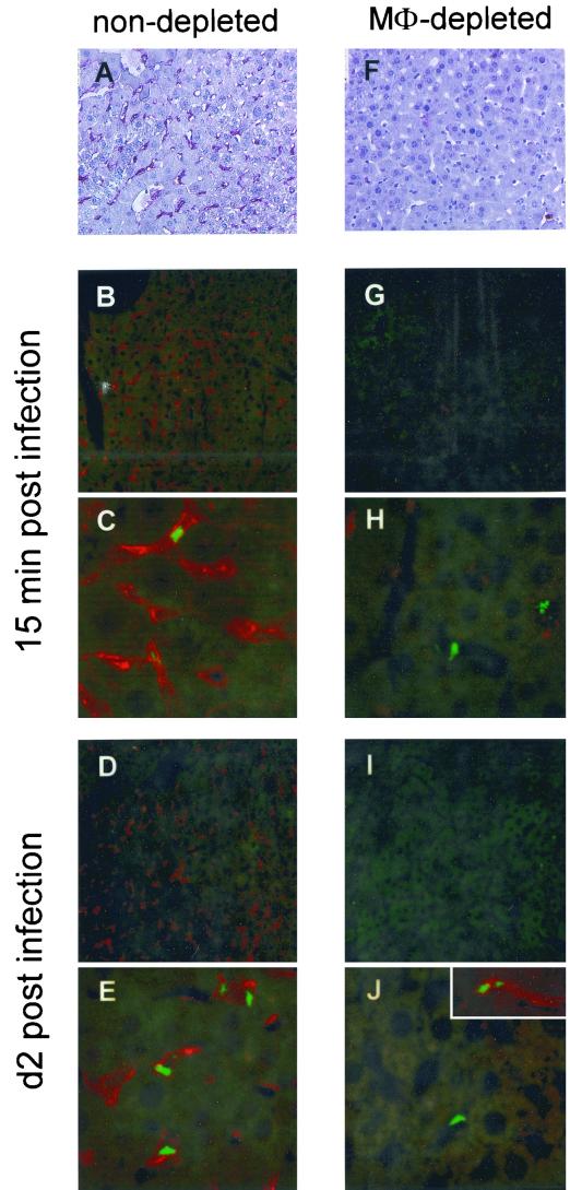
Confocal analysis of liver cell infection in vivo. C57BL/6 mice were either left untreated (A to E) or treated with 1.5 mg (F to J) of clodronate liposomes intraperitoneally at day −3. At day 0, mice were infected intravenously with 106 CFU of rBCG-GFP. At 15 min and 2 days postinfection, liver sections were stained with the rat MAb F4/80 and the secondary PAb goat anti-rat alkaline phosphatase (A and F) or PAb goat anti-rat Cy3.18 (B to E and G to J) in order to detect liver macrophages. Clusters of rBCG-GFP were detected inside the liver macrophages by confocal laser microscopy. Original magnification, ×200 (A, B, D, F, G, and I) and ×630 (C, E, H, and J). Shown is one representative experiment of two similar ones.
Similar articles
-
Intramacrophage growth of Mycobacterium avium during infection of mice.Infect Immun. 1991 Jun;59(6):2207-14. doi: 10.1128/iai.59.6.2207-2214.1991. Infect Immun. 1991. PMID: 2037382 Free PMC article.
-
Short report: modulation of Mycobacterium tuberculosis infection by Plasmodium in the murine model.Am J Trop Med Hyg. 2004 Feb;70(2):144-8. Am J Trop Med Hyg. 2004. PMID: 14993625
-
Mycobacterium tuberculosis strains disrupted in mce3 and mce4 operons are attenuated in mice.J Med Microbiol. 2008 Feb;57(Pt 2):164-170. doi: 10.1099/jmm.0.47454-0. J Med Microbiol. 2008. PMID: 18201981
-
Mycobacterium tuberculosis ECF sigma factor sigC is required for lethality in mice and for the conditional expression of a defined gene set.Mol Microbiol. 2004 Apr;52(1):25-38. doi: 10.1111/j.1365-2958.2003.03958.x. Mol Microbiol. 2004. PMID: 15049808
-
Iron and Mycobacterium tuberculosis infection.J Clin Virol. 2001 Feb;20(3):123-6. doi: 10.1016/s1386-6532(00)00136-0. J Clin Virol. 2001. PMID: 11166659 Review.
Cited by
-
Inflammatory myofibroblastic tumor of the liver due to Mycobacterium tuberculosis in an immunocompetent girl.Pediatr Surg Int. 2009 May;25(5):451-4. doi: 10.1007/s00383-009-2361-7. Epub 2009 Apr 25. Pediatr Surg Int. 2009. PMID: 19396450
-
Ornithine-A urea cycle metabolite enhances autophagy and controls Mycobacterium tuberculosis infection.Nat Commun. 2020 Jul 15;11(1):3535. doi: 10.1038/s41467-020-17310-5. Nat Commun. 2020. Retraction in: Nat Commun. 2022 Oct 18;13(1):6159. doi: 10.1038/s41467-022-33608-y. PMID: 32669568 Free PMC article. Retracted.
-
A ferritin mutant of Mycobacterium tuberculosis is highly susceptible to killing by antibiotics and is unable to establish a chronic infection in mice.Infect Immun. 2012 Oct;80(10):3650-9. doi: 10.1128/IAI.00229-12. Epub 2012 Jul 16. Infect Immun. 2012. PMID: 22802345 Free PMC article.
-
Deficiency of decay-accelerating factor and complement receptor 1-related gene/protein y on murine platelets leads to complement-dependent clearance by the macrophage phagocytic receptor CRIg.Blood. 2008 Aug 15;112(4):1109-19. doi: 10.1182/blood-2008-01-134304. Epub 2008 Jun 4. Blood. 2008. PMID: 18524992 Free PMC article.
References
-
- Abdel-Dayem H M, Naddaf S, Aziz M, Mina B, Turoglu T, Akisik M F, Omar W S, DiFabrizio L, LaBombardi V, Kempf J S. Sites of tuberculous involvement in patients with AIDS. Autopsy findings and evaluation of gallium imaging. Clin Nucl Med. 1997;22:310–314. - PubMed
-
- Arruda S, Bomfim G, Knights R, Huima-Byron T, Riley L W. Cloning of an M. tuberculosis DNA fragment associated with entry and survival inside cells. Science. 1993;261:1454–1457. - PubMed
-
- Borrel A. Tuberculose pulmonaire experimentale. Ann Inst Pasteur (Paris) 1893;7:593–627.
Publication types
MeSH terms
LinkOut - more resources
Full Text Sources
Medical
