

doi: 10.1128/IAI.69.12.7933-7936.2001.
Identification of the sigB operon in Staphylococcus epidermidis: construction and characterization of a sigB deletion mutant
Affiliations
- PMID: 11705980
- PMCID: PMC98894
- DOI: 10.1128/IAI.69.12.7933-7936.2001
Item in Clipboard
Identification of the sigB operon in Staphylococcus epidermidis: construction and characterization of a sigB deletion mutant
Infect Immun.
2001 Dec.
Abstract
The role of the alternative sigma factor sigma(B) in Staphylococcus epidermidis was investigated by the construction, complementation, and characterization of a sigB deletion mutant. Electrophoretic analyses confirmed a profound influence of sigma(B) on the expression of exoproteins and cytoplasmic proteins. Detailed investigation revealed reduced lipase and enhanced protease activity in the sigma(B) mutant. Furthermore, no significant influence of sigma(B) on heterologous biofilm formation or on the activity of the global regulator agr was detected.
Figures
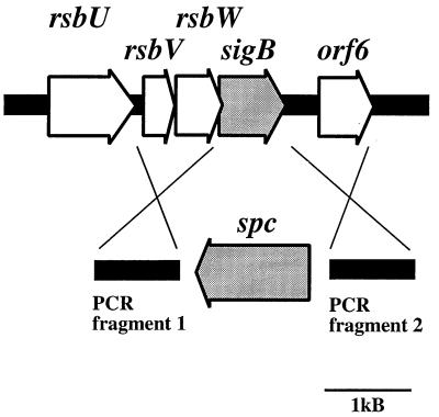
Physical map of the sigB operon of S. epidermidis and construction of a sigB deletion mutant using plasmid pBTΔsigB. Arrows depict open reading frames and indicate their orientation and size. The sigB gene was replaced with the spectinomycin resistance gene (spc) as shown in the lower part of the figure. The spc gene and two PCR-amplified regions flanking sigB were cloned into plasmid pBT2, yielding integration vector pBTΔsigB. The crosses indicate the sites of homologous recombination.
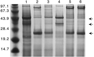
Influence of S. epidermidis ςB on production of exoproteins. Protein profiles of 16-h cultures of the following strains are shown: S. epidermidis sigB deletion mutant S. epidermidis Tü3298ΔsigB (lane 1), S. epidermidis wild-type Tü3298 (lane 2), the complemented mutant S. epidermidis TüΔsigB(pTXsigB) (lane 3), the control strain S. epidermidis TüΔsigB(pTX16) (lane 4), the ςB-overexpressing strain S. epidermidis Tü(pTXsigB) (lane 5), and its corresponding control strain S. epidermidis Tü(pTX16) (lane 6). The proteins were separated on SDS–10% polyacrylamide gels and stained with Coomassie brilliant blue G250. The arrows mark the positions of 27-kDa, 38-kDa, and 44-kDa proteins present or absent in the different strains. The molecular masses (in kilodaltons) of size standard proteins are shown on the left.
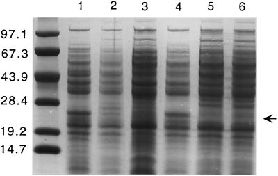
Influence of S. epidermidis ςB on production of cytoplasmic proteins. Protein profiles of 16-h cultures of the following strains are shown: S. epidermidis sigB deletion mutant S. epidermidis Tü3298ΔsigB (lane 1), S. epidermidis wild-type Tü3298 (lane 2), the complemented mutant S. epidermidis TüΔsigB(pTXsigB) (lane 3), the control strain S. epidermidis TüΔsigB(pTX16) (lane 4), the ςB-overexpressing strain S. epidermidis Tü(pTXsigB) (lane 5), and its corresponding control strain S. epidermidis Tü(pTX16) (lane 6). The proteins were separated on SDS–10% polyacrylamide gels and stained with Coomassie brilliant blue G250. The arrow indicates the position of a 27-kDa protein present or absent in the different strains. The molecular masses (in kilodaltons) of size standard proteins are shown on the left.
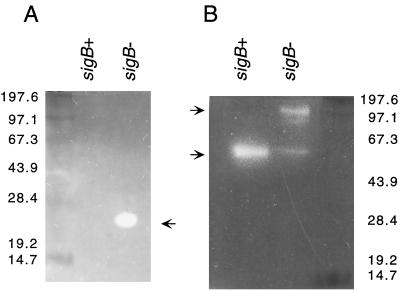
Zymographic analysis of protease (A) and lipase (B) activity. Exoprotein samples of strains S. epidermidis Tü3298 (wild-type; sigB+) and S. epidermidis TüΔsigB (sigB deletion mutant; sigB−) were separated on SDS–10% polyacrylamide gels. Zymographic analysis of the gels was carried out on protease or lipase agarose test plates. Arrows indicate the positions of proteolytic or lipolytic bands. The molecular masses (in kilodaltons) of size standard proteins are shown on the left (A) and right (B).
References
-
- Augustin J, Götz F. Transformation of Staphylococcus epidermidis and other staphylococcal species with plasmid DNA by electroporation. FEMS Microbiol Lett. 1990;54:203–207. - PubMed
-
- Brückner R. Gene replacement in Staphylococcus carnosus and Staphylococcus xylosus. FEMS Microbiol Lett. 1997;151:1–8. - PubMed
Publication types
MeSH terms
Substances
LinkOut - more resources
Full Text Sources
