. 2001 Nov 12;155(4):581-92.
doi: 10.1083/jcb.200106065. Epub 2001 Nov 12.

Yeast Cdc42 functions at a late step in exocytosis, specifically during polarized growth of the emerging bud

Affiliations

Yeast Cdc42 functions at a late step in exocytosis, specifically during polarized growth of the emerging bud

J E Adamo et al. J Cell Biol. .

Abstract

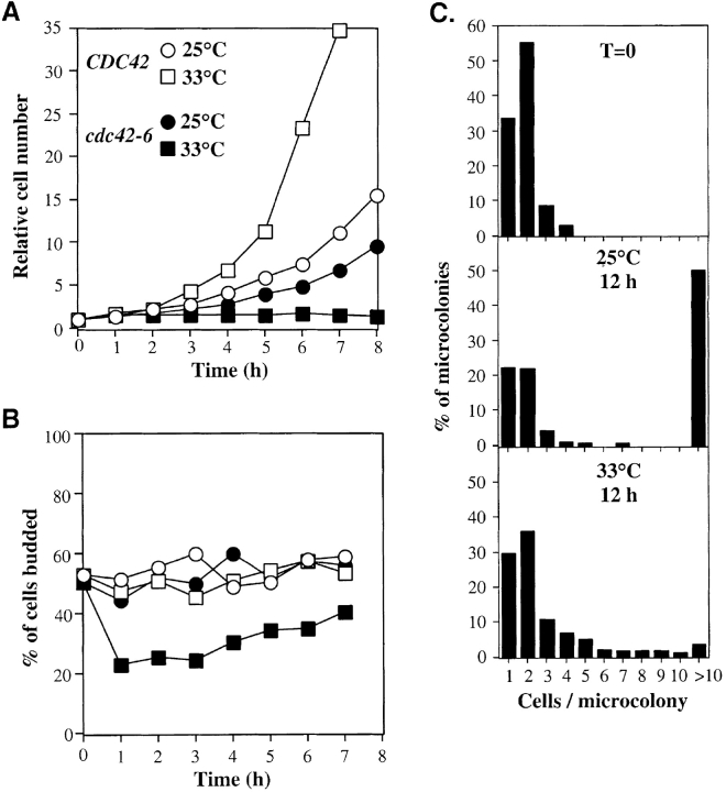
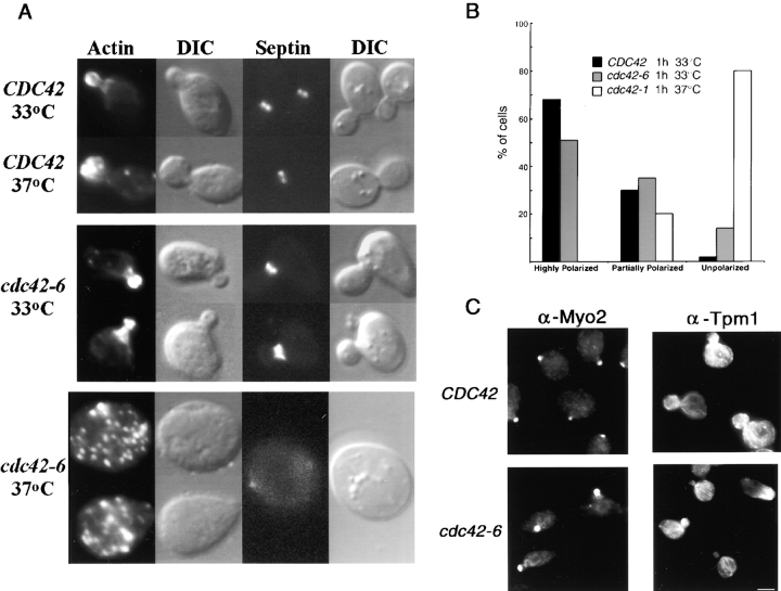
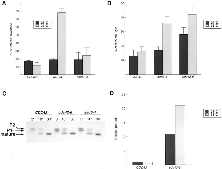
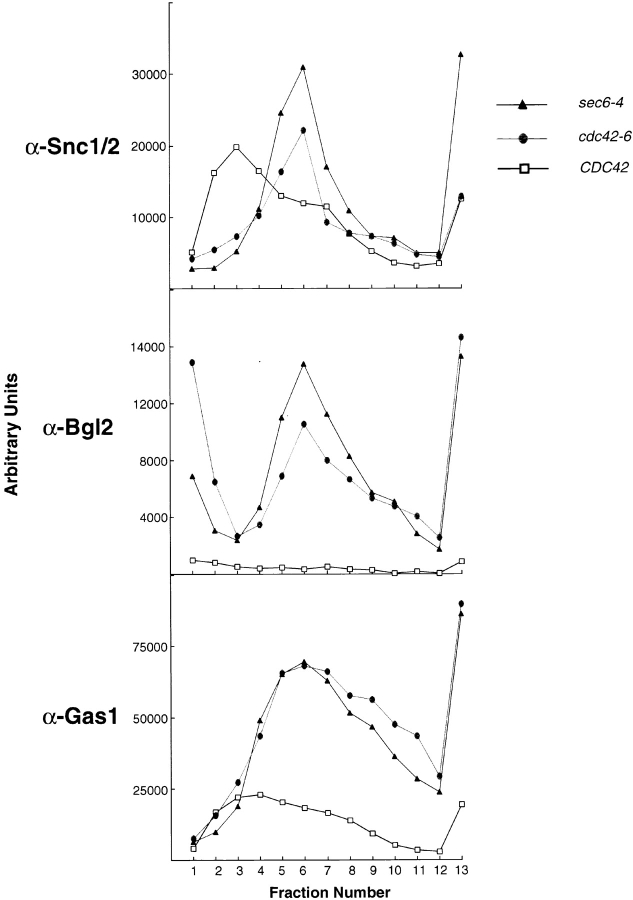
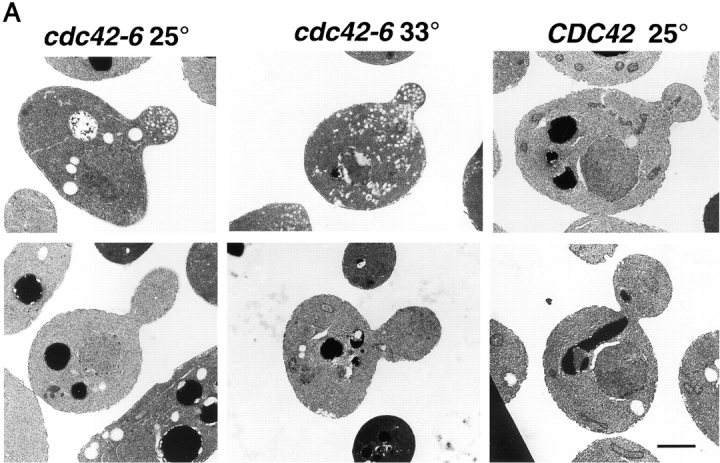
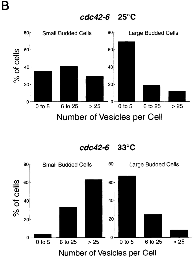
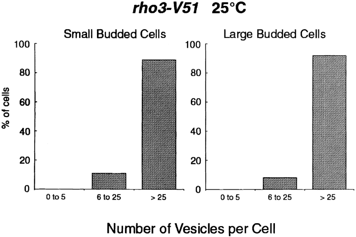
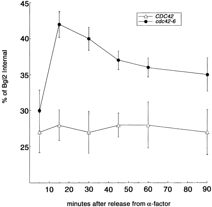
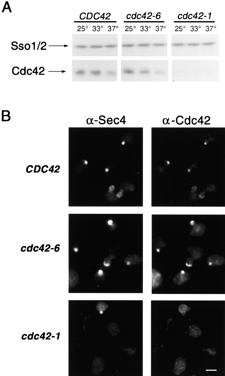
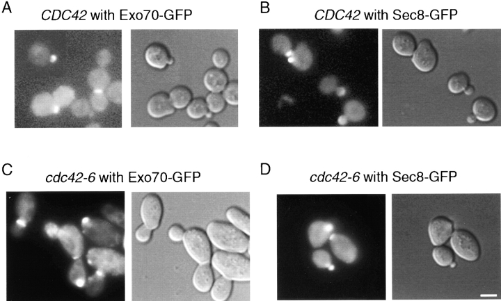
The Rho family GTPase Cdc42 is a key regulator of cell polarity and cytoskeletal organization in eukaryotic cells. In yeast, the role of Cdc42 in polarization of cell growth includes polarization of the actin cytoskeleton, which delivers secretory vesicles to growth sites at the plasma membrane. We now describe a novel temperature-sensitive mutant, cdc42-6, that reveals a role for Cdc42 in docking and fusion of secretory vesicles that is independent of its role in actin polarization. cdc42-6 mutants can polarize actin and deliver secretory vesicles to the bud, but fail to fuse those vesicles with the plasma membrane. This defect is manifested only during the early stages of bud formation when growth is most highly polarized, and appears to reflect a requirement for Cdc42 to maintain maximally active exocytic machinery at sites of high vesicle throughput. Extensive genetic interactions between cdc42-6 and mutations in exocytic components support this hypothesis, and indicate a functional overlap with Rho3, which also regulates both actin organization and exocytosis. Localization data suggest that the defect in cdc42-6 cells is not at the level of the localization of the exocytic apparatus. Rather, we suggest that Cdc42 acts as an allosteric regulator of the vesicle docking and fusion apparatus to provide maximal function at sites of polarized growth.


Figures

Figure 1.
Figure 1.
Phenotype of cdc42-6 mutants. (A and B) CDC42 (open symbols) and cdc42-6 (closed symbols) cells were grown to a density of about 106 cells/ml in YPD at 25°C and split into flasks that were then incubated either at 25°C (circles) or 33°C (squares). Aliquots were fixed with 3.7% formaldehyde at hourly intervals and cell number (A) and the percentage of cells budded (B) were scored. (C) cdc42-6 cells were sonicated briefly and spread onto YPD plates. The number of cell bodies per microcolony was scored immediately after plating (T = 0) or after 12 h at 23 or 33°C, as indicated. An unbudded cell was counted as one cell body, and a budded cell was counted as two cell bodies.
Figure 2.
Figure 2.
Actin cytoskeleton is largely polarized in the cdc42-6 mutant at 33°C. (A) Wild-type (CDC42) and mutant (cdc42-6) cells were grown at 25°C and shifted to the indicated temperature for 4 h before fixation and processing for fluorescence microscopy. F-actin was visualized by staining with rhodamine-conjugated phalloidin and septin distribution was visualized using Cdc12-GFP. DIC images of the same cells are shown to the right. (B) Quantitation of the actin polarity phenotypes. Cells were fixed and then stained with rhodamine-phalloidin for analysis. Cells were considered highly polarized if they contained both polarized cortical actin patches and correctly positioned actin cables. Partially polarized cells met at least one of these two criteria while unpolarized cells met neither. (C) Localization of Myo2 and Tpm1. Immunofluorescence was performed to visualize Tpm1, a specific marker for actin cables (Liu and Bretscher, 1989), and Myo2, whose polarized localization is highly dependent on the presence of actin cables (Pruyne et al., 1998). Cells were shifted for 1 h shift to the restrictive temperature of 33°C before fixation. Bar, 5 μm.
Figure 3.
Figure 3.
Trafficking of Bgl2, invertase, and CPY in the cdc42-6 mutant. (A) The graph depicts the percentage of Bgl2 protein that remains internal in CDC42, cdc42-6, and sec6-4 strains. The percent distribution of Bgl2 was determined by immunoblot analysis using affinity-purified antibodies raised against the COOH terminus of Bgl2. Quantitation of the bands was done using ImageQuant software and the results are presented as a percentage of total Bgl2 that is found internally. (B) The graph depicts the percentage of invertase protein that remains internal in CDC42, cdc42-6, and sec6-4 strains. The percent of internal invertase was determined by enzyme assays and then shown in bar graph form. (C) Transport of the vacuolar protein CPY is not affected in cdc42-6, as it is found exclusively in the mature form, (mCPY, 61 kD). Wild-type CDC42 and the late secretory mutant, sec6-4, are also shown to process CPY fully to the mature form while sec18-1, an ER-to-Golgi mutant, accumulates the earlier forms. (D) Vesicle accumulation was quantitated for wild-type and cdc42-6 cells at both permissive and restrictive (33°C for 1 h) temperatures from randomly chosen fields of cells. Numbers shown were determined by dividing the total number of 80–100-nm vesicles observed per field of cells by the total number of cells present per field.
Figure 4.
Figure 4.
Snc1, Bgl2, and Gas1 are all associated with vesicles that accumulate in cdc42-6 cells. Vesicle gradients of 20 to 40% sorbitol were prepared for each of CDC42, cdc42-6, and sec6-4. Normalized volumes of lysed cells (shifted for 1 h to 37°C for sec6-4 and 33°C for CDC42 and cdc42-6) for each strain were layered onto a gradient before a 1.5 h, 71,000 g spin. The gradients were then collected in 16 fractions and these fractions were subjected to SDS-PAGE analysis and blotted with the indicated antibodies.
Figure 5.
Figure 5.
Vesicle accumulation in small-budded cdc42-6 cells. (A) CDC42 at 25°C and cdc42-6 at 25 and 33°C. An example of both small- (<1 μm) and large-budded (1 μm or greater) cells is shown. Preparation of cells is described in the Materials and methods. Cells were only counted as budded cells if the section clearly went through the neck region between the mother and the bud size is as described in the text. (B) Cells were scored for the number of accumulated vesicles and bar graphs show the percentage of small and large budded cells with vesicles. Approximately 30 cells were counted for cdc42-6 at each temperature. Bar, 1 μm.
Figure 5.
Figure 5.
Vesicle accumulation in small-budded cdc42-6 cells. (A) CDC42 at 25°C and cdc42-6 at 25 and 33°C. An example of both small- (<1 μm) and large-budded (1 μm or greater) cells is shown. Preparation of cells is described in the Materials and methods. Cells were only counted as budded cells if the section clearly went through the neck region between the mother and the bud size is as described in the text. (B) Cells were scored for the number of accumulated vesicles and bar graphs show the percentage of small and large budded cells with vesicles. Approximately 30 cells were counted for cdc42-6 at each temperature. Bar, 1 μm.
Figure 6.
Figure 6.
Vesicle accumulation in rho3-V51 mutants affects both large- and small-budded cells similarly. The rho3-V51 micrographs were reevaluated and vesicle accumulation was now counted with respect to bud size, using the same criteria as in Fig. 4.
Figure 7.
Figure 7.
Bgl2 secretion in synchronized cells traversing the cell cycle. CDC42 and cdc42-6 cells were synchronized using α-factor and then washed and released into fresh media for 5, 15, 30, 45, 60, or 90 min before undergoing a 30-min shift to restrictive temperature (33°C). Cells were then processed as in Fig. 3 for analysis of Bgl2 secretion.
Figure 8.
Figure 8.
Levels of Cdc42 accumulation and localization of Cdc42 and Sec4 in CDC42 , cdc42-6 and cdc42-1 strains. (A) Whole cell lysates were prepared and subjected to SDS-PAGE analysis and then blotted with anti-Cdc42 or anti-Sso2 antibodies. (B) Immunofluorescence probing for Sec4 and Cdc42 localization was done after a shift to the restrictive temperature of 33°C for CDC42 (top), cdc42-6 (middle) and to 37°C for cdc42-1 (bottom). Bar, 5 μm.
Figure 9.
Figure 9.
Localization of Exo70-GFP and Sec8-GFP in CDC42 and cdc42-6 strains. COOH terminally tagged GFP constructs were transformed into wild-type and cdc42-6 cells and examined by fluorescence microscopy. The results for wild-type (A and B) and cdc42-6 cells following a 1-h shift at 33°C (C and D) demonstrate that the polarization of the exocyst complex was unaffected in cdc42-6 cells. DIC and GFP fluorescence images were captured immediately following temperature shift. Similar were results were obtained by addition of formaldehyde directly to the media at the end of the temperature shift. Bar, 5 μm.

References

    1. Adamo, J.E., G. Rossi, and P. Brennwald. 1999. The Rho GTPase Rho3 has a direct role in exocytosis that is distinct from its role in actin polarity. Mol. Biol. Cell. 10:4121–4133. - PMC - PubMed
    1. Adams, A.E., D.I. Johnson, R.M. Longnecker, B.F. Sloat, and J.R. Pringle. 1990. CDC42 and CDC43, two additional genes involved in budding and the establishment of cell polarity in the yeast Saccharomyces cerevisiae. J. Cell Biol. 111:131–142. - PMC - PubMed
    1. Brennwald, P., and P. Novick. 1993. Interactions of three domains distinguishing the Ras-related GTP-binding proteins Ypt1 and Sec4. Nature. 362:560–563. - PubMed
    1. Brennwald, P., B. Kearns, K. Champion, S. Keranen, V. Bankaitis, and P. Novick. 1994. Sec9 is a SNAP-25-like component of a yeast SNARE complex that may be the effector of Sec4 function in exocytosis. Cell. 79:245–258. - PubMed
    1. Drubin, D.G., and W.J. Nelson. 1996. Origins of cell polarity. Cell. 84:335–344. - PubMed

Publication types

MeSH terms

Substances