. 2001 Nov 12;155(4):613-23.
doi: 10.1083/jcb.200012116. Epub 2001 Nov 12.

Toxoplasma gondii myosins B/C: one gene, two tails, two localizations, and a role in parasite division

Affiliations

Toxoplasma gondii myosins B/C: one gene, two tails, two localizations, and a role in parasite division

F Delbac et al. J Cell Biol. .

Abstract

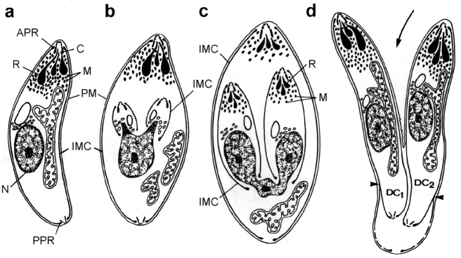
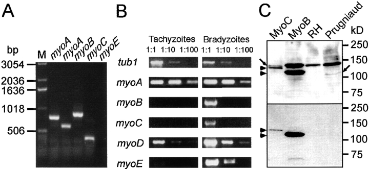
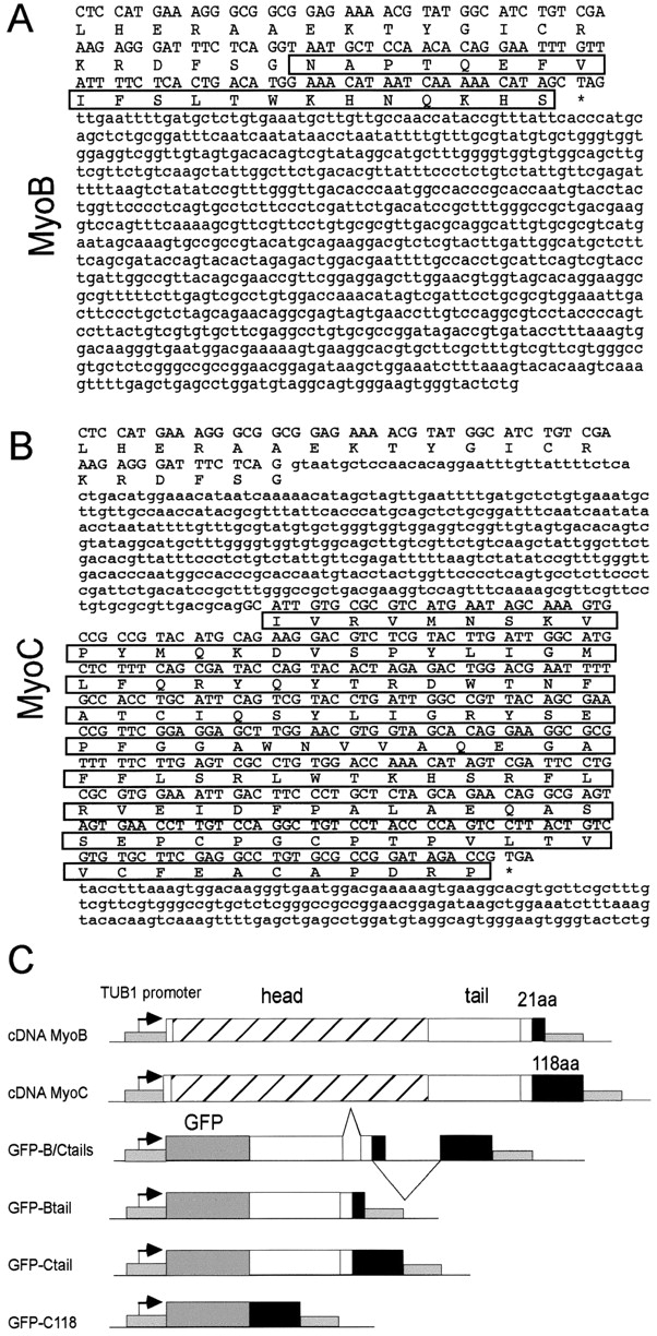
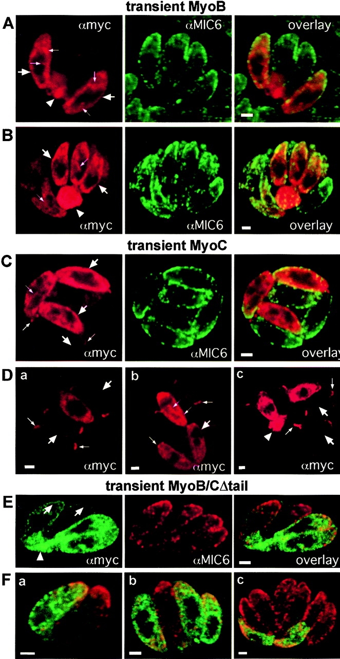
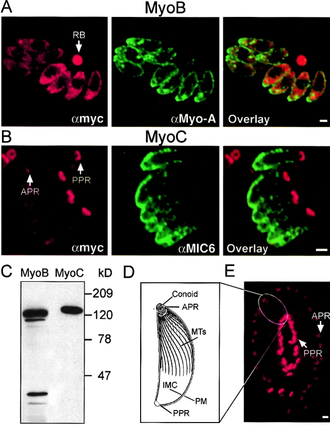
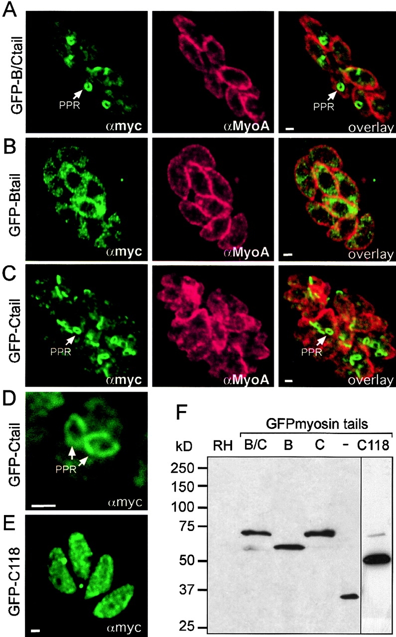
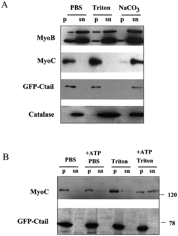
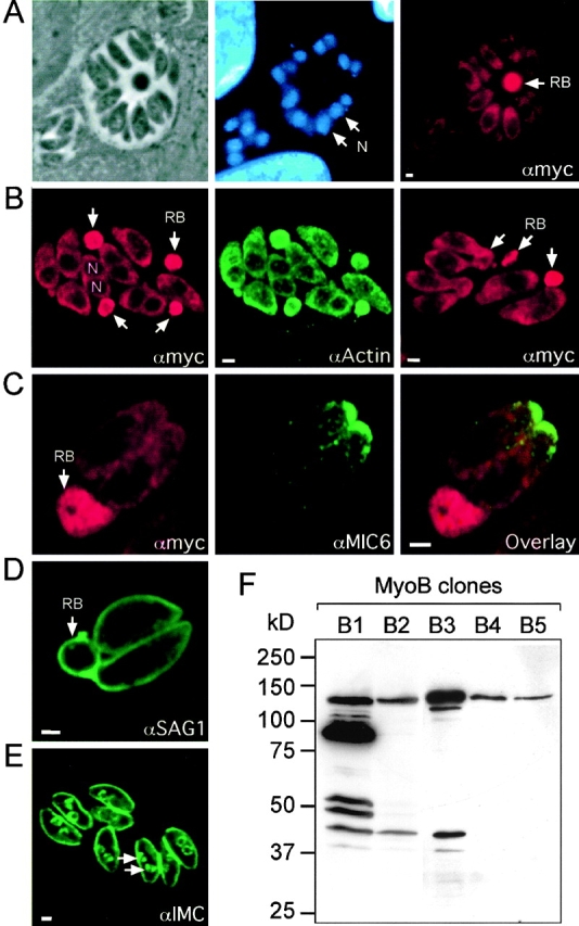
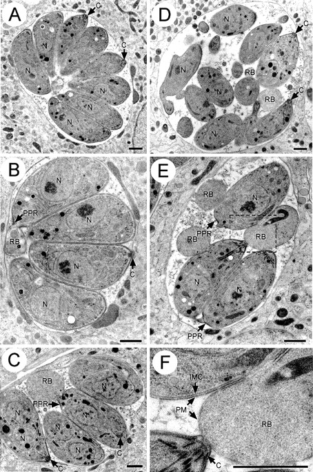
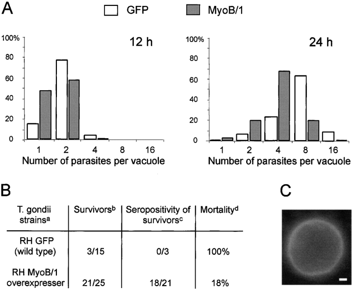
In apicomplexan parasites, actin-disrupting drugs and the inhibitor of myosin heavy chain ATPase, 2,3-butanedione monoxime, have been shown to interfere with host cell invasion by inhibiting parasite gliding motility. We report here that the actomyosin system of Toxoplasma gondii also contributes to the process of cell division by ensuring accurate budding of daughter cells. T. gondii myosins B and C are encoded by alternatively spliced mRNAs and differ only in their COOH-terminal tails. MyoB and MyoC showed distinct subcellular localizations and dissimilar solubilities, which were conferred by their tails. MyoC is the first marker selectively concentrated at the anterior and posterior polar rings of the inner membrane complex, structures that play a key role in cell shape integrity during daughter cell biogenesis. When transiently expressed, MyoB, MyoC, as well as the common motor domain lacking the tail did not distribute evenly between daughter cells, suggesting some impairment in proper segregation. Stable overexpression of MyoB caused a significant defect in parasite cell division, leading to the formation of extensive residual bodies, a substantial delay in replication, and loss of acute virulence in mice. Altogether, these observations suggest that MyoB/C products play a role in proper daughter cell budding and separation.


Figures

Figure 1.
Figure 1.
Endodyogeny in T. gondii. Schematic representation of the different stages of division within a mother cell (adapted from Kepka and Scholtyseck, 1970; Frankelia species). Cell division starts with the formation of the two daughter conoids, the IMCs, and the microtubule baskets (b). It is followed by the entrapment of the apical secretory organelles, micronemes, and rhoptries (c). The IMC of the mother cell (a) somehow gives rise to the daughter cells' membrane complexes, which progress toward the posterior pole while the nucleus divides. Final separation occurs by budding through the mother cell plasma membrane and by constriction at the posterior end (d). APR, apical polar ring; C, conoid; M, micronemes; N, nucleus; PM, plasma membrane; PPR, posterior polar ring; R, rhoptries.
Figure 2.
Figure 2.
Comparison of the expression of T. gondii myosin transcripts and proteins between tachyzoites and bradyzoites. (A) RT-PCR amplification of T. gondii myosin transcripts from total tachyzoite RNA with two distinct sets of primers specific to MyoA and one set of primers specific for MyoB, MyoC, and MyoE. (B) Semiquantitative RT-PCR analysis on total RNA prepared from tachyzoites cultivated in vitro and bradyzoites encysted in vivo. After first strand synthesis, the cDNAs of MyoA, MyoB, MyoC, MyoD, and MyoE, as well as the tubulin mRNA, TUB1 (used as a control), were detected using appropriate primer pairs. Serial dilutions of the cDNAs were 1:10 and 1:100. The size of the PCR products were as expected from the respective cDNA sequence. (C) Western blot analysis of lysates prepared from extracellular RH, the persistent strain Prugniaud, RHMyoC, and RHMyoB. In the upper half, the membrane was incubated with the antiserum raised against peptides B/C1 and B/C2. The monoclonal anti-myc was used in the lower half.
Figure 3.
Figure 3.
The T. gondii MyoB/C gene is alternatively spliced to produce the two class XIV myosins B and C. (A and B) Sequences of the MyoB/C gene encompassing the two alternatively spliced COOH-terminal exons and 3′-UTR. When the last intron remains unspliced, the long transcript generates MyoB, which contains 21 specific amino acids at the COOH terminus. Splicing of the last intron produces a shorter transcript coding for MyoC, which contains 118 specific amino acids. (C) Schematic representation of some constructs used in this study. All expression vectors contain a HXGPRT selection marker controlled by the DHFR-TS flanking sequences. The expression cassette is controlled by the TUB1 promoter and SAG1 3′UTR sequences and contains a myc-7xHis tag fused after the start codon as previously described (Hettmann et al., 2000). GFP–B/C corresponds to a fusion of GFP with the genomic sequence encompassing both MyoB and MyoC coding tail exons. The different domains are not drawn to scale.
Figure 4.
Figure 4.
Uneven segregation of MyoB/C motor proteins during cell division. Double IFA and confocal microscopy analysis of parasites 24 h after transfection with MyoB (A and B), MyoC (C and D), or MyoB/CΔtail (E and F) constructs. Anti-MIC6 antibodies were used as markers to detect all the parasites present within a given vacuole. MyoB and MyoC (in red) and MyoB/CΔtail (in green) were detected with anti-myc antibodies. The immunofluorescence overlays showed the uneven segregation of the myosin after cell division. Transient expression of the three constructs induced the formation of residual bodies (A, B, Dc, E, arrowheads). The myosins significantly accumulated at the periphery of the cells (A–E, big arrows). In dividing parasites, MyoB (A and B) and MyoC (C and Db) were enriched in distinct structures or lamellae that appeared to extend from the periphery in between the daughter cells (small arrows). In addition, MyoC was concentrated at the poles of the parasites (C and D, small arrows). Bars, 1 μm.
Figure 5.
Figure 5.
Determination of the subcellular localization of MyoB and MyoC. Transgenic parasites expressing myc–MyoB (A) or myc–MyoC (B and E) were analyzed by IFA and confocal microscopy after staining with anti-myc (A, B, and E) and either anti-MyoA (A) or anti-MIC6 (B) antibodies. MIC6 is a microneme marker defining the apical pole of the parasite. (C) Western blot analysis of parasites expressing myc–MyoB or myc–MyoC with anti-myc antibodies. (D) Schematic representation of some major morphological features of a tachyzoite (adapted from Nichols and Chiappino, 1987). (E) A parasitophorous vacuole containing a rosette of 32 tachyzoites. Note the homogeneous orientation of the parasites with their apical poles pointing outwards. APR, anterior polar ring; MTs, microtubules; PM, plasma membrane; PPR, posterior polar ring. Bars, 1 μm.
Figure 6.
Figure 6.
Determination of the subcellular localization of GFP fusions. (A) Transgenic parasites expressing the different GFP fusion constructs with the tails of MyoB (B), MyoC (C and D), MyoB/C (A), or the COOH-terminal 118 residues specific to MyoC (E) were analyzed by confocal immunofluorescence microscopy after staining with anti-myc (A–E) and anti-MyoA (A–C) antibodies. (D) A parasite in the process of cell division showed two ring structures in the middle of the dividing mother cell. (F) Western blot analysis of wild-type parasites (RH) and transgenic parasites expressing GFP alone, GFP–Btail, GFP–Ctail, GFP–B/Ctail, or GFP–C118. PPR, posterior polar ring. Bars, 1 μm.
Figure 7.
Figure 7.
Distribution of recombinant MyoB and MyoC, studied by subcellular fractionation. Cells expressing myc–MyoB and myc–MyoC were lysed in the presence or absence of 10 mM ATP under different conditions, and separated by high speed centrifugations into soluble (sn) and particulate (p) fractions. Cells were lysed either in PBS, PBS with 2% Triton X-100, 0.1 M Na2CO3 (pH 11.5), or 1 M NaCl. The distribution of myc–MyoB, myc–MyoC, and GFP–Ctail were detected using the anti-myc antibodies. The distribution of endogenous catalase was used as a control and determined simultaneously by immunoblotting with anticatalase polyclonal antibodies.
Figure 8.
Figure 8.
Phenotypic analysis of parasites overexpressing MyoB. Detection of residual bodies in intracellular parasites expressing myc–MyoB (clone MyoB/1) by confocal microscopy. (A) The residual bodies detected with anti-myc antibodies did not stain with DAPI. (B) Colocalization of MyoB (polyclonal anti-myc) with actin illustrated their coenrichment in residual bodies. (C) Residual bodies were not significantly stained with the microneme marker anti-TgMIC6. In contrast, they stained with antibodies recognizing the glycosyl phosphatidylinositol–anchored surface antigen SAG1 (D), but were not detected by antibodies recognizing a protein of the IMC (E). The latter marker visualized the high frequency of dividing parasites in the population by detecting the presence of newly formed daughter cell IMCs (E, arrows). (F) Western blot analysis of independent recombinant T. gondii clones expressing MyoB at different levels. N, nucleus; RB, residual body. Bars, 1 μm.
Figure 9.
Figure 9.
Ultrastructural analysis of the residual bodies induced by MyoB overexpression. Thin section electron micrographs of wild-type parasites (A and B) and parasites overexpressing myc–MyoB (C–F). Wild-type parasites divide and form regular rosettes (A and B), whereas the vacuoles of transgenic parasites are highly disorganized (C–E). The boxed region in E is presented at higher magnification in F. Residual bodies are clearly distinguished from parasites, as they lack the IMC and are surrounded only by the plasma membrane (C–F). In wild-type parasites, residual bodies such as the one seen in B were rare. C, conoid; N, nucleus; PM, plasma membrane; PPR, posterior polar ring; RB, residual body. Bars, 1 μm.
Figure 10.
Figure 10.
Overexpression of MyoB delays cell division and reduces virulence in mice. (A) Parasites expressing GFP or MyoB were allowed to invade host cells at 37°C for 10 min; the cells were then further incubated for 12 and 24 h. For each condition, 100 vacuoles were analyzed, and the number of parasites per vacuole is presented in a histogram. (B) Overexpression of MyoB allowed mice to survive acute infection with T. gondii RH. Summary of two independent experiments: a group of five mice was inoculated with the two parasite lines in the first experiment. Groups of 10 mice (RHGFP) and 20 mice (RHMyoB overexpresser) were inoculated per parasite line in the second experiment. (Ba) The RHMyoB strain was compared with RH expressing GFP. (Bb) Numerators correspond to the number of surviving mice 4 wk after infection; denominators indicate the total number of mice inoculated in two independent experiments. (Bc) Numerators represent numbers of surviving mice that were positive for T. gondii serology; denominators represent the total number of mice surviving in the two separate experiments. (Bd) The percentage was corrected according to the real infection rate; the seronegative mice were not taken into account. (C) Isolation of cysts in brains of infected mice 2 mo after intraperitoneal inoculation with mutant parasites overexpressing MyoB. The cysts were stained specifically with the Dolichos biflorus FITC–labeled lectin, which recognizes the cell wall of T. gondii cysts. Bar, 10 μm.

Similar articles

Cited by

References

    1. Ajioka, J.W., J.C. Boothroyd, B.P. Brunk, A. Hehl, L. Hiller, I.D. Manger, G.C. Overton, M. Marrra, D. Roos, K.-L. Wan, et al. 1998. Gene discovery by EST sequencing in Toxoplasma gondii reveals sequences restricted to the Apicomplexa. Genome Res. 8:18–28. - PubMed
    1. Bezanilla, M., J.M. Wilson, and T.D. Pollard. 2000. Fission yeast myosin-II isoforms assemble into contractile rings at distinct times during mitosis. Curr. Biol. 10:397–400. - PubMed
    1. Boothroyd, J.C., M. Black, S. Bonnefoy, A. Hehl, L.J. Knoll, I.D. Manger, E. Ortega-Barria, and S. Tomavo. 1997. Genetic and biochemical analysis of development in Toxoplasma gondii. Philos. Trans. R. Soc. Lond. B Biol. Sci. 352:1347–1354. - PMC - PubMed
    1. Ding, M., C. Clayton, and D. Soldati. 2000. Toxoplasma gondii catalase: are there peroxisomes in Toxoplasma? J. Cell Sci. 113:2409–2419. - PubMed
    1. Dobrowolski, J., and L.D. Sibley. 1997. The role of the cytoskeleton in host cell invasion by Toxoplasma gondii. Behring Inst. Mitt. 99:90–96. - PubMed

Publication types

MeSH terms

Associated data