. 2001 Nov 12;155(4):625-35.
doi: 10.1083/jcb.200103128. Epub 2001 Nov 12.

Myosin V exhibits a high duty cycle and large unitary displacement

Affiliations

Myosin V exhibits a high duty cycle and large unitary displacement

J R Moore et al. J Cell Biol. .

Abstract

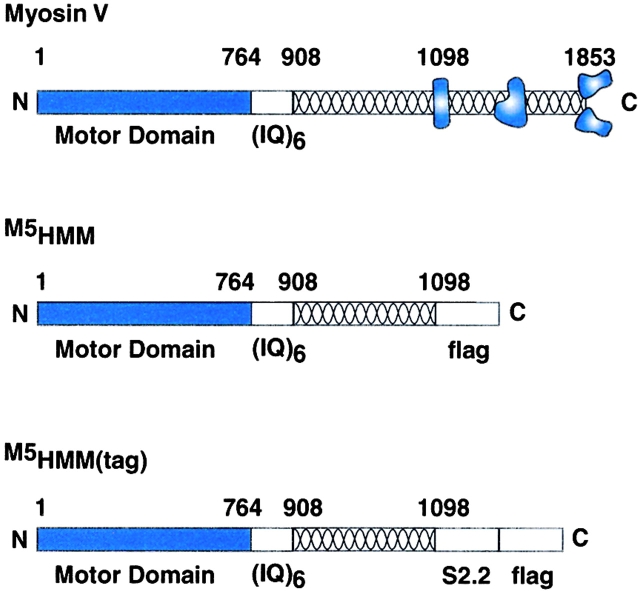
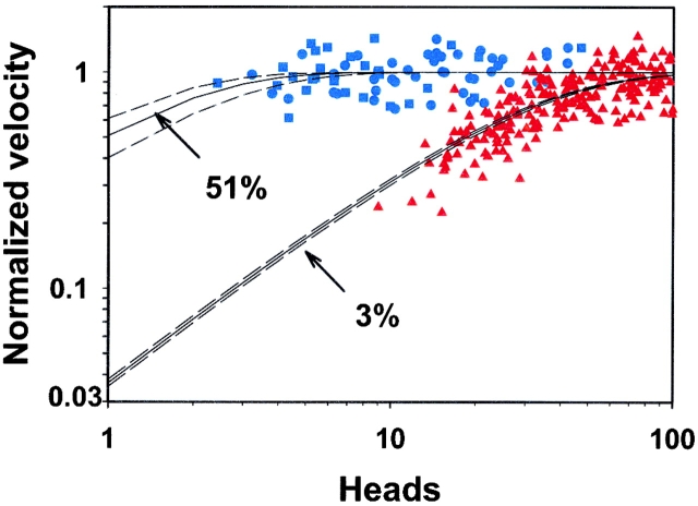
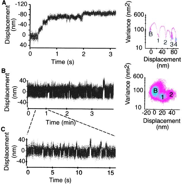
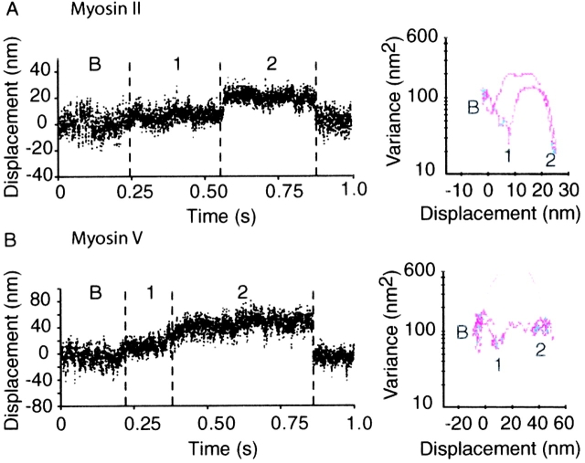
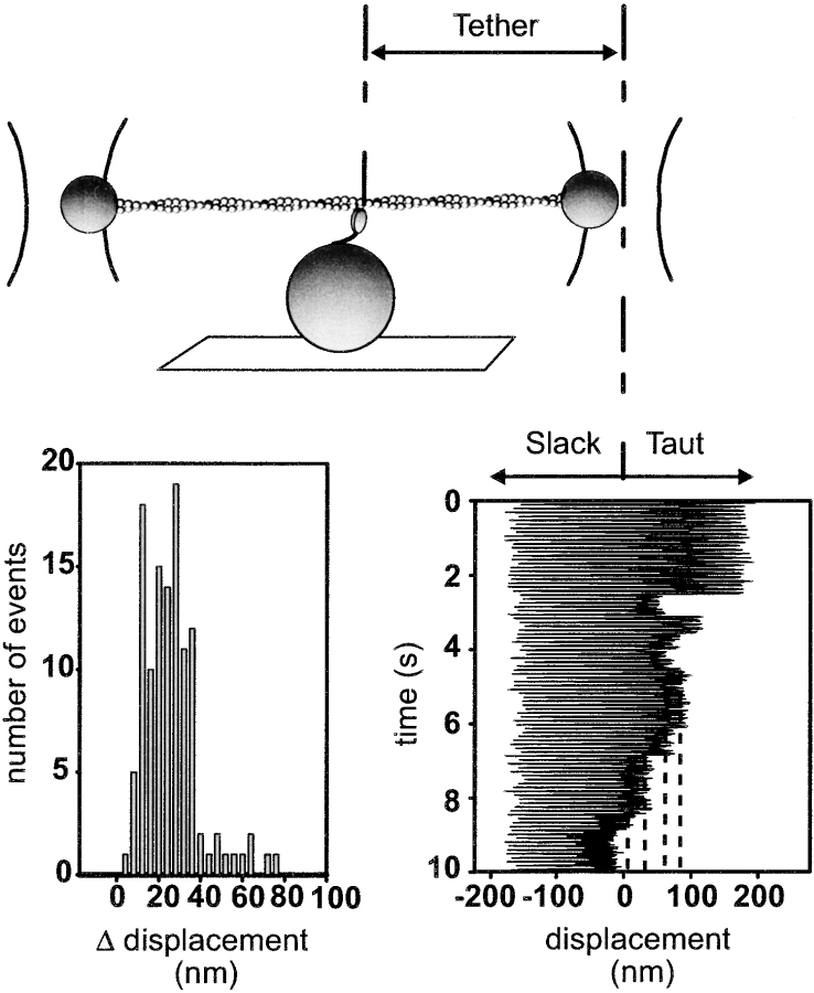
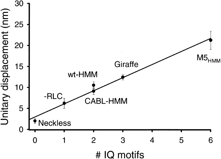
Myosin V is a double-headed unconventional myosin that has been implicated in organelle transport. To perform this role, myosin V may have a high duty cycle. To test this hypothesis and understand the properties of this molecule at the molecular level, we used the laser trap and in vitro motility assay to characterize the mechanics of heavy meromyosin-like fragments of myosin V (M5(HMM)) expressed in the Baculovirus system. The relationship between actin filament velocity and the number of interacting M5(HMM) molecules indicates a duty cycle of > or =50%. This high duty cycle would allow actin filament translocation and thus organelle transport by a few M5(HMM) molecules. Single molecule displacement data showed predominantly single step events of 20 nm and an occasional second step to 37 nm. The 20-nm unitary step represents the myosin V working stroke and is independent of the mode of M5(HMM) attachment to the motility surface or light chain content. The large M5(HMM) working stroke is consistent with the myosin V neck acting as a mechanical lever. The second step is characterized by an increased displacement variance, suggesting a model for how the two heads of myosin V function in processive motion.


Figures

Figure 1.
Figure 1.
Schematic representation comparing the sequences of the expressed myosin V constructs to wild-type myosin V. We constructed two COOH terminally truncated myosin V constructs, M5HMM and M5HMM(tag), where amino acids 1099–1853 were removed. Note that removal of the residues, which include the tail region of the molecule, results in a M5HMM protein that resembles the proteolytic HMM fragment of vertebrate smooth and skeletal muscle. M5HMM and M5HMM(tag) are identical except that M5HMM(tag) contains the S2.2 epitope for specific attachment to the motility surface (described in Materials and methods).
Figure 2.
Figure 2.
SDS-PAGE of the expressed constructs used in this study. (Lane 1) The M5HMM construct expressed with calmodulin only. (Lane 2) The M5HMM construct expressed with calmodulin and LC1sa. Asterisks indicate several minor proteolytic cleavage products that react with anti-FLAG antibody, indicating that they are derived from the COOH terminus of the M5HMM heavy chain. An arrowhead indicates the complementary proteolytic cleavage product that does not react with the anti-FLAG antibody. Note that these cleavages are suppressed in the presence of the tighter binding LC1sa light chain.
Figure 3.
Figure 3.
The ionic strength dependence of M5 HMM in vitro motility. Normalized filament velocity (mean ±SEM, n > 10 filaments) as a function of added potassium chloride for M5HMM expressed with LC1sa (•) and calmodulin (▴) (M5HMM loading concentration of 10–25 μg/ml). Normalized filament velocity as a function of added potassium chloride for smooth muscle myosin II (♦) from Harris and Warshaw (1992) for comparison (loading concentration of 100 μg/ml). Note the reduction in actin filament velocity at potassium chloride concentrations <125 mM for both M5HMM and myosin II. Also note the plateau at physiological ionic strengths and greater (between 125 mM and 325 mM potassium chloride) for M5HMM. Velocities were normalized to the velocity at 25 mM potassium chloride. A similar dependence of actin filament velocity was observed regardless of light chain content.
Figure 4.
Figure 4.
The dependence of actin filament velocity on the number of interacting M5 HMM heads. Normalized filament velocity as a function the number of M5HMM heads available to interact with each filament at a loading concentration of 5 (▪) and 1 μg/ml (•). Note the independence of M5HMM-driven actin filament velocity on the number of interacting heads. The number of available heads was calculated via surface ATPase assays (described in Materials and methods). For comparison, normalized filament velocity as a function the number of smooth muscle myosin heads available to interact with each filament (▴; data from Harris and Warshaw, 1993) at a loading concentration of 10 μg/ml. (Bottom solid line) A least squares fit of the myosin II data to Eq. 1, revealing a 3% duty cycle for the myosin II data. (Top solid line) A least squares fit of the M5HMM data to Eq. 1, revealing a 51% duty cycle. Dashed lines represent the range of the fit determined by standard error of the estimate.
Figure 5.
Figure 5.
Laser trap data time series of single M5 HMM molecules. (A) Representative time series for M5HMM at a flow cell loading of 1 μg/ml. For clarity, a 3-s portion of the total ∼140-s time series is shown. The corresponding MV histogram for this 3-s time series is shown with the baseline population indicated by a “B.” The event populations are numbered consecutively and are separated from the baseline by an increase in mean displacement and a reduction in system variance. (B) Representative 220-s time series for M5HMM at a flow cell loading of 0.1 μg/ml. The corresponding MV histogram for the entire time series is also shown. Note that the variance of the second event population is between baseline and the first event population. (C) To illustrate event size and duration more clearly, a 15-s portion of the time series in B is also shown.
Figure 8.
Figure 8.
Double step events. (A) Representative “double step” event for smooth muscle myosin II taken at higher myosin surface densities where presumably more than one molecule is available to interact with the actin filament. Note that the second step is characterized by a similar duration and lower variance than the first step. (B) Representative “double step” event for myosin V, which gives rise to the intermediate variance 37-nm steps (summarized in Table III). Note the higher variance and longer duration of the second step. “B” denotes the baseline population, and event populations are numbered to highlight at the respective mean and variance for each particular population.
Figure 6.
Figure 6.
Compliance-corrected step measurements. The experimental configuration for the oscillation experiments (described in Materials and methods). An example trace for M5HMM at 100 μM ATP is also shown (bottom right). Note that when myosin binds the sinusoidal oscillation of the trapped bead is clipped. The difference between clipped levels (dashed lines) provides a measure of the compliance-corrected displacement. The histogram of step increments indicates a displacement of ∼23 nm (bottom left).
Figure 7.
Figure 7.
Determination of event duration. Plot of MV histogram event volumes versus window width for a 127-s M5HMM(tag) displacement time series. A fit of Eq. 2 to the data yields an average event duration of 104 ms.
Figure 9.
Figure 9.
Unitary step size is a linear function of relative lever arm length. Unitary displacements for smooth muscle myosin II–HMM neck length mutants and M5HMM are plotted as mean ± SEM against the number of IQ motifs. These data were obtained from Warshaw et al. (2000) except for the addition of the M5HMM data, which is the mean of all the M5HMM constructs used in this study (Table I). WT-HMM or CABL-HMM contain two IQ motifs. Zero, one, three, and six IQ motifs were assigned to Neckless, -RLC, Giraffe, and the M5HMM, respectively. The linear regression for the data suggest that the neck region of myosin acts as a rigid lever arm up to 6IQ motifs. Furthermore, the regression does not pass through zero but intersects the y-axis, suggesting that lever may extend into the motor domain.
Figure 10.
Figure 10.
The relationship between the double step events and hypothetical states in the walking cycle of a double-headed myosin V molecule. In this model, a myosin V molecule walks from left to right along an actin filament (solid lines). The helical repeat of actin is indicated by tick marks, and dashed lines indicate 20- and 40-nm displacements from the site of initial attachment. After attachment, the myosin V molecule undergoes its working stroke (states 1–2) with the attachment of the second head following very rapidly (state 3). The working stroke and double-headed attachment results in the low variance first step. After the second head's powerstroke, release of the rear head discharges the intramolecular strain, bringing the rear head toward the next actin-binding site. Under normal processive movement, myosin V would then bind to the next actin-binding site (states 3–5), thus giving rise to the 40-nm walking stride. However, the expressed M5HMM did not demonstrate processivity but stalled during its second step at an intermediate displacement level, having high position variance (state 4) resulting from single-headed attachment.

Similar articles

Cited by

References

    1. Anson, M., M.A. Geeves, S.E. Kurzawa, and D.J. Manstein. 1996. Myosin motors with artificial lever arms. EMBO J. 15:6069–6074. - PMC - PubMed
    1. Baker, J.E., I. Brust-Mascher, S. Ramachandran, L.E. LaConte, and D.D. Thomas. 1998. A large and distinct rotation of the myosin light chain domain occurs upon muscle contraction. Proc. Natl. Acad. Sci. USA. 95:2944–2949. - PMC - PubMed
    1. Baker, J.P., and M.A. Titus. 1998. Myosins: matching functions with motors. Curr. Opin. Cell Biol. 10:80–86. - PubMed
    1. De La Cruz, E.M., A.L. Wells, S.S. Rosenfeld, E.M. Ostap, and H.L. Sweeney. 1999. The kinetic mechanism of myosin V. Proc. Natl. Acad. Sci. USA. 96:13726–13731. - PMC - PubMed
    1. De La Cruz, E.M., H.L. Sweeney, and E.M. Ostap. 2000. a. ADP inhibition of myosin V ATPase activity. Biophys. J. 79:1524–1529. - PMC - PubMed

Publication types