. 2001 Nov 12;155(4):649-59.
doi: 10.1083/jcb.200108080. Epub 2001 Nov 12.

Phosphatidylserine (PS) induces PS receptor-mediated macropinocytosis and promotes clearance of apoptotic cells

Affiliations

Phosphatidylserine (PS) induces PS receptor-mediated macropinocytosis and promotes clearance of apoptotic cells

P R Hoffmann et al. J Cell Biol. .

Abstract

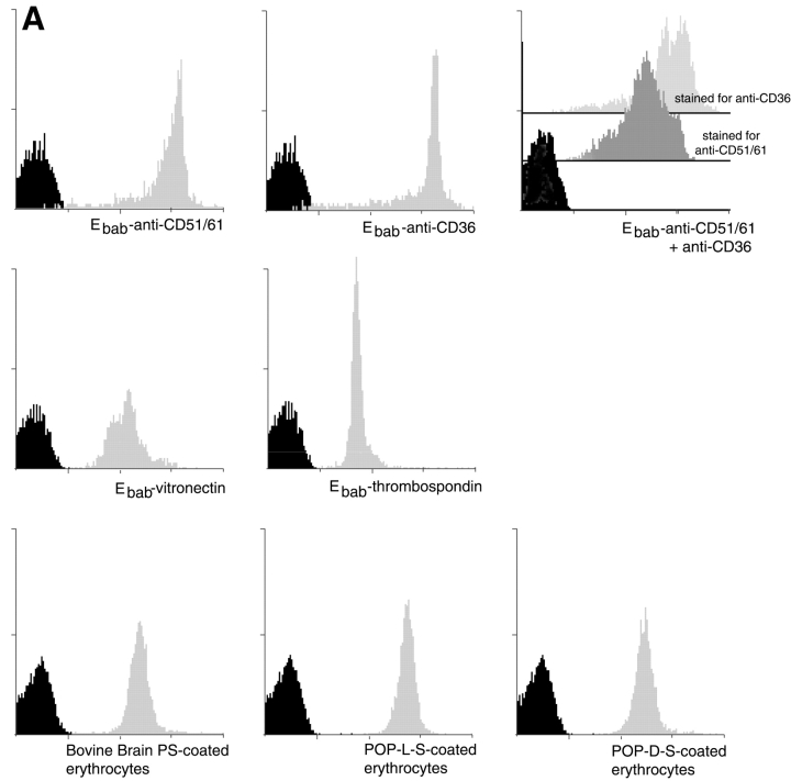
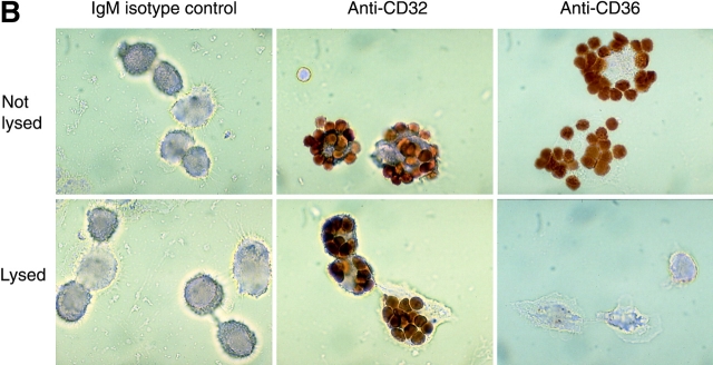
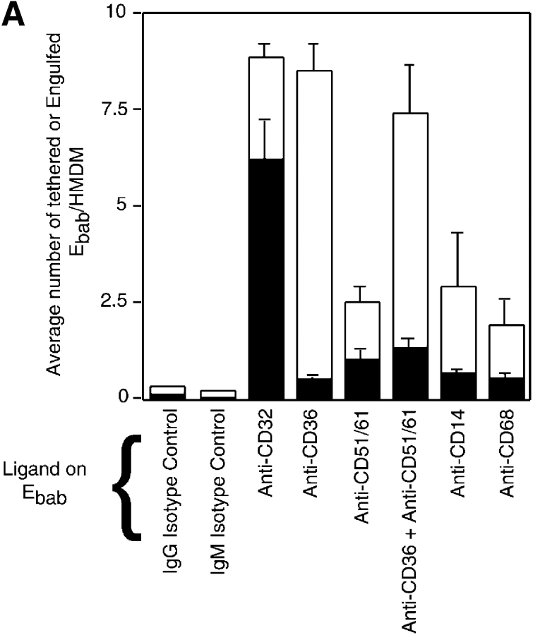
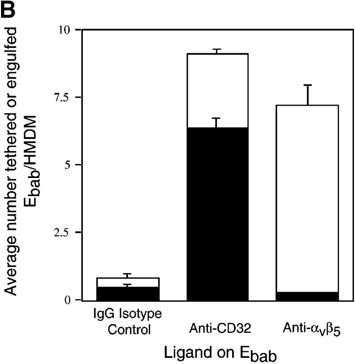
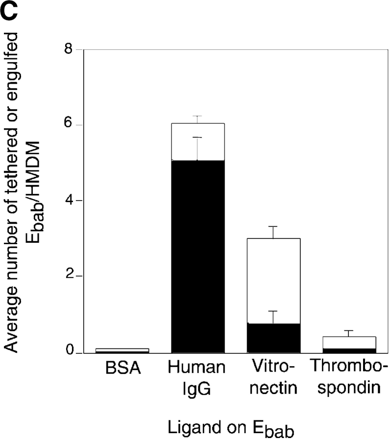
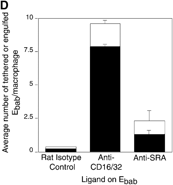
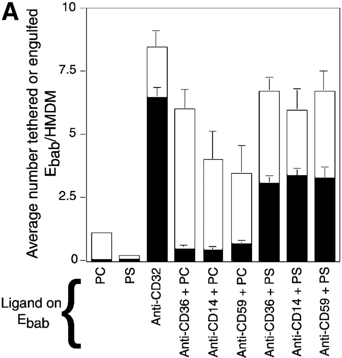
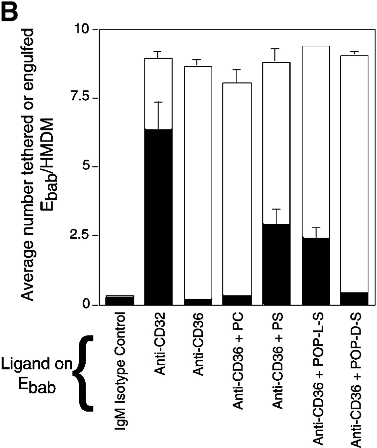
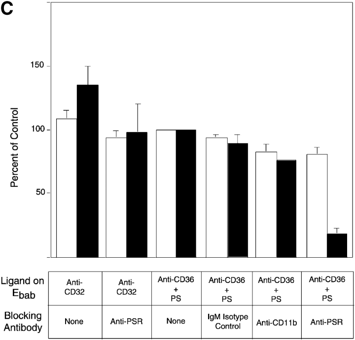
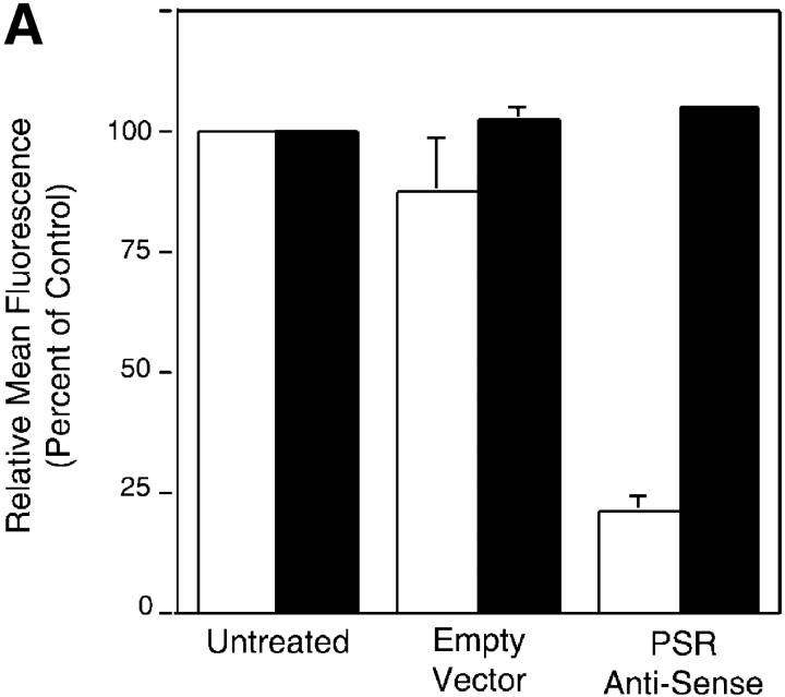
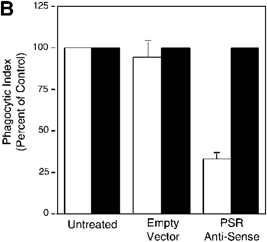
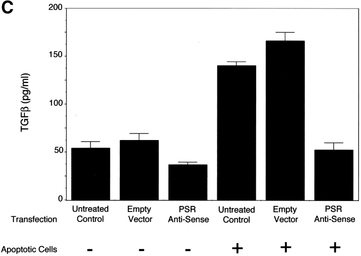
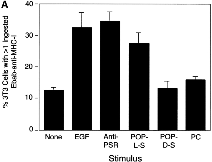
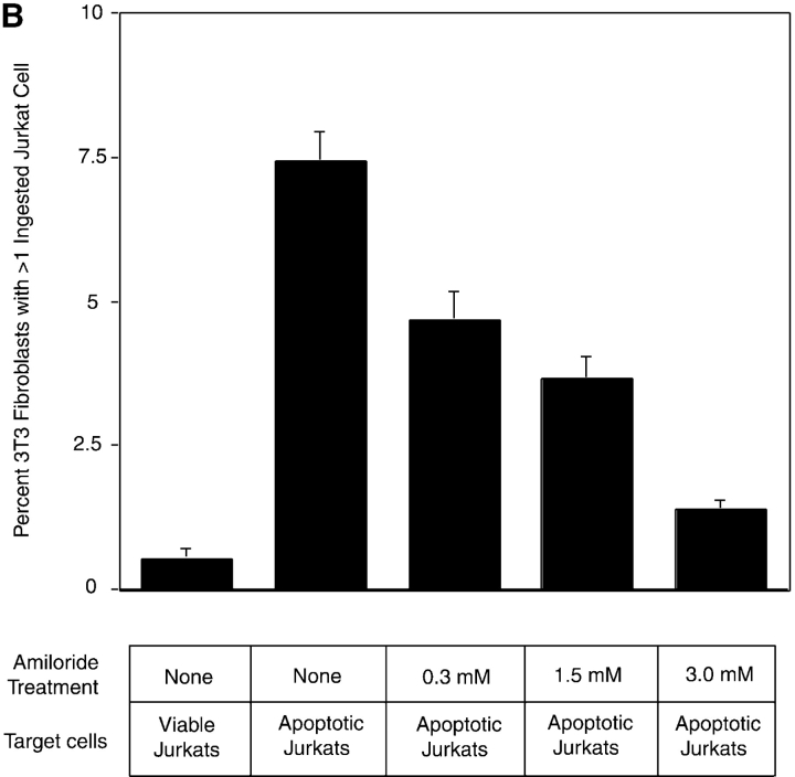
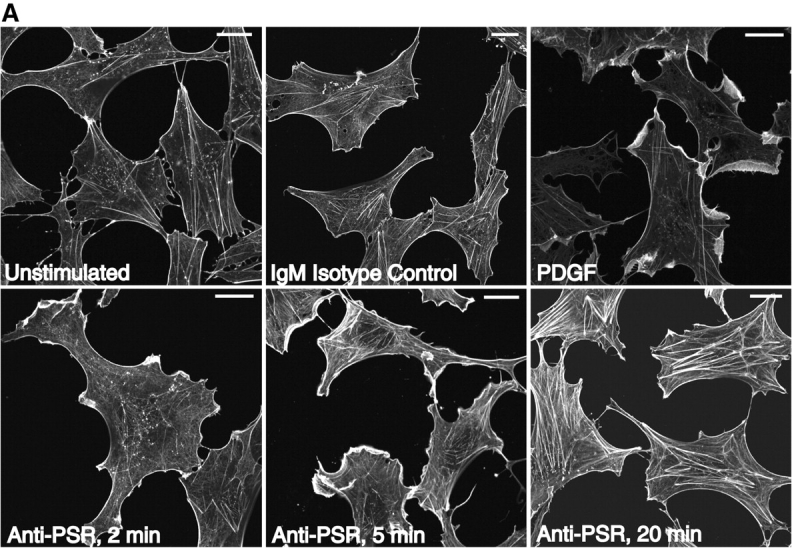
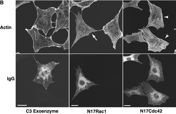
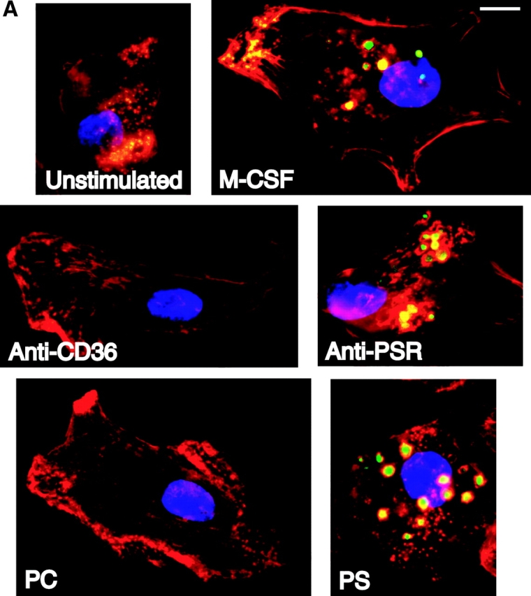
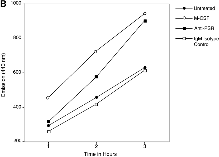
Efficient phagocytosis of apoptotic cells is important for normal tissue development, homeostasis, and the resolution of inflammation. Although many receptors have been implicated in the clearance of apoptotic cells, the roles of these receptors in the engulfment process have not been well defined. We developed a novel system to distinguish between receptors involved in tethering of apoptotic cells versus those inducing their uptake. Our results suggest that regardless of the receptors engaged on the phagocyte, ingestion does not occur in the absence of phosphatidylserine (PS). Further, recognition of PS was found to be dependent on the presence of the PS receptor (PSR). Both PS and anti-PSR antibodies stimulated membrane ruffling, vesicle formation, and "bystander" uptake of cells bound to the surface of the phagocyte. We propose that the phagocytosis of apoptotic cells requires two events: tethering followed by PS-stimulated, PSR-mediated macropinocytosis.


Figures

Figure 1.
Figure 1.
Erythrocytes can be coated with antibodies, protein ligands, or aminophospholipids targeting specific phagocytic receptors. Binding and uptake of erythrocytes by macrophages can then be assessed quantitatively. (A) Cytofluorographs demonstrate the efficient coating of erythrocytes with antibodies, protein ligands, or different forms of PS. Cy3-conjugated antibodies were used to detect either biotinylated antibodies or biotinylated proteins on the surface of erythrocytes. Phycoerythrin–annexin V was used to detect various forms of PS on the surface of erythrocytes. Histogram in black demonstrates unstained erythrocytes. (B) Light microscopy was used to accurately assess the binding or uptake of Ebab by HMDM. Erythrocytes coated with either an irrelevant IgM isotype control (anti-TNP), anti-CD32, or anti-CD36 were incubated with HMDM for 30 min. After washing away nonadherent Ebab, distilled H2O was used to lyse the Ebab bound but not engulfed by the HMDM. This figure illustrates the high level of both tethering and engulfment of Ebab–anti-CD32 by HMDM, whereas Ebab–anti-CD36 were mostly tethered but not engulfed.
Figure 1.
Figure 1.
Erythrocytes can be coated with antibodies, protein ligands, or aminophospholipids targeting specific phagocytic receptors. Binding and uptake of erythrocytes by macrophages can then be assessed quantitatively. (A) Cytofluorographs demonstrate the efficient coating of erythrocytes with antibodies, protein ligands, or different forms of PS. Cy3-conjugated antibodies were used to detect either biotinylated antibodies or biotinylated proteins on the surface of erythrocytes. Phycoerythrin–annexin V was used to detect various forms of PS on the surface of erythrocytes. Histogram in black demonstrates unstained erythrocytes. (B) Light microscopy was used to accurately assess the binding or uptake of Ebab by HMDM. Erythrocytes coated with either an irrelevant IgM isotype control (anti-TNP), anti-CD32, or anti-CD36 were incubated with HMDM for 30 min. After washing away nonadherent Ebab, distilled H2O was used to lyse the Ebab bound but not engulfed by the HMDM. This figure illustrates the high level of both tethering and engulfment of Ebab–anti-CD32 by HMDM, whereas Ebab–anti-CD36 were mostly tethered but not engulfed.
Figure 2.
Figure 2.
Many proposed phagocytic receptors promote tethering, but not engulfment. Ebab targeting various receptors on the surface of HMDM were used to investigate the role of the receptors in binding versus uptake. All results represent mean ± SEM from three or more experiments. (A) Ebab coated with antibodies against several phagocytic receptors, either individually or in combination, tethered to HMDM, but were not efficiently engulfed. (B) Ebab coated with antibodies against the αvβ5 integrin also demonstrated tethering to HMDM with little uptake. (C) Ebab coated with vitronectin or thrombospondin, natural ligands for αvβ3 integrin or αvβ3/CD36, respectively, were not efficiently engulfed by HMDM. (D) Ebab coated with anti-SRA antibodies, tethered to murine macrophages, but were not efficiently engulfed compared with positive controls. ▪, Engulfed; □, tethered.
Figure 2.
Figure 2.
Many proposed phagocytic receptors promote tethering, but not engulfment. Ebab targeting various receptors on the surface of HMDM were used to investigate the role of the receptors in binding versus uptake. All results represent mean ± SEM from three or more experiments. (A) Ebab coated with antibodies against several phagocytic receptors, either individually or in combination, tethered to HMDM, but were not efficiently engulfed. (B) Ebab coated with antibodies against the αvβ5 integrin also demonstrated tethering to HMDM with little uptake. (C) Ebab coated with vitronectin or thrombospondin, natural ligands for αvβ3 integrin or αvβ3/CD36, respectively, were not efficiently engulfed by HMDM. (D) Ebab coated with anti-SRA antibodies, tethered to murine macrophages, but were not efficiently engulfed compared with positive controls. ▪, Engulfed; □, tethered.
Figure 3.
Figure 3.
Presence of PS on the surface of tethered Ebab results in their uptake in a stereo-specific manner. Erythrocytes coated with PS (or PC, as a control) were incubated with HMDM. All results represent mean ± SEM. from three or more experiments. (A) Addition of PS, but not PC, to erythrocytes resulted in little tethering or uptake by HMDM. Addition of PS to various Ebab induced their engulfment by HMDM, whereas PC had no effect. (B) To address the stereo specificity of PS-mediated Ebab engulfment, POP-L-S or POP-D-S was added to the erythrocyte membrane. Addition of POP-L-S induced uptake of tethered Ebab by HMDM to similar levels observed with bovine brain PS, whereas POP-D-S did not. (C) Preincubation of HMDM with anti-PSR antibodies blocked the engulfment of PS-coated Ebab, but minimally affected engulfment of Ebab via FcγR. Preincubation of HMDM with other IgM antibodies did not block engulfment of PS-coated Ebab. ▪, Engulfed; □, tethered.
Figure 3.
Figure 3.
Presence of PS on the surface of tethered Ebab results in their uptake in a stereo-specific manner. Erythrocytes coated with PS (or PC, as a control) were incubated with HMDM. All results represent mean ± SEM. from three or more experiments. (A) Addition of PS, but not PC, to erythrocytes resulted in little tethering or uptake by HMDM. Addition of PS to various Ebab induced their engulfment by HMDM, whereas PC had no effect. (B) To address the stereo specificity of PS-mediated Ebab engulfment, POP-L-S or POP-D-S was added to the erythrocyte membrane. Addition of POP-L-S induced uptake of tethered Ebab by HMDM to similar levels observed with bovine brain PS, whereas POP-D-S did not. (C) Preincubation of HMDM with anti-PSR antibodies blocked the engulfment of PS-coated Ebab, but minimally affected engulfment of Ebab via FcγR. Preincubation of HMDM with other IgM antibodies did not block engulfment of PS-coated Ebab. ▪, Engulfed; □, tethered.
Figure 4.
Figure 4.
TGF-β secretion and PS-dependent engulfment of targeted erythrocytes and apoptotic cells is mediated by PSR. (A) Transfection of NIH 3T3 fibroblasts with vectors containing PSR antisense DNA resulted in a decrease of PSR expression to 25% of control levels, as measured by flow cytometry. Antisense transfection did not alter the levels of other phagocytic receptors, shown here by flow cytometric analysis of CD51 (αv integrin) expression. Transfection with sense PSR (unpublished data) or empty vector controls did not alter levels of PSR expression. □, PSR expression; ▪, αv expression. (B) The decreased expression of PSR induced by antisense transfection (Fig. 3 A) resulted in a decrease of apoptotic cell engulfment compared with control levels. However, antisense PSR DNA transfection did not alter the ability of these cells to engulf latex beads. Data were normalized to control (unmanipulated) cells. All results represent mean ± SEM from at least three experiments. □, Apoptotic cell uptake; ▪, latex bead uptake. (C) PSR is essential for TGF-β release by phagocytes during uptake of apoptotic cells. NIH 3T3 fibroblasts showed decreased production of TGF-β after an overnight feeding of apoptotic cells, as compared with unmanipulated fibroblasts and fibroblasts transfected with empty vector controls. All results represent mean ± SEM from at least two experiments.
Figure 4.
Figure 4.
TGF-β secretion and PS-dependent engulfment of targeted erythrocytes and apoptotic cells is mediated by PSR. (A) Transfection of NIH 3T3 fibroblasts with vectors containing PSR antisense DNA resulted in a decrease of PSR expression to 25% of control levels, as measured by flow cytometry. Antisense transfection did not alter the levels of other phagocytic receptors, shown here by flow cytometric analysis of CD51 (αv integrin) expression. Transfection with sense PSR (unpublished data) or empty vector controls did not alter levels of PSR expression. □, PSR expression; ▪, αv expression. (B) The decreased expression of PSR induced by antisense transfection (Fig. 3 A) resulted in a decrease of apoptotic cell engulfment compared with control levels. However, antisense PSR DNA transfection did not alter the ability of these cells to engulf latex beads. Data were normalized to control (unmanipulated) cells. All results represent mean ± SEM from at least three experiments. □, Apoptotic cell uptake; ▪, latex bead uptake. (C) PSR is essential for TGF-β release by phagocytes during uptake of apoptotic cells. NIH 3T3 fibroblasts showed decreased production of TGF-β after an overnight feeding of apoptotic cells, as compared with unmanipulated fibroblasts and fibroblasts transfected with empty vector controls. All results represent mean ± SEM from at least two experiments.
Figure 5.
Figure 5.
PS- and PSR-mediated uptake of apoptotic cells occurs by a process akin to macropinocytosis. (A) To separately examine the tethering and engulfment events, Ebab were first allowed to tether to MHC-I on the phagocyte surface for 15 min, followed by stimulation with various forms of PS, anti-PSR antibody, or EGF (positive control) for 45 min. Ebab–anti-MHC-I were engulfed by fibroblasts when stimulated with EGF, anti-PSR, or POP-L-S, but not when stimulated with POP-D-S stereoisomer or PC. Results represent mean ± SEM from three experiments. (B) Amiloride, a specific inhibitor of macropinocytosis, inhibited the engulfment of apoptotic Jurkat T cells by NIH 3T3 fibroblasts at concentrations as low as 0.3 mM. Results represent mean ± SEM from three experiments.
Figure 5.
Figure 5.
PS- and PSR-mediated uptake of apoptotic cells occurs by a process akin to macropinocytosis. (A) To separately examine the tethering and engulfment events, Ebab were first allowed to tether to MHC-I on the phagocyte surface for 15 min, followed by stimulation with various forms of PS, anti-PSR antibody, or EGF (positive control) for 45 min. Ebab–anti-MHC-I were engulfed by fibroblasts when stimulated with EGF, anti-PSR, or POP-L-S, but not when stimulated with POP-D-S stereoisomer or PC. Results represent mean ± SEM from three experiments. (B) Amiloride, a specific inhibitor of macropinocytosis, inhibited the engulfment of apoptotic Jurkat T cells by NIH 3T3 fibroblasts at concentrations as low as 0.3 mM. Results represent mean ± SEM from three experiments.
Figure 6.
Figure 6.
Anti-PSR antibody stimulates membrane ruffling in Swiss 3T3 fibroblasts. (A) Anti-PSR antibodies induce membrane ruffling in Swiss 3T3 fibroblasts within 2 min of antibody stimulation. Membrane ruffling is optimally induced by 5 min, and stress fiber formation is observed by 20 min (bottom). These results are not seen with an IgM isotype control antibody (top, center) or in unstimulated cells (top, left). PDGF-induced membrane ruffling at 10 min is shown as a positive control (top, right). (B) Quiescent Swiss 3T3 fibroblasts were co-microinjected with rat IgG and C3 exoenzyme (a specific inhibitor of RhoA), N17Cdc42, or N17Rac, and stimulated with IgM anti-PSR for 10 min. Cells were fixed and stained for injected antibody (bottom) and actin (top). Representative injected cells for each experiment are shown. Microinjection of C3 exoenzyme did not inhibit anti-PSR–induced membrane ruffling (left), although it inhibited FCS-induced stress fiber formation (not shown). (Center) N17Rac1 inhibited anti-PSR–induced membrane ruffling and resulted in formation of filopodia (arrow). (Right) Cells injected with N17Cdc42 also did not ruffle in response to anti-PSR stimulation, and had enhanced stress fiber formation (arrowheads). Bars: (A) 2 μm; (B) 20 μm.
Figure 6.
Figure 6.
Anti-PSR antibody stimulates membrane ruffling in Swiss 3T3 fibroblasts. (A) Anti-PSR antibodies induce membrane ruffling in Swiss 3T3 fibroblasts within 2 min of antibody stimulation. Membrane ruffling is optimally induced by 5 min, and stress fiber formation is observed by 20 min (bottom). These results are not seen with an IgM isotype control antibody (top, center) or in unstimulated cells (top, left). PDGF-induced membrane ruffling at 10 min is shown as a positive control (top, right). (B) Quiescent Swiss 3T3 fibroblasts were co-microinjected with rat IgG and C3 exoenzyme (a specific inhibitor of RhoA), N17Cdc42, or N17Rac, and stimulated with IgM anti-PSR for 10 min. Cells were fixed and stained for injected antibody (bottom) and actin (top). Representative injected cells for each experiment are shown. Microinjection of C3 exoenzyme did not inhibit anti-PSR–induced membrane ruffling (left), although it inhibited FCS-induced stress fiber formation (not shown). (Center) N17Rac1 inhibited anti-PSR–induced membrane ruffling and resulted in formation of filopodia (arrow). (Right) Cells injected with N17Cdc42 also did not ruffle in response to anti-PSR stimulation, and had enhanced stress fiber formation (arrowheads). Bars: (A) 2 μm; (B) 20 μm.
Figure 7.
Figure 7.
Anti-PSR antibodies and PS stimulate macropinocytosis. (A) HMDM were stimulated with M-CSF (as a positive control), antibodies to the PSR or CD36, PS, or PC. Vesicle formation was visualized using LY (green). Nuclei were stained with DAPI (blue), and cells were stained for actin with rhomadine–phalloidin (red). Macrophages stimulated with either anti-PSR antibodies or PS formed vesicles similar to those formed after stimulation with M-CSF, a known inducer of macropinocytosis. Representative images are shown. (B) Accumulation of Cascade blue was quantified by fluorimetry. MEM were untreated or stimulated with M-CSF, anti-PSR antibody, or an IgM isotype control antibody, in the presence of dye. At the designated time point, cells were washed, lysed, and fluorescence of the lysate was measured. Similar results were obtained on four separate occasions. Bar, 5 μm.
Figure 7.
Figure 7.
Anti-PSR antibodies and PS stimulate macropinocytosis. (A) HMDM were stimulated with M-CSF (as a positive control), antibodies to the PSR or CD36, PS, or PC. Vesicle formation was visualized using LY (green). Nuclei were stained with DAPI (blue), and cells were stained for actin with rhomadine–phalloidin (red). Macrophages stimulated with either anti-PSR antibodies or PS formed vesicles similar to those formed after stimulation with M-CSF, a known inducer of macropinocytosis. Representative images are shown. (B) Accumulation of Cascade blue was quantified by fluorimetry. MEM were untreated or stimulated with M-CSF, anti-PSR antibody, or an IgM isotype control antibody, in the presence of dye. At the designated time point, cells were washed, lysed, and fluorescence of the lysate was measured. Similar results were obtained on four separate occasions. Bar, 5 μm.

Comment in

Similar articles

Cited by

References

    1. Albert, M.L., S.F. Pearce, L.M. Francisco, B. Sauter, P. Roy, R.L. Silverstein, and N. Bhardwaj. 1998. Immature dendritic cells phagocytose apoptotic cells via alphavbeta5 and CD36, and cross-present antigens to cytotoxic T lymphocytes. J. Exp. Med. 188:1359–1368. - PMC - PubMed
    1. Albert, M.L., J.I. Kim, and R.B. Birge. 2000. alphavbeta5 integrin recruits the CrkII-Dock180-rac1 complex for phagocytosis of apoptotic cells. Nat. Cell Biol. 2:899–905. - PubMed
    1. Balasubramanian, K., and A.J. Schroit. 1998. Characterization of phosphatidylserine-dependent beta2-glycoprotein I macrophage interactions. Implications for apoptotic cell clearance by phagocytes. J. Biol. Chem. 273:29272–29277. - PubMed
    1. Balasubramanian, K., J. Chandra, and A.J. Schroit. 1997. Immune clearance of phosphatidylserine-expressing cells by phagocytes. The role of beta2-glycoprotein I in macrophage recognition. J. Biol. Chem. 272:31113–31117. - PubMed
    1. Bretscher, M.S., and C. Aguado-Velasco. 1998. Membrane traffic during cell locomotion. Curr. Opin. Cell Biol. 10:537–541. - PubMed

Publication types