. 2001 Nov;127(3):777-91.

Developmental and thermal regulation of the maize heat shock protein, HSP101

Affiliations

Developmental and thermal regulation of the maize heat shock protein, HSP101

T E Young et al. Plant Physiol. 2001 Nov.

Abstract

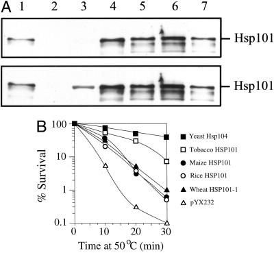
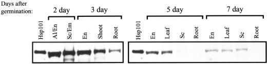
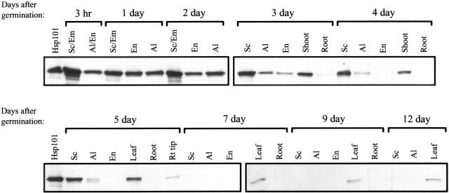
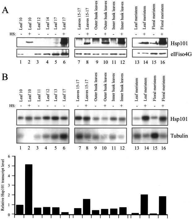
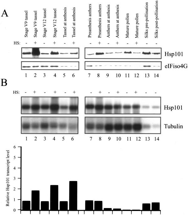
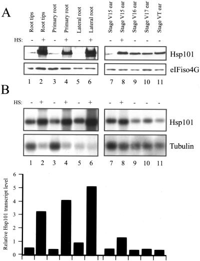
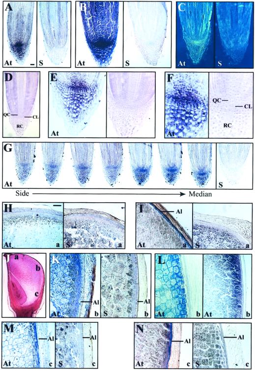
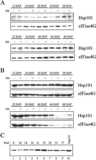
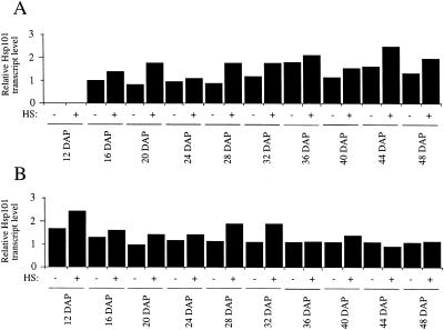
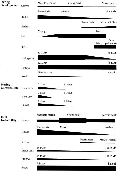
The plant heat stress protein, Hsp101, and the yeast ortholog, Hsp104, are required to confer thermotolerance in plants and yeast (Saccharomyces cerevisiae), respectively. In addition to its function during stress, Hsp101 is developmentally regulated in plants although its function during development is not known. To determine how the expression of Hsp101 is regulated in cereals, we investigated the Hsp101 expression profile in developing maize (Zea mays). Hsp101 protein was most abundant in the developing tassel, ear, silks, endosperm, and embryo. It was less abundant in the vegetative and floral meristematic regions and was present at only a low level in the anthers and tassel at anthesis, mature pollen, roots, and leaves. As expected, heat treatment resulted in an increase in the level of Hsp101 protein in several organs. In expanding foliar leaves, husk leaves, the tassel at the premeiosis stage of development, or pre-anthesis anthers, however, the heat-mediated increase in protein was not accompanied by an equivalent increase in mRNA. In contrast, the level of Hsp101 transcript increased in the tassel at anthesis following a heat stress without an increase in Hsp101 protein. In other organs such as the vegetative and floral meristematic regions, fully expanded foliar leaves, the young ear, and roots, the heat-induced increase in Hsp101 protein was accompanied by a corresponding increase in Hsp101 transcript level. However, anthers at anthesis, mature pollen, developing endosperm, and embryos largely failed to mount a heat stress response at the level of Hsp101 protein or mRNA, indicating that Hsp101 expression is not heat inducible in these organs. In situ RNA localization analysis revealed that Hsp101 mRNA accumulated in the subaleurone and aleurone of developing kernels and was highest in the root cap meristem and quiescent center of heat-stressed roots. These data suggest an organ-specific control of Hsp101 expression during development and following a heat stress through mechanisms that may include posttranscriptional regulation.


Figures

Figure 1
Figure 1
Alignment of Hsp101 from monocot and dicot species. The complete sequence for maize Hsp101 (accession no. AF133840) is shown and only those positions within OsHsp101 (rice; accession no. AF332981), TaHsp101-1 and TaHsp101-2 (wheat; accession nos. AF083344 and AF097363, respectively), NtHsp101 (tobacco; accession no. AF083343), GmHsp101 (soybean; accession no. L35272), and AtHsp101 (Arabidopsis; accession no. U13949) that differ from the maize ortholog are indicated. Identity with maize Hsp101 is indicated by dots, whereas gaps introduced to maintain alignment are indicated by dashes. The start of each of the five domains is indicated by an arrow. Conserved signature sequences (Schirmer et al., 1996) within each domain are indicated above the maize amino acid sequence where non-conserved or hydrophobic amino acids are indicated with an “x” or “h,” respectively, and the roman numeral or letter designations by which they are known (Schirmer et al., 1996) appears above each conserved signature sequence.
Figure 2
Figure 2
Analysis of tobacco, maize, rice, and wheat Hsp101 expression and thermotolerance function in yeast. Each Hsp101 cDNA was introduced into a yeast expression vector under the control of the TPI promoter and the constructs introduced into the hsp104 yeast mutant, SL304A. A, Yeast extract from 6 × 10⁶ exponentially growing cells was resolved on a 10% (w/v) SDS-PAGE gel, transferred to nitrocellulose membrane, probed using anti-wheat Hsp101 antibodies (top), and then probed with anti-yeast Hsp104 antibodies (bottom). Detection in each case used peroxidase-linked secondary antibody and chemiluminescence. Lane 1, Purified wheat Hsp101; lane 2, yeast containing pYX232; lane 3, yeast expressing Hsp104; lane 4, yeast expressing tobacco Hsp101; lane 5, yeast expressing wheat Hsp101-1; lane 6, yeast expressing maize Hsp101; and lane 7, yeast expressing rice Hsp101. The bands below the full-length Hsp101 represent degradation products. B, SL304A, containing the tobacco, maize, rice, and wheat Hsp101-1 cDNAs or yeast Hsp104 under the control of the TPI promoter, was grown to an early exponential stage in synthetic dextrose medium prior to assaying for thermotolerance. The expression vector, pYX232, was used as a negative control. The percentage of survival at 50oC was plotted as a function of the length of the heat treatment. The results shown are representative for these constructs under the conditions employed.
Figure 3
Figure 3
Presence of Hsp101 in wheat seedling tissues. Wheat seedlings were grown for the times indicated above the panels and the seedlings dissected into endosperm (En), aleurone (Al), embryo (Em), scutellum (Sc), shoot, and root tissue. Five micrograms of soluble protein extracted from each tissue was resolved using SDS-PAGE, transferred to nitrocellulose membrane, and incubated with anti-Hsp101 antibodies. Detection used peroxidase-linked secondary antibody and chemiluminescence. Purified wheat Hsp101 was included as a control in the first lane of each panel.
Figure 4
Figure 4
Presence of Hsp101 in germinating maize seedling tissues. Maize seedlings were grown for the times indicated above the panels and the seedlings dissected into endosperm (En), aleurone (Al), embryo (Em), scutellum (Sc), shoot, leaf, and root or root tip tissue. Note that the scutellum tissue includes the embryonic axis for the 3-h, 1- and 2-d germinated kernels (Sc/Em), whereas the endosperm tissue includes the aleurone layer for the 3-h germinated kernels (Al/En). Five micrograms of soluble protein extracted from each tissue was resolved using SDS-PAGE, transferred to nitrocellulose membrane, and incubated with anti-Hsp101 antibodies. Purified wheat Hsp101 was included as a control in the first lane of each left panel.
Figure 5
Figure 5
Developmental and heat-regulated expression of Hsp101 in maize foliar and husk leaves. A, The level of maize Hsp101 protein was determined by western analysis in the organs indicated above each lane. Total protein was isolated, transferred to nitrocellulose membrane following resolution using SDS-PAGE, and probed for Hsp101 or eIFiso4G. Whether an organ was heat stressed (HS) at 41°C for 1 h or maintained at 21°C is indicated above each lane. B, The level of maize Hsp101 mRNA was determined by northern analysis in the organs indicated above each lane. Poly(A+) mRNA was isolated, transferred to nylon membrane following resolution on a 7% (w/v) formaldehyde/MOPS [3-(N-morpholino)-propanesulfonic acid] gel, and probed with maize Hsp101 or maize tubulin antisense RNA. The level of Hsp101 mRNA was quantitated by densitometry, normalized to the ribosomal RNA and tubulin mRNA, and expressed in the histograms relative to the level of Hsp101 mRNA in the fully expanded, foliar leaf 10 that was set at a value of 1. Numbering of maize leaves begins with the first leaf that emerges from the kernel. All foliar leaves were collected at the same time from six plants to eliminate plant-to-plant variation. The husk leaves were collected at silking stage.
Figure 6
Figure 6
Developmental and heat-regulated expression of Hsp101 in maize tassel, anthers, pollen, and silks. A, The level of maize Hsp101 protein was determined by western analysis in the organs indicated above each lane. The western analysis was performed as described in Figure 5 and in the same experiment. Whether an organ was heat stressed (HS) at 41°C for 1 h or maintained at 21°C is indicated above each lane. B, The level of maize Hsp101 mRNA was determined by northern analysis in the organs indicated above each lane. The northern analysis was performed as described in Figure 5 and in the same experiment. The level of Hsp101 mRNA was quantitated by densitometry, normalized to the ribosomal RNA and tubulin mRNA, and expressed in the histograms relative to the level of Hsp101 mRNA in the fully expanded, foliar leaf 10 (as shown in Fig. 5) that was set at a value of 1. The preanthesis anthers were taken 1 week prior to anthesis. The post-pollination silks were taken 1 week following pollination. The organs were collected from six plants to eliminate plant-to-plant variation.
Figure 7
Figure 7
Developmental and heat-regulated expression of Hsp101 in maize roots and ear. A, The level of maize Hsp101 protein was determined by western analysis in the organs indicated above each lane. The western analysis was performed as described in Figure 5 and in the same experiment. Whether an organ was heat-stressed (HS) at 41°C for 1 h or maintained at 21°C is indicated above each lane. B, The level of maize Hsp101 mRNA was determined by northern analysis in the organs indicated above each lane. The northern analysis was performed as described in Figure 5 and in the same experiment. The level of Hsp101 mRNA was quantitated by densitometry, normalized to the ribosomal RNA and tubulin mRNA, and expressed in the histograms relative to the level of Hsp101 mRNA in the fully expanded, foliar leaf 10 (as shown in Fig. 5) that was set at a value of 1. The roots used for this analysis were taken from a 4-week-old plant and the terminal 5 mm was collected. Root tips represented the terminal 2 mm of root tissue. The organs were collected from six plants to eliminate plant-to-plant variation.
Figure 8
Figure 8
In situ localization of Hsp101 mRNA during maize root and kernel development. Sections of root (A–G) and kernels (H–N) were hybridized with riboprobes containing digoxygenin. Hybridization was detected as a blue precipitate by staining with nitroblue tetrazolium. A, Median section of a 10-d-old, heat-stressed (all heat-treatments performed at 41oC for 1 h) root probed with a low concentration of antisense (At) Hsp101 RNA to detect Hsp101 mRNA or sense (S) RNA to serve as a control. B, Median section of a 10-d-old, heat-stressed root probed with a high concentration of antisense or sense RNA. C, Median section of a non-stressed root in which the positive signal (At) is represented by yellow staining region in this inverted image. D, Median section of 10-d-old root stained with safranin O to show the QC, CL, and RC. E, Median section of 10-d-old, heat-stressed root tip probed with antisense Hsp101 RNA (left) or stained with safranin O (right). F, Median section at high magnification of 10-d-old, heat-stressed root tip probed with antisense Hsp101 RNA (left) or stained with safranin O (right). G, Serial sections from the side to the median section of a 12-d-old, heat-stressed root probed with antisense RNA (first seven images) to detect Hsp101 mRNA or sense RNA (right) to serve as a control. H, Median section of the crown region of a 26-d after pollination (DAP) developing kernel probed with antisense Hsp101 (At) RNA to detect Hsp101 mRNA or sense (S) RNA to serve as a control. I, Median section of the crown region of an 18-DAP developing kernel probed with antisense Hsp101 (At) or sense (S) RNA. J, Median section of an 18-DAP kernel stained with safranin O to illustrates the regions (labeled a, b, or c) analyzed in H through N. K, Median section (region b) of a 26-DAP kernel probed with antisense Hsp101 (At) or sense (S) RNA. L, Median section (region b) of an 18-DAP kernel probed with antisense α-zein RNA at a high (left) or low (right) magnification. M, Median section (region c) of a 26-DAP kernel probed with antisense Hsp101 (At) or sense (S) RNA. N, Median section (region c) of an 18-DAP kernel probed with antisense Hsp101 (At) or sense (S) RNA. The bar in A represents 100 mm and in H represents 350 mm.
Figure 9
Figure 9
The level of Hsp101 protein is developmentally regulated during seed development in maize and wheat. Hsp101 protein in maize embryo (A) or endosperm (B) from kernels collected at DAP or from whole wheat seed (C) collected at selected days after flowering (DAF) was measured by western analysis. Total protein was resolved using SDS-PAGE, transferred to nitrocellulose membrane, and incubated with anti-Hsp101 antibodies or reprobed with anti-eIFiso4G antibodies. A and B, The heat-stress treatment (HS) is indicated above the lanes. The 12-DAP samples represent whole kernels. C, Purified wheat Hsp101 (lane 10) was included as a control.
Figure 10
Figure 10
Hsp101 transcript level is not significantly induced in developing maize endosperm and embryo following a heat stress. Hsp101 transcript abundance in maize embryo (A) or endosperm (B) from kernels collected at time points following pollination was measured by northern analysis and normalized to maize tubulin RNA. The level of Hsp101 RNA is indicated relative to that in non-stressed 16-DAP embryos, which is given a value of 1. DAP and the heat stress treatment (HS) are indicated below the histograms. The 12-DAP samples represent whole kernels.
Figure 11
Figure 11
Summary of the relative expression of Hsp101 protein in maize during development, germination, and following a heat stress. The change in the level of Hsp101 protein during the development of those organs analyzed in Figures 4 through 7 and 9 are illustrated where changes in the thickness of a bar represents an estimate of the degree of change in the level of Hsp101.

Similar articles

Cited by

References

    1. Becker J, Craig EA. Heat-shock proteins as molecular chaperones. Eur J Biochem. 1994;219:11–23. - PubMed
    1. Bradford MM. A rapid and sensitive method for the quantitation of microgram quantities of protein utilizing the principle of protein-dye binding. Anal Biochem. 1976;72:248–254. - PubMed
    1. Browning KS, Lax SR, Ravel JM. Identification of two messenger RNA cap binding proteins in wheat germ: evidence that the 28-kDa subunit of eIF-4B and the 26-kDa subunit of eIF-4F are antigenically distinct polypeptides. J Biol Chem. 1987;262:11228–11232. - PubMed
    1. Caliebe A, Soill J. News in chloroplast protein import. Plant Mol Biol. 1999;39:641–645. - PubMed
    1. Caplan AJ. Hsp90's secrets unfold: new insights from structural and functional studies. Trends Cell Biol. 1999;9:262–268. - PubMed

Publication types

MeSH terms

LinkOut - more resources