. 2001 Nov;127(3):803-16.

The Arabidopsis SOMATIC EMBRYOGENESIS RECEPTOR KINASE 1 gene is expressed in developing ovules and embryos and enhances embryogenic competence in culture

Affiliations

The Arabidopsis SOMATIC EMBRYOGENESIS RECEPTOR KINASE 1 gene is expressed in developing ovules and embryos and enhances embryogenic competence in culture

V Hecht et al. Plant Physiol. 2001 Nov.

Erratum in

  • Plant Physiol 2002 Jan;128(1):314

Abstract

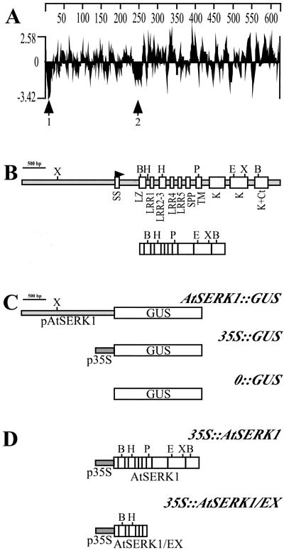
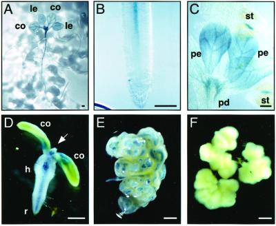
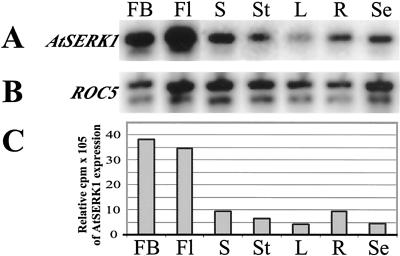
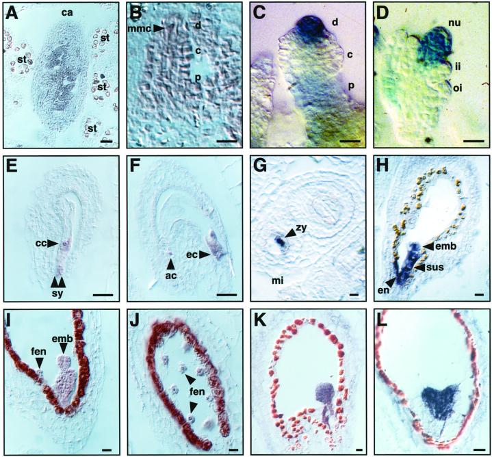
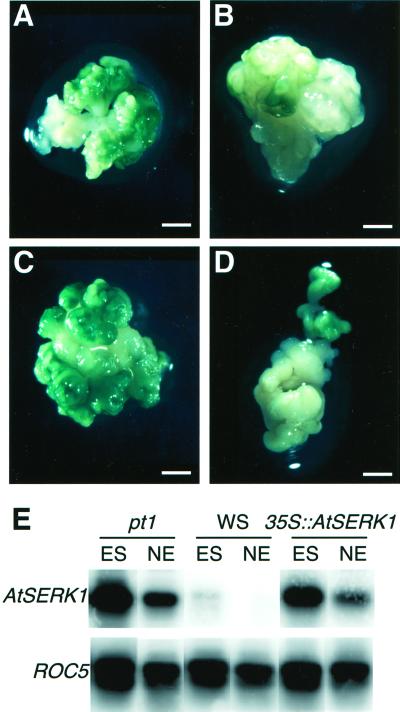
We report here the isolation of the Arabidopsis SOMATIC EMBRYOGENESIS RECEPTOR-LIKE KINASE 1 (AtSERK1) gene and we demonstrate its role during establishment of somatic embryogenesis in culture. The AtSERK1 gene is highly expressed during embryogenic cell formation in culture and during early embryogenesis. The AtSERK1 gene is first expressed in planta during megasporogenesis in the nucellus [corrected] of developing ovules, in the functional megaspore, and in all cells of the embryo sac up to fertilization. After fertilization, AtSERK1 expression is seen in all cells of the developing embryo until the heart stage. After this stage, AtSERK1 expression is no longer detectable in the embryo or in any part of the developing seed. Low expression is detected in adult vascular tissue. Ectopic expression of the full-length AtSERK1 cDNA under the control of the cauliflower mosaic virus 35S promoter did not result in any altered plant phenotype. However, seedlings that overexpressed the AtSERK1 mRNA exhibited a 3- to 4-fold increase in efficiency for initiation of somatic embryogenesis. Thus, an increased AtSERK1 level is sufficient to confer embryogenic competence in culture.


Figures

Figure 1
Figure 1
Description of the Arabidopsis SERK1 gene and protein. A, Kyte-Doolitle hydrophylicity plot of AtSERK1 protein. Arrows 1 and 2 indicate the two hydrophobic regions of the protein. B, Genomic organization of AtSERK1 gene in Arabidopsis. Up, Genomic DNA; down, cDNA. White boxes indicate exons and gray boxes indicate non-coding regions. SS, Signal sequence; SPP, Pro-rich domain containing the SPP motif; TM, transmembrane domain; K, kinase domain; Ct, C terminus. C, Constructs used for expression analysis. The AtSERK1 2-kb promoter was fused to the GUS reporter gene in AtSERK1::GUS construct. As positive control, the cauliflower mosaic virus (CaMV) 35S promoter was fused to GUS 35S::GUS, and as negative control, the GUS gene was promoterless 0::GUS. D, Constructs used for overexpression analysis. 35S::AtSERK1 and 35S::AtSERK1-EX are fusions between AtSERK1 complete or partial cDNA to the 35S promoter. Restriction sites: B, BamHI; E, EcoRI; H, HindIII; P, PstI; and X, XbaI.
Figure 2
Figure 2
AtSERK1 expression pattern postembryonically and in embryogenic cultures. GUS expression of plants containing the AtSERK1::GUS construct was followed throughout plant life (A–C) and during induction of embryogenic cultures in amp1 seedlings (D–F). A, Seedling 15 d after germination (DAG). B, Root tip of the seedling shown in A. C, Flower of an adult plant. D, 4 DAG seedlings germinated in presence of 2, 4-D; arrow indicates the SAM. E, Embryogenic callus at 28 DAG. F, Nonembryogenic callus after 40 DAG. Bar, 1 mm; co, cotyledon; h, hypocotyl; le, primary leaves; pd, pedicel; pe, petal; r, root; st, stamen.
Figure 3
Figure 3
AtSERK1 expression during plant development determined by semiquantitative RT-PCR. A, AtSERK1 expression pattern at 30 cycles. B, ROC5 expression pattern at 30 cycles. C, Relative expression of AtSERK1. FB, Flower buds; Fl, opened flowers containing embryos from stages 0 through 7; S, siliques containing embryos from stages 7 through 20; St, stems; L, rosette leaves; R, roots; Se, seedlings 7 DAG.
Figure 4
Figure 4
Expression of the AtSERK1 in developing ovules and seeds. Expression pattern determined by ISH or by GUS staining (GUS). A, Transversal section of a flower bud containing young ovule primordia (ISH). B, Ovule primordia at stage 2-II, arrow indicates dividing MMC (ISH). C, Ovule primordia at stage 2-IV (GUS). D, Ovule primordia at stage 3-I (GUS). E, Mature embryo sac showing expression in the egg cell and the antipodal cells (ISH). F, Mature embryo sac showing expression in the synergids and the central cell (ISH). G, Transversal section of a developing seed containing an embryo at stage 2 (ISH). H, Longitudinal section of a developing seed containing an embryo at stage 4 (ISH). I, Longitudinal section of a developing seed containing an embryo at stage 5 (ISH). J, Longitudinal section of the same developing seed as in (I) showing the free endosperm nuclei (ISH). K, Longitudinal section of a developing seed containing an embryo at stage 8 (ISH). L, Longitudinal section of a developing seed containing an embryo at heart stage (ISH). Bars: A through D, 10 μm; E through L, 20 μm. ac, Antipodal cell; c, central; d, distal; cc, central cell; ec, egg cell; emb, embryo proper; en, endothelium; fen, free endosperm nuclei; ii, inner integument; mi, micropylar pole; nu, nucleus; oi, outer integument; p, proximal; st, stamen; su, suspensor; sy, synergids; zy, zygote.
Figure 5
Figure 5
The effect of ectopic AtSERK1 expression on embryogenic potential of seedlings. A through D, Embryogenic callus 4 weeks after initiation. A, Embryogenic callus of a amp1 culture. B, Nonembryogenic callus of a Wassilewskija (WS) wild-type culture. C, Embryogenic callus of a 35S::AtSERK1culture. D, Embryogenic callus of a 35S::AtSERK1-EX culture. E, AtSERK1 expression determined by RT-PCR after 30 cycles of callus from amp1, WS wild type, and 35S::AtSERK1 4 weeks after embryogenic culture initiation as shown in Figure 4, I through K. ROC5 expression is shown as internal control. ES, Embryogenic structures; NE, nonembryogenic structures. Bar, A through D, 1 mm.

References

    1. Albrecht C, Geurts R, Lapeyrie F, Bisseling T. Endomycorrhizae and rhizobial Nod factors both require SYM8 to induce the expression of the early nodulin genes PsENOD5 and PsENOD12A. Plant J. 1998;15:605–614. - PubMed
    1. Altschul SF, Gish W, Miller W, Myers EW, Lipman DJ. Basic alignment search tool. J Mol Biol. 1990;215:403–410. - PubMed
    1. Baudino S, Hansen S, Brettschneider R, Hecht V, Dresselhaus T, Lörz H, Dumas C, Rogowsky P. Molecular characterization of two novel maize LRR receptor-like kinases, which belong to the SERK gene family. Planta. 2001;213:1–10. - PubMed
    1. Bechtold N, Ellis J, Pelletier G. In planta Agrobacterium mediated transfer by infiltration of adult Arabidopsis thaliana plants. C R Acad Sci Paris. 1993;316:1194–1199.
    1. Becker D, Kemper E, Schell J, Masterson R. New plant binary vectors with selectable markers located proximal to the left T-DNA border. Plant Mol Biol. 1992;20:1195–1197. - PubMed

Publication types