. 2001 Nov;127(3):842-51.

Diversity of Arabidopsis genes encoding precursors for phytosulfokine, a peptide growth factor

Affiliations

Diversity of Arabidopsis genes encoding precursors for phytosulfokine, a peptide growth factor

H Yang et al. Plant Physiol. 2001 Nov.

Abstract

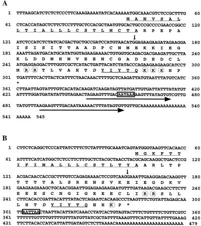
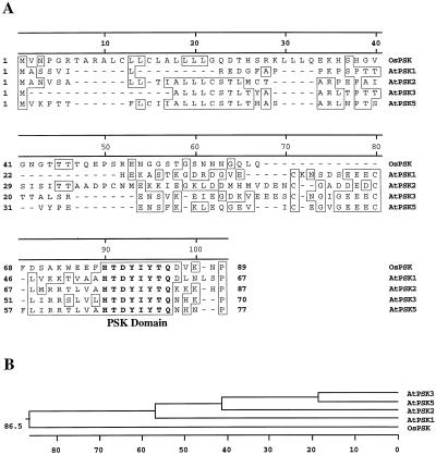
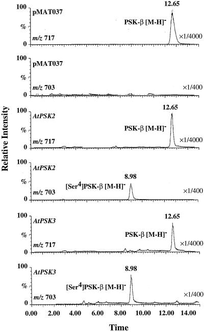
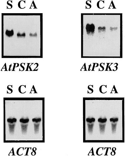
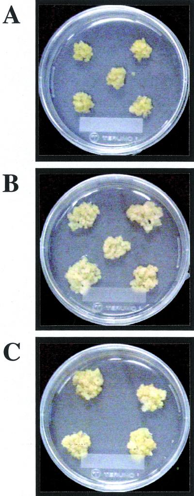
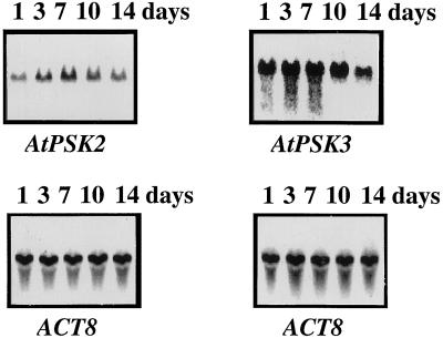
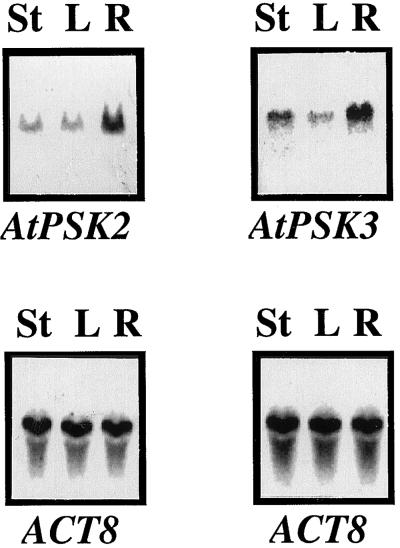
Phytosulfokine-alpha (PSK-alpha), a unique plant peptide growth factor, was originally isolated from conditioned medium of asparagus (Asparagus officinalis) mesophyll cell cultures. PSK-alpha has several biological activities including promoting plant cell proliferation. Four genes that encode precursors of PSK-alpha have been identified from Arabidopsis. Analysis of cDNAs for two of these, AtPSK2 and AtPSK3, shows that both of these genes consist of two exons and one intron. The predicted precursors have N-terminal signal peptides and only a single PSK-alpha sequence located close to their carboxyl termini. Both precursors contain dibasic processing sites flanking PSK, analogous to animal and yeast prohormones. Although the PSK domain including the sequence of PSK-alpha and three amino acids preceding it are perfectly conserved, the precursors bear very limited similarity among Arabidopsis and rice (Oryza sativa), suggesting a new level of diversity among polypeptides that are processed into the same signaling molecule in plants, a scenario not found in animals and yeast. Unnatural [serine-4]PSK-beta was found to be secreted by transgenic Arabidopsis cells expressing a mutant of either AtPSK2 or AtPSK3 cDNAs, suggesting that both AtPSK2 and AtPSK3 encode PSK-alpha precursors. AtPSK2 and AtPSK3 were expressed demonstrably not only in cultured cells but also in intact plants, suggesting that PSK-alpha may be essential for plant cell proliferation in vivo as well as in vitro. Overexpression of either precursor gene allowed the transgenic calli to grow twice as large as the controls. However, the transgenic cells expressing either antisense cDNA did not dramatically decrease mitogenic activity, suggesting that these two genes may act redundantly.


Figures

Figure 1
Figure 1
Nucleotide and deduced amino acid sequences of AtPSK2 and AtPSK3 cDNAs. The deduced amino acid sequences with single-letter abbreviations are shown below the nucleotide sequences of AtPSK2 cDNA (A) and AtPSK3 cDNA (B). The positions of introns are shown with vertical arrows. The repeats are indicated with horizontal arrows. The most likely sequences for a polyadenylation signal are boxed. The potential N-terminal signal sequences are underlined, and the PSK-α sequence is underscored with double lines. The Asp residues near PSK-α are printed in italic, and the putative bordering processing sites are circled. The nucleotide sequences reported in this paper have been deposited in the GenBank, EMBL, and DDBJ nucleotide sequence databases (accession nos. AB029344, AB052752, AB029820, and AB050627).
Figure 2
Figure 2
Amino acid sequence comparison of Arabidopsis and rice PSK precursors. The alignment (A) and phylogenetic tree (B) were generated using the MegAlign program. Single-letter abbreviations for the amino acid residues are used. Gaps are shown as dashes (−). Identical amino acid residues are boxed. The amino acid sequence of the PSK domain is in bold.
Figure 3
Figure 3
DNA gel blot showing the presence of AtPSK2 and AtPSK3 genes. Total DNA (10 μg) isolated from Arabidopsis culture cells was digested with BamHI (lane 1), EcoRI (lane 2), HindIII (lane 3), or XbaI (lane 4) and subjected to DNA gel-blot analysis using 32P-labeled AtPSK2 (A) or AtPSK3 cDNA (B) at 50°C. Marker lengths are indicated on the left in kilobases.
Figure 4
Figure 4
Mass chromatograph-obtained LC/MS analyses. The transgenic cells harboring the empty vector, the mutated AtPSK2 cDNA, or the mutated AtPSK3 cDNA were cultured in B5 liquid medium for 2 weeks to prepare CM. PSK-α and its analogs containing in the CM were concentrated by two steps of column chromatograph and subjected to LC/MS analysis with selected ion monitoring at m/z 703 ([M-H] of [Ser-4]PSK-β) and m/z 717 ([M-H] of PSK-β). The peaks eluting at 8.9 and 12.65 were [Ser-4]PSK-β and PSK-β, respectively.
Figure 5
Figure 5
Changes in AtPSK2 and AtPSK3 transcripts in transgenic cells. Twenty-microgram aliquots of total RNA extracted from sense transgenic cells (lane S), control (lane C), or antisense transgenic cells (lane A) cultured for 14 d were separated on 1.2% (w/v) agarose gels, transferred to nylon membranes, and allowed to hybridize with 32P-labeled full-length AtPSK2 or AtPSK3 cDNAs, as described in “Materials and Methods.” The blots were reprobed with an Arabidopsis actin cDNA to indicate equal loading of RNA.
Figure 6
Figure 6
Comparison of growth of control and transgenic calli. Control or transgenic cells were transplanted into fresh B5 medium containing 0.5 mg L−1 of 2,4-D, 0.05 mg L−1 of KIN, and 0.8% (w/v) agar. Photographs were taken 2 weeks after culture at 25°C under 16-h-light/8-h-dark cycle. This experiment was repeated for three times and four or five lines of each construct were shown. A, Transgenic calli harboring empty pMAT037 alone. B, Transgenic calli harboring the sense AtPSK2 chimeric gene. C, Transgenic calli harboring the sense AtPSK3 chimeric gene.
Figure 7
Figure 7
Persistent expression of AtPSK2 and AtPSK3 in cultured Arabidopsis cells. Twenty micrograms of total RNA extracted from Arabidopsis cells cultured for 1 (lane 1), 3 (lane 3), 7 (lane 7), 10 (lane 10), or 14 (lane 14) d were separated on 1.2% (w/v) agarose gels, transferred to nylon membranes, and allowed to hybridize with 32P-labeled full-length AtPSK2 or AtPSK3 cDNAs, as described in “Materials and Methods.” The blots were reprobed with an Arabidopsis actin cDNA to indicate equal loading of RNA.
Figure 8
Figure 8
Tissue specificity of AtPSK2 and AtPSK3 expression. Twenty micrograms of total RNA prepared from stems (lane S), leaves (lane L), and roots (lane R) of 28-d-old Arabidopsis plants were separated on 1.2% (w/v) agarose gels, transferred to nylon membranes, and allowed to hybridize with 32P-labeled full-length AtPSK2 or AtPSK3 cDNAs, as described in “Materials and Methods.” The blots were reprobed with an Arabidopsis actin cDNA to indicate equal loading of RNA.

References

    1. Brand U, Fletcher JC, Hobe M, Meyerowitz EM, Simon R. Dependence of stem cell fate in Arabidopsis on a feedback loop regulated by CLV3 activity. Science. 2000;289:617–619. - PubMed
    1. Chomczynski P. A reagent for the single-step simultaneous isolation of RNA, DNA and proteins from cell and tissue samples. BioTechniques. 1993;15:532–536. - PubMed
    1. Clark SE, Williams RW, Meyerowitz EM. The CLAVATA1 gene encodes a putative receptor kinase that controls shoot and floral meristem size in Arabidopsis. Cell. 1997;89:575–585. - PubMed
    1. Dorner AJ, Kaufman RJ. Analysis of synthesis, processing, and secretion of proteins expressed in mammalian cells. Methods Enzymol. 1990;185:577–598. - PubMed
    1. Fletcher JC, Brand U, Running MP, Simon R, Meyerowitz EM. Signaling of cell fate decisions by CLAVATA3 in Arabidopsis shoot meristems. Science. 1999;283:1911–1914. - PubMed

Publication types

MeSH terms