. 2001 Nov 15;20(22):6265-76.
doi: 10.1093/emboj/20.22.6265.

Functional and physical interactions of the adaptor protein complex AP-4 with ADP-ribosylation factors (ARFs)

Affiliations

Functional and physical interactions of the adaptor protein complex AP-4 with ADP-ribosylation factors (ARFs)

M Boehm et al. EMBO J. .

Abstract

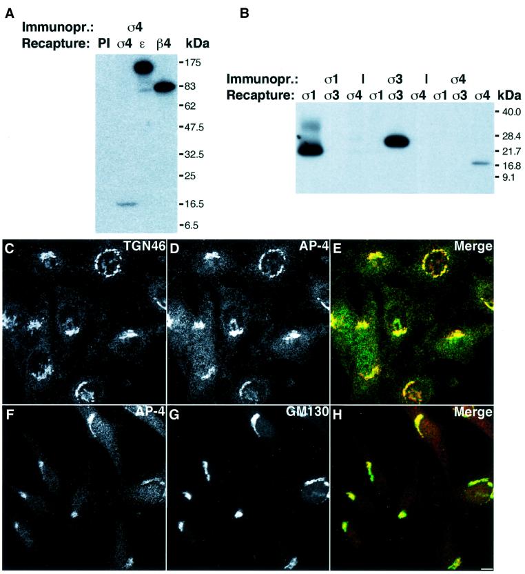
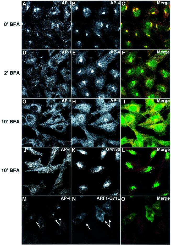
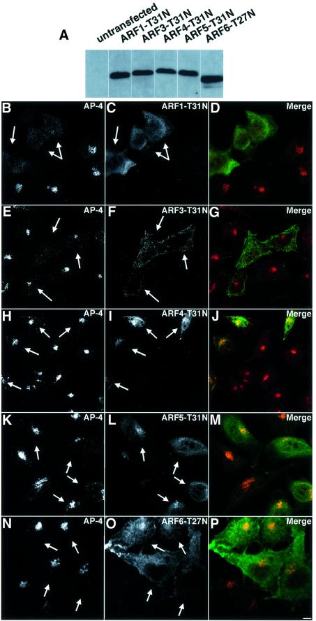
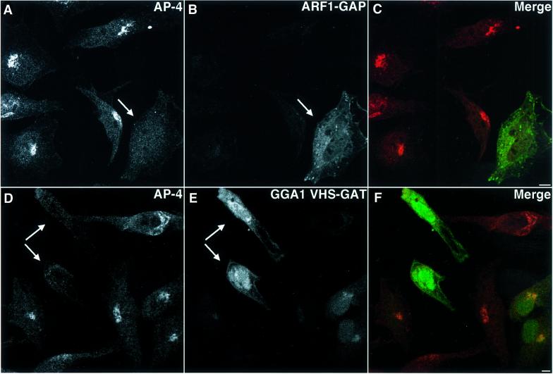
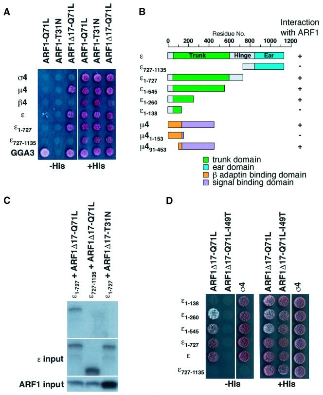
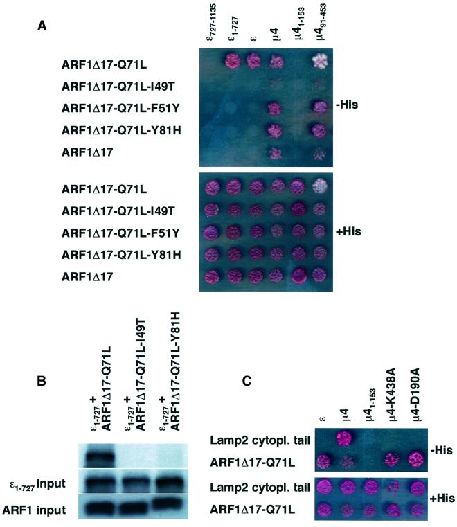
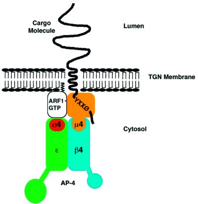
AP-4 is a member of the family of heterotetrameric adaptor protein (AP) complexes that mediate the sorting of integral membrane proteins in post-Golgi compartments. This complex consists of four subunits (epsilon, beta4, mu4 and sigma4) and localizes to the cytoplasmic face of the trans-Golgi network (TGN). Here, we show that the recruitment of endogenous AP-4 to the TGN in vivo is regulated by the small GTP-binding protein ARF1. In addition, we demonstrate a direct interaction of the epsilon and mu4 subunits of AP-4 with ARF1. epsilon binds only to ARF1-GTP and requires residues in the switch I and switch II regions of ARF1. In contrast, mu4 binds equally well to the GTP- and GDP-bound forms of ARF1 and is less dependent on switch I and switch II residues. These observations establish AP-4 as an ARF1 effector and suggest a novel mode of interaction between ARF1 and an AP complex involving both constitutive and regulated interactions.


Figures

None
Fig. 1. The new anti-σ4 antibody specifically recognizes AP-4 in immunoprecipitation and immunofluorescence experiments and does not crossreact with σ1 or σ3. (A) Native immunoprecipitation (Immunopr.) of AP-4 by the anti-σ4 antibody followed by denaturing recapture using pre-immune serum (PI), the anti-σ4 antibody or previously described anti-ε and anti-β4 antibodies. (B) Native immunoprecipitation (Immunopr.) of AP-1, AP-3 and AP-4 using anti-σ1, anti-σ3 and anti-σ4 antibodies, respectively, followed by denaturing recapture using the anti-σ1, anti-σ3 or anti-σ4 antibody. (CH) HeLa cells were grown in DMEM, 10% fetal calf serum (FCS) and fixed with 2% H2CO–PBS. Cells were labeled with rabbit anti-σ4 (D and F), sheep anti-TGN46 (C) or mouse anti-GM130 (G), followed by Cy3-conjugated anti-sheep Ig (C), Alexa 488-conjugated anti-rabbit Ig (D), Cy3-conjugated anti-rabbit Ig (F) and Alexa 488-conjugated anti-mouse Ig (G). Bar (C–H), 5 µm.
None
Fig. 2. BFA redistributes AP-4 to the cytosol. ARF1-Q71L expression attenuates the BFA-induced effect. (AL) HeLa cells were grown in DMEM, 10% FCS, and BFA was added to a final concentration of 5 µg/ml. Cells were fixed with 2% H2CO–PBS immediately (A–C), after 2 min (D–F) or after 10 min (G–L) of incubation at 37°C. Cells were stained with mouse anti-γ primary, Alexa 568-conjugated anti-mouse secondary antibody (A, D and G), rabbit anti-σ4 primary, Alexa 488-conjugated anti-rabbit secondary antibody (B, E and H), rabbit anti-σ4 primary, Cy3-conjugated anti-rabbit secondary antibody (J) or mouse anti-GM130 primary, Alexa 488-conjugated anti-mouse secondary antibody (K). (MO) HeLa cells were transiently transfected with pXS-ARF1-Q71L-HA and after a 24 h incubation, BFA (5 µg/ml) was added to the cells. After 20 min, cells were fixed and stained using the anti-σ4 antibody (Cy3) for AP-4 and an anti-HA antibody (Alexa 488) for ARF1-Q71L. (M) and (N) Transfected cells are indicated by arrows. Bar, 5 µm.
None
Fig. 3. Class I ARF proteins specifically affect the AP-4 TGN localization. HeLa cells were transiently transfected with pXS-ARF1-T31N-HA (BD), pXS-ARF3-T31N-HA (EG), pXS-ARF4-T31N-HA (HJ), pXS-ARF5-T31N-HA (KM) or pXS-ARF6-T27N-HA (NP) and incubated for 24 h. (A) Post-nuclear supernatants were prepared and immunoblotting was performed using an anti-HA antibody. (B–P) After 24 h, cells were fixed and double labeled for immunofluorescence microscopy using the anti-σ4 antibody (Cy3) for (B), (E), (H), (K) and (N) and an anti-HA (Alexa 488) antibody for (C), (F), (I), (L) and (O) to detect AP-4 localization and ARF expression, respectively. Transfected cells are indicated by arrows. Bar (B–P), 5 µm.
None
Fig. 4. AP-4 localization is affected by the expression of ARF1-GAP and GGA1 VHS-GAT. HeLa cells were transiently transfected with an ARF1-GAP expression plasmid (AC) or a GGA1 YFP-VHS-GAT expression plasmid (DF). After 24 h cells were fixed and stained using the anti-σ4 [Cy3 (A and D)] and anti-His6 antibodies [Alexa 488 (B)]. Transfected cells are indicated by arrows. Bars, 5 µm.
None
Fig. 5. Interaction analyses between ARF1 and AP-4 subunits and truncation analyses of the ε–ARF1 interaction. (A and D) HF7C yeast strain was transfected with constructs expressing the indicated proteins and co-transformants were spotted on plates with (right) or without (left) histidine. Interaction of proteins was assessed by growth on the plate lacking histidine. (B) Bar diagram summarizing the results depicted in (A), (D) and Figure 6A. (C) In vitro transcribed/translated 35S-labeled ε and ARF-myc constructs were mixed, incubated, and ARF-myc was immunoprecipitated using an anti-myc antibody. Top, the co-precipitation of ε1–727 was detected by autoradiography. Middle and bottom, labeled ε and ARF1 constructs, respectively, which were used as input.
None
Fig. 6. Interaction analyses of ε adaptin and µ4 adaptin with switch mutants of ARF1. (A and C) HF7C yeast strain was transfected with constructs expressing the indicated proteins and co-transformants were spotted on plates with (bottom) or without (top) histidine. Growth of colonies on the plate lacking histidine indicates an interaction of the proteins. (B) In vitro transcribed/translated 35S-labeled ε and ARF-myc constructs were mixed, incubated, and ARF-myc was immunoprecipitated using an anti-myc antibody. Top, the co-precipitation of ε1–727 was detected by autoradiography. Middle and bottom, labeled ε1–727 and ARF1 constructs, respectively, which were used as input.
None
Fig. 7. Model for the interaction of AP-4 with ARF1 and a trans membrane cargo molecule bearing a YXXØ-based tyrosine-based sorting motif in its cytosolic tail.

References

    1. Aguilar R.C., Ohno,H., Roche,K.W. and Bonifacino,J.S. (1997) Functional domain mapping of the clathrin-associated adaptor medium chains µ1 and µ2. J. Biol. Chem., 272, 27160–27166. - PubMed
    1. Aguilar R.C., Boehm,M., Gorshkova,I., Crouch,R.J., Tomita,K., Saito,T., Ohno,H. and Bonifacino,J.S. (2001) Signal-binding specificity of the µ4 subunit of the adaptor protein complex AP-4. J. Biol. Chem., 276, 13145–13152. - PubMed
    1. Antonny B., Beraud-Dufour,S., Chardin,P. and Chabre,M. (1997) N-terminal hydrophobic residues of the G-protein ADP-ribosylation factor-1 insert into membrane phospholipids upon GDP to GTP exchange. Biochemistry, 36, 4675–4684. - PubMed
    1. Aoe T., Cukierman,E., Lee,A., Cassel,D., Peters,P.J. and Hsu,V.W. (1997) The KDEL receptor, ERD2, regulates intracellular traffic by recruiting a GTPase-activating protein for ARF1. EMBO J., 16, 7305–7316. - PMC - PubMed
    1. Austin C., Hinners,I. and Tooze,S.A. (2000) Direct and GTP-dependent interaction of ADP-ribosylation factor 1 with clathrin adaptor protein AP-1 on immature secretory granules. J. Biol. Chem., 275, 21862–21869. - PubMed

Publication types

MeSH terms