. 2001 Nov 15;20(22):6347-58.
doi: 10.1093/emboj/20.22.6347.

Proteomic and functional evidence for a P2X7 receptor signalling complex

Affiliations

Proteomic and functional evidence for a P2X7 receptor signalling complex

M Kim et al. EMBO J. .

Abstract

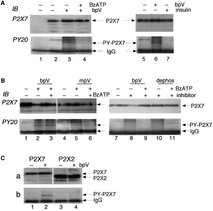
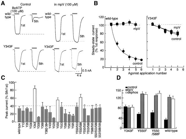
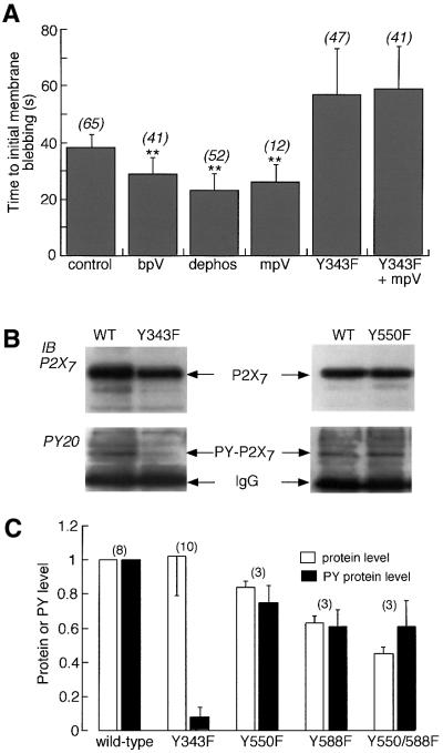
P2X receptors are ATP-gated ion channels in the plasma membrane, but activation of the P2X7 receptor also leads to rapid cytoskeletal re-arrangements such as membrane blebbing. We identified 11 proteins in human embryonic kidney cells that interact with the rat P2X7 receptor, by affinity purification followed by mass spectroscopy and immunoblotting [laminin alpha3, integrin beta2, beta-actin, alpha-actinin, supervillin, MAGuK, three heat shock proteins, phosphatidylinositol 4-kinase and receptor protein tyrosine phosphatase-beta (RPTPbeta)]. Activation of the P2X7 receptor resulted in its dephosphorylation. Whole-cell recordings from cells expressing P2X7 receptors showed that this markedly reduced subsequent ionic currents and it also slowed membrane bleb formation. By mutagenesis, we identified Tyr(343) in the putative second transmembrane domain as the site of phosphorylation. Thus, we have identified a P2X7 receptor signalling complex, some members of which may initiate cytoskeletal rearrangements following receptor activation. Others, such as RPTPbeta, might exert feedback control of the channel itself through its dephosphorylation.


Figures

None
Fig. 1. Proteomic characterization of P2X7 receptor protein complex. (A) Protein staining of SDS–PAGE membrane extracts from HEK cells stably expressing P2X7 receptors. Lane 1 is total membrane extract; lanes 2 and 3 are negative controls showing membrane extracts bound to γ-bind G-Sepharose and after immunoprecipitation with an anti-P2X2 Ab, respectively. Lane 4 shows protein staining after immunoprecipiteads show position of bands excised for mass spectroscopy. IgG bands were identified by N-terminal amino acid sequencing. Right-hand panel shows MALDI-TOF spectrum of the 70 kDa protein band after digestion with trypsin; ‘T’ labels peptide from trypsin autolysis. The remaining peaks match peptides from Hsp70. (B) Blotting with antibodies to β-actin, α-actinin, integrin β2, P2X7 receptor, Hsp70, Hsc71, Hsp90, supervillin, laminin α3 and MAGuK-p55 (as indicated) from membrane fractions (a) from HEK293 cells stably expressing P2X7 receptors or (b) from HEK293 cells transiently expressing P2X7-EE receptor. In each case, lane 1 is solubilized membrane fraction, lane 2 is negative control with mouse IgG, and lane 3 is from IP with ecto-P2X7 or EE Ab. (C) Blots with anti-RPTPβ Ab. Lane 1 is membrane fraction, lane 2 is IP with ecto-P2X7 Ab from P2X7-expressing cells (stable transfection), lane 3 is IP with anti-EE Ab from P2X7-EE-expressing cells (transient transfection), and lane 4 is negative control with mouse IgG. (D) Co-expression of P2X7-EE and PI4K-HA cDNA (lanes 3 and 4). Lane 1, P2X7-EE cDNA only; lane 2, PI4K-HA cDNA only. Lanes 1 and 3, IP with anti-EE Ab; lanes 2 and 4, IP with anti-HA Ab; membranes blotted with anti-P2X7 or anti-HA as indicated.
None
Fig. 2. Tyrosine phosphorylation of P2X7 receptor. (A) Membrane extracts from untransfected (lane 1, negative control) or P2X7 receptor-expressing HEK293 cells were immunoprecipitated with ecto-P2X7 Ab, and phosphotyrosine incorporation into the P2X7 protein was detected by probing with anti-phosphotyrosine Ab PY20. The blot was then stripped and re-probed with C-terminal anti-P2X7 Ab to confirm that the phosphotyrosine bands (lanes 3, 4 and 6) were the P2X7 receptor. No tyrosine phosphorylation was detected in control P2X7-expressing cells (lanes 2 and 5) but after 10 min incubation with the tyrosine phosphatase inhibitor bpV (100 µM) phosphotyrosine was clearly detected (lanes 3 and 6). The level was much reduced if the P2X7 receptor was activated for 10 min with BzATP (100 µM) prior to incubation with bpV (lane 4). (B) Similar experiments using phosphatase inhibitors mpV (100 µM, lanes 5 and 6) or 3,4-dephostatin (100 µM, lanes 10 and 11) also show tyrosine phosphorylation of P2X7 subunit in the presence of the phosphatase inhibitors (lanes 5 and 10), which is reduced when BzATP is added prior to application of phosphatase inhibitor (lane 11), but not when BzATP is added after the phosphatase inhibitor (lanes 3 and 6). (C) Tyrosine phosphorylation occurs on P2X7 receptor but not P2X2 receptor. Similar experiment to others performed on HEK cells transiently transfected with P2X7-EE (lanes 1 and 2) or P2X2-EE (lanes 3 and 4) receptors. Immunoprecipitation was with anti-EE Ab in the absence or presence of bpV as indicated. Anti-PY20 blotting detected phosphotyrosine-P2X7 after bpV treatment (b, lane 2) but no tyrosine phosphorylation of P2X2 receptor (b, lanes 3 and 4). Stripping and re-probing with C-terminal anti-P2X7 (a, lanes 1 and 2) or anti-P2X2 (a, lanes 3 and 4) confirms tyrosine phosphorylation of P2X7 but not P2X2 receptor even though there is greater expression of P2X2 protein.
None
Fig. 3. Functional evidence for use-dependent tyrosine dephosphorylation of P2X7 receptor. (A) Currents in response to successive applications of 100 µM BzATP (4 s duration, indicated by bars) at 2 min intervals; note the marked change in kinetics of the current. (B) Currents recorded from another cell in response to successive applications of 100 µM BzATP in the presence of the tyrosine phosphatase inhibitor bpV (100 µM). (C) Summary of results from all experiments as illustrated in (A) and (B) (n = 43 for each point in control and 7–13 for each point in any of the phosphatase inhibitors). (D) Similar experiments performed on P2X2-expressing HEK cells. Minimal run-down occurs with successive stimuli and is unaltered by bpV (100 µM; n = 6–12 for each point). All results were obtained from HEK cells stably expressing P2X7 (A–C) or P2X2 (D) receptors.
None
Fig. 4. Mutation of Tyr343 prevents run-down of current and effect of phosphatase inhibitors. (A) Currents recorded from wild type (upper traces) and Y343F (lower traces) in response to BzATP (100 µM). Left traces are from cells in normal solution and right traces are cells in mpV (100 µM). (B) Current amplitude at end of 4 s duration application of BzATP as a function of agonist application number for all experiments with HEK cells transiently transfected with wild type (left; n = 12–24) and Y343F (right; n = 6 each) P2X7 receptor. (C) Summary of tyrosine mutagenesis of P2X7 receptor. Histogram shows BzATP-evoked current as a percentage of amplitude of 5th/1st application; only Y343F and Y550F or Y550/588F (white bars) show significantly less run-down than wild type. (D) Phosphatase inhibitors (bpV and dephostatin) have no effect on Y343F but affect Y550F and Y550/588F to the same extent as at the wild-type receptor; data are plotted as in (C); n = 3 for each mutant, 13 for wild type.
None
Fig. 5. P2X7 receptor signalling to the cytoskeleton is modulated by phosphorylation of Tyr343. (A) Time to initial membrane blebbing in response to application of 100 µM BzATP. Phosphatase inhibitors (100 µM) significantly decreased time to initial blebbing (p < 0.0001) in wild type but not in Y343F mutant. Numbers in parentheses are number of cells. (B) Tyrosine phosphorylation of P2X7 receptor is markedly reduced in Y343F mutant. Immunoblots for wild type and Y550F or wild type and Y343F are shown. In each case anti-EE Ab was used for immunoprecipitation from cells transiently transfected with P2X7-EE receptor. Tyrosine phosphorylation was then detected with anti-PY-20 Ab, after which the membrane was stripped and re-probed with anti-P2X7 Ab. In all cases bpV (100 µM) was present for 10 min prior to membrane extraction. Tyrosine phosphorylation of wild type and Y550F was observed, but not Y343F. (C) Densitometric analysis of wild-type and mutant P2X7 receptors. Each protein band (P2X7, open bars; tyrosine phosphorylated P2X7, closed bars) was scanned using GeneGenius and raw data were normalized to that of wild type.
None
Fig. 6. Schematic summary of P2X7 receptor signalling complex. (A) Depiction of P2X7 receptor and interacting proteins identified. Laminin is a heterotrimeric extracellular matrix molecule; the α3 subunit is found in laminin-5, laminin-6 and laminin-7 (Colognato and Yurchenko, 2000). RPTPβ indicates the 230 kDa form of RPTPβ; the extracellular part of the molecule has an N-terminal carbonic anhydrase domain and 16 fibronectin type 3 domains, and the intracellular part has one or two tyrosine phosphatase catalytic domains (Fischer et al., 1991). Integrin β2 is a subunit of hetero dimeric integrin receptors Mac1 (cd11b, αMβ2) and LFA (cd11a, αLβ2); its cytoplasmic C-terminus can bind to α-actinin (Sampath et al., 1998). α-actinin indicates α-actinin 4, an actin-bundling protein. This forms a reciprocal homodimer, with the two N-terminal actin binding domains at either end. It also has two calponin homology domains, four spectrin repeats and two EF hands. Supervillin is also an F-actin bundling protein that associates with the plasma membrane; it has multiple actin-binding domains and a villin/gelsolin homology domain (Wulfkuhle et al., 1999). PI4K is phosphatidylinositol 4-kinase 230; its domains include an N-terminus SH3 domain, two proline-rich regions, two leucine-zippers, a pleckstrin homology domain and the C-terminal catalytic domain (Gehrmann and Heilmeyer, 1998). MAGuK is membrane-associated guanylate kinase P55 subfamily 3; it contains PDZ, SH3 and GUK domains (Dimitratos et al., 1999). Hsp90 is heat shock protein 90-β. Hsp70 is heat shock protein 70 kDa 1. Hsc71 is heat shock cognate 71 kDa protein. See Table I and text for further information. (B) Depiction of the second hydrophobic domain of the P2X7 receptor (Thr325–Asn356) as a membrane-spanning α-helix. Residues are coloured as follows: blue, hydrophobicity index >2.5 (probably lipid facing); red, polar or charged (protein- or pore-facing); black, non-polar. Some residues are partially hidden; the complete sequence is TGGKFDIIQLVVYIGSTLSYFGLATVCIDLIIN. The first two-thirds of the helix might span the membrane and expose Tyr343 (arrow) at the inner juxtamembrane region. Residues conserved among all P2X subunits are shown in bold (Gly328, G329, Lys330, Phe331, Gly345 and Asp353).

References

    1. Bohnsack J.F., Akiyama,S.K., Damsky,C.H., Knape,W.A. and Zimmerman,G.A. (1990) Human neutrophil adherence to laminin in vitro. Evidence for a distinct neutrophil integrin receptor for laminin. J. Exp. Med., 171, 1221–1237. - PMC - PubMed
    1. Bronte V., Macino,B., Zambon,A., Rosato,A., Mandruzzato,S., Zanovello,P. and Collavo,D. (1996) Protein tyrosine kinase inhibitors and phosphatases control apoptosis induced by extracellular adenosine 5′-triphosphate. Biochem. Biophys. Res. Commun., 218, 344–351. - PubMed
    1. Caplan A.J. (1999) Hsp90’s secrets unfold: new insights from structural and functional studies. Trends Cell Biol., 9, 262–268. - PubMed
    1. Cohn Z.A. and Parks,E. (1967) The regulation of pinocytosis in mouse macrophages. III. The induction of vesicle formation by nucleosides and nucleotides. J. Exp. Med., 125, 457–466. - PMC - PubMed
    1. Collo G., Neidhart,S., Kawashima,E., Kosco-Vilbois,M., North,R.A. and Buell,G. (1997) Tissue distribution of the P2X7 receptor. Neuropharmacology, 36, 1277–1283. - PubMed

Publication types

MeSH terms