. 2001 Nov 15;20(22):6394-403.
doi: 10.1093/emboj/20.22.6394.

Stepwise activation of the immunoglobulin mu heavy chain gene locus

Affiliations

Stepwise activation of the immunoglobulin mu heavy chain gene locus

D Chowdhury et al. EMBO J. .

Abstract

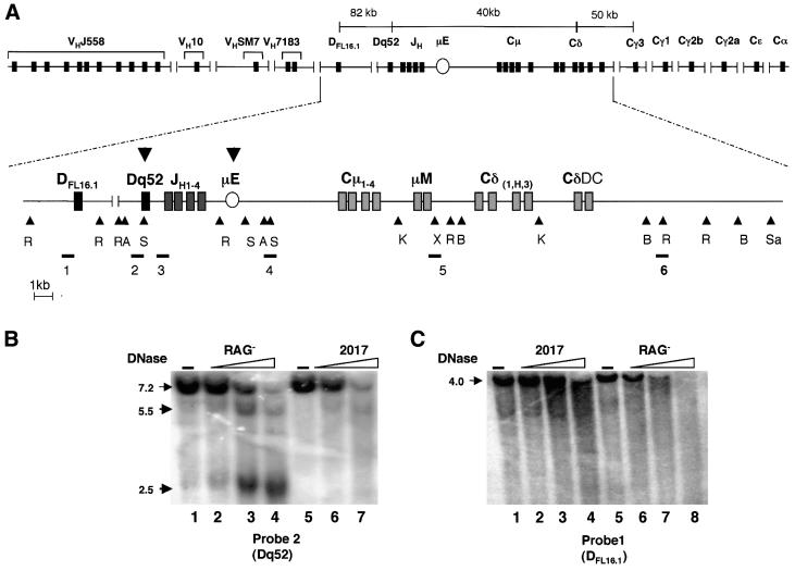
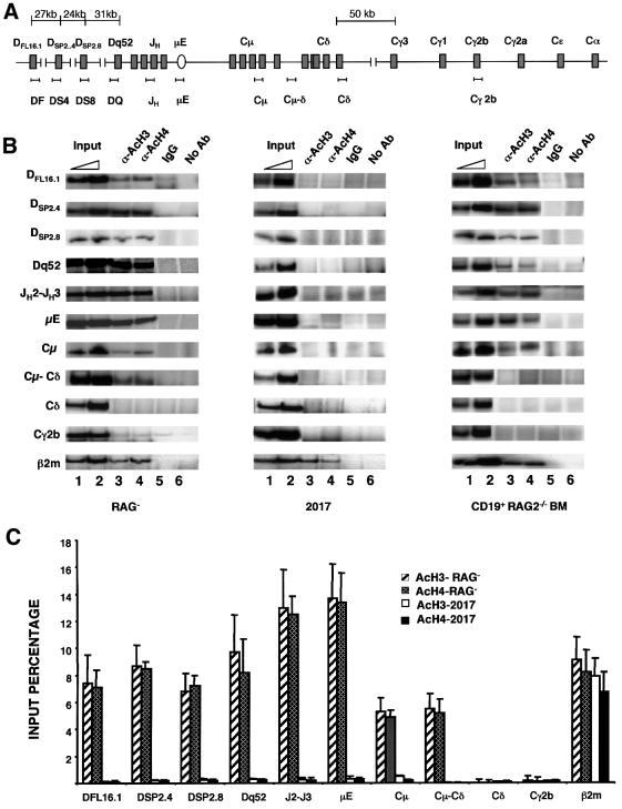
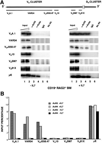
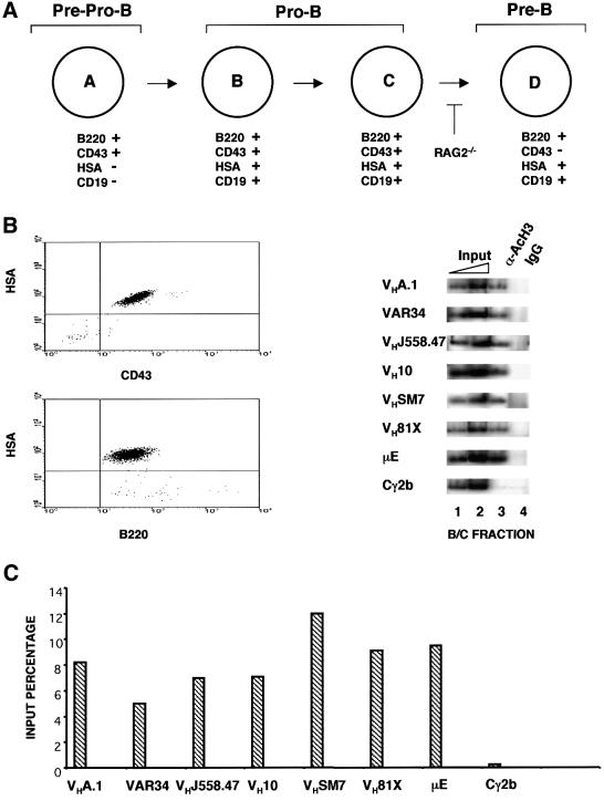
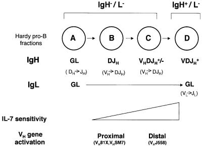
The immunoglobulin heavy chain (IgH) gene locus spans several megabases. We show that IgH activation during B-cell differentiation, as measured by histone acetylation, occurs in discrete, independently regulated domains. Initially, a 120 kb domain of germline DNA is hyperacetylated, that extends from D(FL16.1), the 5'-most D(H) gene segment, to the intergenic region between Cmu and Cdelta. Germline V(H) genes were not hyperacetylated at this stage, which accounts for D(H) to J(H) recombination occurring first during B-cell development. Subsequent activation of the V(H) locus happens in at least three differentially regulated domains: an interleukin-7-regulated domain consisting of the 5' J558 family, an intermediate domain and the 3' V(H) genes, which are hyperacetylated in response to DJ(H) recombination. These observations lead to mechanisms for two well-documented phenomena in B-cell ontogeny: the sequential rearrangement of D(H) followed by V(H) gene segments, and the preferential recombination of D(H)-proximal V(H) genes in pro-B cells. We suggest that stepwise activation may be a general mechanism by which large segments of the genome are prepared for expression.


Figures

None
Fig. 1. DNase I-hypersensitive site analysis of the murine IgH gene locus. (A) Schematic representation of the IgH locus with DNase I-hypersensitive sites. The complete locus including VH gene segments and all heavy chain isotypes is shown on the top line. The second line is a detailed schematic of part of the locus starting from the 5′ DFL16.1 gene segment to 7 kb downstream of the Cδ membrane exon. The restriction endonuclease sites EcoRI (R), AseI (A), SacI (S), KpnI (K), SalI (Sa), XhoI (X) and BglII (B) were used for Southern blot analysis of DNase I-treated genomic DNA. Probes used in the study are labeled 1–6, and are shown below their corresponding hybridizing fragments. The position of the intronic µ enhancer is marked with an oval µE. The nuclease-hypersenstive sites identified are indicated by the bold arrowheads. (B and C) DNase I sensitivity assays. Nuclei from RAG (pro-B) cells or 2017 (pro-T) cells were treated with increasing concentrations of DNase I as indicated, and purified genomic DNA analyzed by Southern blotting. DNA from untreated nuclei is shown in lanes 1 and 5. Restriction enzymes and probes used were as follows: (B) AseI, probe 2; (C) EcoRI, probe 1. DNase-hypersensitive sites are indicated by the bold arrows. The bands at 2.5 and 5.5 kb correspond to hypersensitive sites at the Dq52 region and the µE, respectively. Data shown are from one out of four independent experiments.
None
Fig. 2. Histone acetylation in the IgH locus. (A) Schematic representation of the DH–Cµ locus with approximate locations of the primer sets used in chromatin immunoprecipitation assays. Sequences of the primers are provided in Table I. (B) Chromatin immunoprecipitation assays using pro-B (RAG) and pro-T (2017) cell lines, and primary pro-B cells from RAG2-deficient bone marrow. Formaldehyde-cross-linked chromatin prepared from cells as described in Materials and methods was incubated with anti-acetylated histone H3 (α-AcH3) or anti-acetylated histone H4 (α-AcH4) antibodies. Control immunoprecipitations were carried out using a 2-fold excess of non-specific rabbit IgG (lane 5) or no antibody (lane 6). Antibody-bound DNA was collected by adsorption to protein A–agarose, uncross-linked and amplified by PCR with the primer sets indicated in (A). The 3′ primer was radiolabeled for quantitation, and the products were visualized after fractionation through 6% polyacrylamide gels. Phosphoimager analysis was used to detect and quantitate reaction products. Lanes marked Input (lanes 1 and 2) correspond to DNA purified from chromatin before immunoprecipitation. The amount of input DNA used as the template in the PCR was one-tenth (lane 1) or one-fifth (lane 2) that used for immunoprecipitations (lanes 3–6). Primers hybridizing to the β2-microglobulin gene were used as a positive control. Results shown are representative of one out of three independent experiments. (C) Data in (B) of the RAG and 2017 cell lines were quantitated by phosphoimager analysis and are represented graphically. Results shown are an average of three independent experiments, with the error bars representing the standard deviation. The input percentage was calculated taking the subsaturating PCR product of the input DNA (lane1) and also taking into account that the input had 10-fold less DNA template per PCR.
None
Fig. 3. IL-7-dependent histone acetylation of VHJ558 gene segments. (A) Schematic representation of the murine VH gene locus showing the approximate location of gene segments analyzed in this study (adapted from Haines and Brodeur, 1998). CD19+ pro-B bone marrow cells from RAG2–/– mice were cultured for 4 days with or without IL-7 (20 ng/ml). Chromatin immunoprecipitation assays were done as described using primers from the VH region as shown in the top panel. The µ enhancer region (µE) was used as a positive control. Representative data from one of two experiments is shown. (B) The results in (A) were quantified by phosphoimager analysis and are represented as a proportion of the input DNA that was immunoprecipitated.
None
Fig. 4. Acetylation of the DH-proximal VH genes in pro-B- and pre-B-cell lines. (A) Chromatin immunoprecipitation assays were done using Abelson murine leukemia virus-transformed pro-B (RAG) and pre-B cell lines (22D6 and 38B9). The IgH locus is in germline configuration in RAG cells, whereas 22D6 and 38B9 cells contain DJH recombination on both alleles. Representative data from one of two experiments are shown. (B) Phosphoimager analysis and graphical representation of the data in (A).
None
Fig. 5. Histone acetylation of VH genes in primary bone marrow pro-B cells. (A) Hardy classification of early B-cell development listing some of the cell surface markers used to distinguish the different cell populations (Hardy et al., 1991). DJH recombination is seen in fraction B cells and VDJ recombination is seen in fraction C cells. (B and C) The B/C fraction pro-B cells were purified from bone marrow of wild-type BALB/c mice by magnetic depletion of erythroid, myeloid and T cells followed by sorting for the B220+ CD43+ HSA+ cells. Chromatin immunoprecipitation assays were carried out using the primers shown in Figure 3A. Phosphoimager quantitation was done as described in the legend to Figure 2. Representative data from one of two experiments using two preparation of bone marrow cells are shown.
None
Fig. 6. Mechanism of ordered VH recombination. Pro-B cell subsets as defined by Hardy are shown in the circles. Genomic structures of the Ig loci are shown in bold below each circle (GL = germline). Presumed ongoing rearrangements in each subset are indicated in parentheses below the genomic structure. Recombination at the IgH locus is initiated in late A fraction cells; our observations suggest that only the DH–Cµ part of the locus is accessible to recombinase at this stage, resulting in DH to JH recombination. Proximal VH gene families are activated on DJH recombined alleles as outlined in the text. We propose that distal VH genes, that are activated by IL-7, lag behind the proximal VHs because IL-7 responsiveness is gained gradually during B-cell development based on Wei et al. (2000) (indicated by a triangle). Preferential rearrangement of proximal VH genes is therefore a consequence of the late activation of the 5′ VH genes via the IL-7 receptor.

References

    1. Akashi K., Kondo,M. and Weissman,I.L. (1998) Role of interleukin-7 in T-cell development from hematopoietic stem cells. Immunol. Rev., 165, 13–28. - PubMed
    1. Alessandrini A. and Desiderio,S.V. (1991) Coordination of immunoglobulin DJH transcription and D-to-JH rearrangement by promoter–enhancer approximation. Mol. Cell. Biol., 11, 2096–2107. - PMC - PubMed
    1. Angelin-Duclos C. and Calame,K. (1998) Evidence that immunoglobulin VH–DJ recombination does not require germ line transcription of the recombining variable gene segment. Mol. Cell. Biol., 18, 6253–6264. - PMC - PubMed
    1. Banerjee A. and Rothman,P. (1998) IL-7 reconstitutes multiple aspects of v-Abl-mediated signaling. J. Immunol., 161, 4611–4617. - PubMed
    1. Braunstein M., Rose,A.B., Holmes,S.G., Allis,C.D. and Broach,J.R. (1993) Transcriptional silencing in yeast is associated with reduced nucleosome acetylation. Genes Dev., 7, 592–604. - PubMed

Publication types

MeSH terms