. 2001 Nov 15;20(22):6485-98.
doi: 10.1093/emboj/20.22.6485.

Hyperactivation of the yeast DNA damage checkpoint by TEL1 and DDC2 overexpression

Affiliations

Hyperactivation of the yeast DNA damage checkpoint by TEL1 and DDC2 overexpression

M Clerici et al. EMBO J. .

Abstract

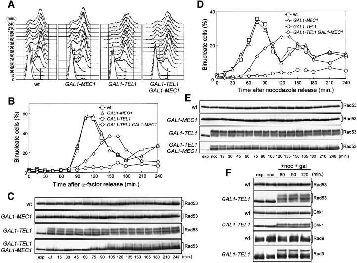
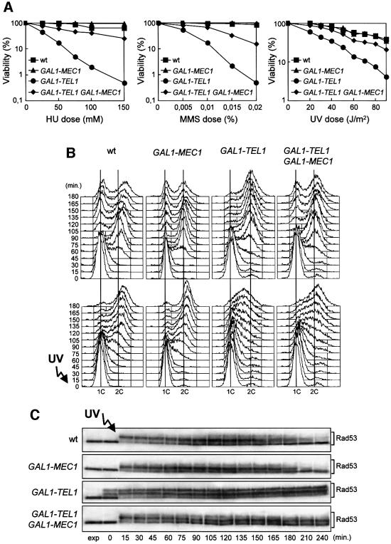
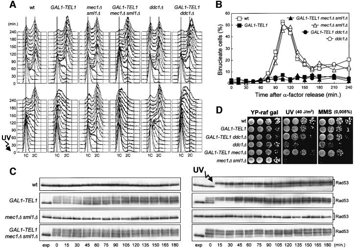
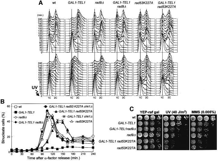
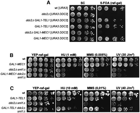
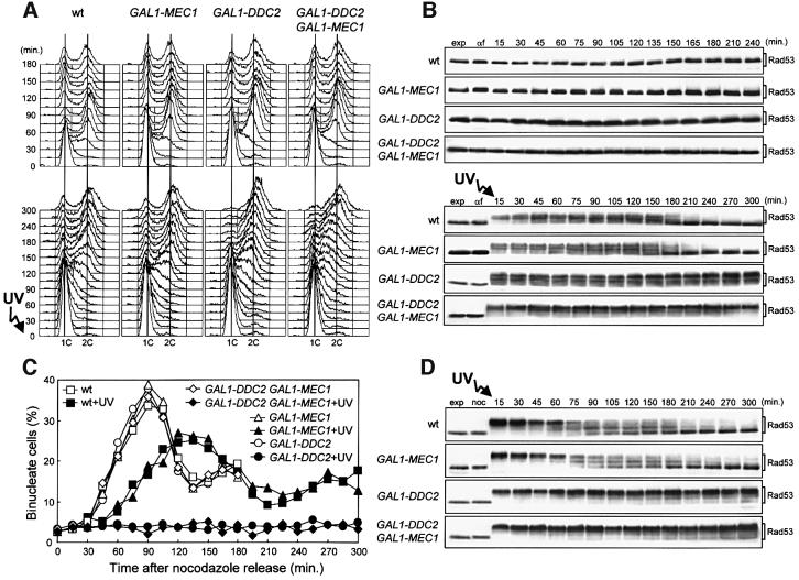
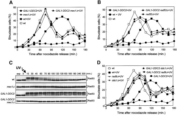
The evolutionarily conserved yeast Mec1 and Tel1 protein kinases, as well as the Mec1-interacting protein Ddc2, are involved in the DNA damage checkpoint response. We show that regulation of Tel1 and Ddc2-Mec1 activities is important to modulate both activation and termination of checkpoint-mediated cell cycle arrest. In fact, overproduction of either Tel1 or Ddc2 causes a prolonged cell cycle arrest and cell death in response to DNA damage, impairing the ability of cells to recover from checkpoint activation. This cell cycle arrest is independent of Mec1 in UV-irradiated Tel1-overproducing cells, while it is strictly Mec1 dependent in similarly treated DDC2-overexpressing cells. The Rad53 checkpoint kinase is instead required in both cases for cell cycle arrest, which correlates with its enhanced and persistent phosphorylation, suggesting that unscheduled Rad53 phosphorylation might prevent cells from re-entering the cell cycle after checkpoint activation. In addition, Tel1 overproduction results in transient nuclear division arrest and concomitant Rad53 phosphorylation in the absence of exogenous DNA damage independently of Mec1 and Ddc1.


Figures

None
Fig. 1. Effects of TEL1 overexpression during an unperturbed cell cycle. Strains were as follows: wild type (K699), GAL1–MEC1 (YLL516), GAL1–TEL1 (DMP3539/10D) and GAL1–TEL1 GAL1–MEC1 (DMP3539/9D). (A–C) Cell cultures, growing logarithmically in YEP-raf, were synchronized in G1 with α-factor in the presence of galactose (2 h). Release from α-factor block was performed at time zero (C, αf) by transferring cell cultures to fresh YEP-raf-gal medium. Samples were taken at the times indicated after release into the cell cycle to determine the DNA content by fluorescence-activated cell sorter (FACS) analysis (A), the kinetics of nuclear division by direct visualization by propidium iodide staining (B) and the amount and phosphorylation of Rad53 by western blotting of protein extracts with anti-Rad53 antibodies (C). (D and E) Cell cultures, growing logarithmically in YEP-raf, were synchronized in G2 with nocodazole in the presence of galactose (2 h). Release from nocodazole block was performed at time zero (E, noc) by transferring cell cultures to fresh YEP-raf-gal medium. Samples were taken at the times indicated after release from nocodazole to determine the kinetics of nuclear division (D) and Rad53 levels and phosphorylation (E) as described in (B) and (C), respectively. (F) Cell cultures of wild-type (DMP3598/21D) and GAL1–TEL1 (DMP3598/6A) strains were arrested with nocodazole in YEP-raf medium (noc) and resuspended in YEP-raf-gal medium containing 15 µg/ml nocodazole (+noc +gal). Protein extracts were prepared from samples withdrawn at the times indicated and analyzed by western blotting. The slowly migrating protein species specifically reacting with anti-Rad53, anti-MYC (Chk1) and anti-Rad9 antibodies represent phosphorylated forms of the corresponding proteins (Sanchez et al., 1996; Vialard et al., 1998, 1999). exp, exponentially growing cells.
None
Fig. 2. Response to DNA damage of TEL1-overexpressing cells. Strains were as follows: wild type (K699), GAL1–MEC1 (YLL516), GAL1–TEL1 (DMP3539/10D) and GAL1–TEL1 GAL1–MEC1 (DMP3539/9D). (A) Dose–response killing curves were determined by plating serial dilutions of YEP-raf exponentially growing cell cultures on YEP-raf-gal plates with or without MMS or HU at the concentrations indicated. One set of YEP-raf-gal plates was exposed at the UV doses indicated. Plates were incubated at 25°C and colony-forming units were counted after 3 days. (B and C) Cell cultures growing logarithmically in YEP-raf were synchronized in G1 with α-factor in the presence of galactose (2 h). Cells were released from the α-factor block at time zero in YEP-raf-gal, or were UV irradiated (40 J/m2) prior to release in YEP-raf-gal. Samples of untreated and UV-irradiated cultures were taken at the times indicated after α-factor release to analyze the DNA content by FACS (B) and to determine the level and phosphorylation of Rad53 by western blot analysis of protein extracts from the UV-treated cell cultures with anti-Rad53 antibodies (C). exp, exponentially growing cells.
None
Fig. 3. Effects of TEL1 overexpression in the absence of Mec1 or Ddc1. Strains were as follows: wild type (K699), GAL1–TEL1 (DMP3539/10D), mec1Δ sml1Δ (YLL490), GAL1–TEL1 mec1Δ sml1Δ (DMP3562/2A), ddc1Δ (YLL244) and GAL1–TEL1 ddc1Δ (DMP3575/4A). (A–C) Cell cultures growing logarithmically in YEP-raf were synchronized in G1 with α-factor in the presence of galactose (2 h) and released from the α-factor block at time zero in YEP-raf-gal, or were UV irradiated (40 J/m2) prior to release in YEP-raf-gal. Samples were collected at the times indicated after α-factor release to analyze the DNA content of untreated (top) and UV-treated (bottom) cell cultures by FACS (A), to score the untreated cell cultures for the percentage of binucleate cells by propidium iodide staining (B) and to analyze protein extracts from the untreated (left) and UV-treated (right) cell cultures by western blotting using anti-Rad53 antibodies (C). exp, exponentially growing cells. (D) Serial dilutions of YEP-raf exponentially growing cell cultures were spotted on YEP-raf-gal plates with or without MMS at the concentration indicated. One YEP-raf-gal plate was UV irradiated (UV).
None
Fig. 4. Cell cycle delay caused by high levels of Tel1 involves Rad53, Rad9 and Chk1. Strains were as follows: wild type (K699), GAL1–TEL1 (DMP3539/10D), rad9Δ (YLL157), GAL1–TEL1 rad9Δ (DMP3575/6B), rad53K227A (DMP3479/2A), GAL1–TEL1 rad53K227A (DMP3479/2B), GAL1–TEL1 chk1Δ (DMP3611/6D) and GAL1–TEL1 rad53K227A chk1Δ (DMP3611/3B). (A and B) Cell cultures growing logarithmically in YEP-raf were synchronized in G1 with α-factor in the presence of galactose (2 h) and released from the α-factor block at time zero in YEP-raf-gal or were UV irradiated (40 J/m2) prior to release in YEP-raf-gal. Samples were collected at the times indicated after α-factor release to analyze the DNA content of untreated (top) and UV-treated (bottom) cell cultures by FACS (A) and to score the untreated cell cultures for the percentage of binucleate cells by propidium iodide staining (B). (C) Serial dilutions of YEP-raf exponentially growing cell cultures were spotted on YEP-raf-gal plates with or without MMS. One YEP-raf-gal plate was UV irradiated (UV).
None
Fig. 5. Genetic interactions between DDC2, MEC1 and TEL1. Strains were as follows: wild type [URA3 YCplac33] (YLL827), ddc2Δ [URA3 DDC2] (YLL275), ddc2Δ GAL1–TEL1 [URA3 DDC2] (DMP3475/3D), GAL1–TEL1 [URA3 DDC2] (YLL943), ddc2Δ [LEU2 GAL1–MEC1] [URA3 DDC2] (YLL930), wild type [LEU2 GAL1–MEC1] [URA3 DDC2] (YLL944), GAL1–MEC1 (YLL516), ddc2Δ sml1Δ (DMP2995/1B), GAL1–MEC1 ddc2Δ sml1Δ (DMP3532/8A), GAL1–TEL1 (DMP3539/10D) and GAL1–TEL1 ddc2Δ sml1Δ (DMP3602/9C). Serial dilutions of YEP-raf exponentially growing cell cultures were spotted on Synthetic Complete (SC)-glucose and 5-Fluoro-orotic acid (5-FoA)-containing SC-raf-gal plates (A) or on YEP-raf-gal plates with or without MMS or HU at the concentrations indicated (B and C). One YEP-raf-gal plate was UV irradiated (UV).
None
Fig. 6. DDC2 overexpression leads to prolonged G2/M cell cycle arrest after checkpoint activation. Strains were as follows: wild type [URA3 YCplac33] (YLL827), wild type [URA3 GAL1–MEC1] (YLL826), GAL1–DDC2 [URA3 YCplac33] (YLL837) and GAL1–DDC2 [URA3 GAL1–MEC1] (YLL836). (A and B) Cell cultures growing logarithmically in YEP-raf were synchronized with α-factor. Galactose was added 2.5 h before α-factor addition. α-factor-synchronized cells were released from the block at time zero [(B), αf] in YEP-raf-gal or were UV irradiated (40 J/m2) prior to release in YEP-raf-gal. Samples of untreated (top) and UV-treated (bottom) cell cultures were collected at the times indicated after α-factor release to analyze the DNA content by FACS (A) and protein extracts by western blotting, using anti-Rad53 antibodies (B). (C and D) Cell cultures growing logarithmically in YEP-raf were synchronized with nocodazole. Galactose was added 2 h before nocodazole addition. Nocodazole-synchronized cells were released from the block at time zero [(D), noc] in YEP-raf-gal or were UV irradiated (50 J/m2) prior to release in YEP-raf-gal. Samples of untreated and UV-treated cell cultures were collected at the times indicated after nocodazole release to score for the percentage of binucleate cells by propidium iodide staining (C) and to analyze protein extracts from the UV-treated cell cultures by western blotting, using anti-Rad53 antibodies (D). exp, exponentially growing cells. In all the experiments, samples were withdrawn from the UV-treated cultures at times zero and 120 min, and appropriate dilutions were plated on YEPD plates to score for colony-forming units (see text for the percentage of cell survival).
None
Fig. 7. Requirement of checkpoint genes for the Ddc2-dependent DNA damage-induced cell cycle arrest. Strains were as follows: wild type (K699), GAL1–DDC2 (YLL279.2), mec1Δ sml1Δ (YLL490), GAL1–DDC2 mec1Δ sml1Δ (DMP3462/2B), rad53Δ sml1Δ (YLL509), GAL1–DDC2 rad53Δ sml1Δ (DMP3356/4A), rad9Δ (YLL157), GAL1–DDC2 rad9Δ (DMP3392/4A), ddc1Δ (YLL244) and GAL1–DDC2 ddc1Δ (DMP3463/10C). Cell cultures growing logarithmically in YEP-raf were synchronized with nocodazole. Galactose was added 2 h before nocodazole addition and nocodazole-synchronized cells were released from the block at time zero in YEP-raf-gal or were UV irradiated (50 J/m2) prior to release in YEP-raf-gal. (A, B and D) Untreated and UV-treated cell cultures were scored at the times indicated for the percentage of binucleate cells by propidium iodide staining. (C) Protein extracts from the UV-treated cell cultures were analyzed by western blotting, using anti-Rad53 antibodies. exp, exponentially growing cells.

References

    1. Bentley N.J., Holtzman,D.A., Flaggs,G., Keegan,K.S., Demaggio,A., Ford,J.C., Hoekstra,M. and Carr,A.M. (1996) The Schizosaccharo myces pombe rad3 checkpoint gene. EMBO J., 15, 6641–6651. - PMC - PubMed
    1. Caspari T., Dahlen,M., Kanter-Smoler,G., Lindsay,H.D., Hofman,K., Papadimitriou,K., Sunnerhagen,P. and Carr,A.M. (2000) Characterization of Schizosaccharomyces pombe Hus1: a PCNA-related protein that associates with Rad1 and Rad9. Mol. Cell. Biol., 20, 1254–1262. - PMC - PubMed
    1. Chabes A., Domkin,V. and Thelander,L. (1999) Yeast Sml1, a protein inhibitor of ribonucleotide reductase. J. Biol. Chem., 274, 36679–36683. - PubMed
    1. D’Amours D. and Jackson,S.P. (2001) The yeast Xrs2 complex functions in S phase checkpoint regulation. Genes Dev., 15, 2238–2249. - PMC - PubMed
    1. Desany B.A., Alcasabas,A.A., Bachant,J.B. and Elledge,S.J. (1998) Recovery from DNA replicational stress is the essential function of the S-phase checkpoint pathway. Genes Dev., 12, 2956–2970. - PMC - PubMed

Publication types

MeSH terms

Substances

Grants and funding