. 2001 Nov 20;98(24):13560-5.
doi: 10.1073/pnas.241371398. Epub 2001 Nov 13.

Crystal structure of thermostable DNA photolyase: pyrimidine-dimer recognition mechanism

Affiliations

Crystal structure of thermostable DNA photolyase: pyrimidine-dimer recognition mechanism

H Komori et al. Proc Natl Acad Sci U S A. .

Erratum in

  • Proc Natl Acad Sci U S A 2002 Jan 22;99(2):1098

Abstract

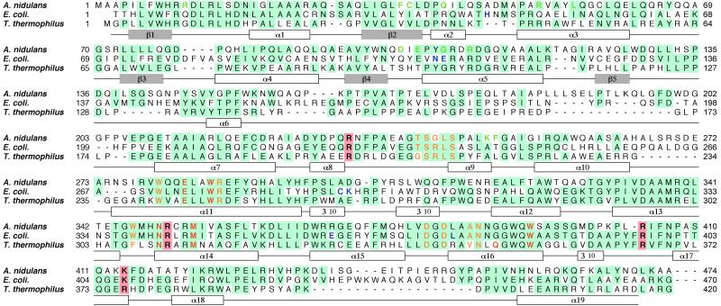
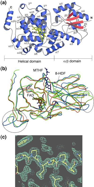
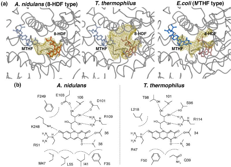
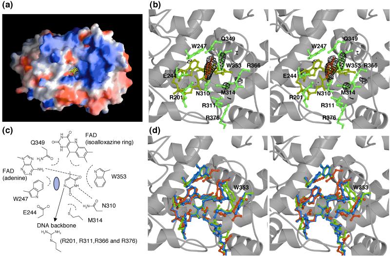
DNA photolyase is a pyrimidine-dimer repair enzyme that uses visible light. Photolyase generally contains two chromophore cofactors. One is a catalytic cofactor directly contributing to the repair of a pyrimidine-dimer. The other is a light-harvesting cofactor, which absorbs visible light and transfers energy to the catalytic cofactor. Photolyases are classified according to their second cofactor into either a folate- or deazaflavin-type. The native structures of both types of photolyases have already been determined, but the mechanism of substrate recognition remains largely unclear because of the lack of structural information regarding the photolyase-substrate complex. Photolyase from Thermus thermophilus, the first thermostable class I photolyase found, is favorable for function analysis, but even the type of the second cofactor has not been identified. Here, we report the crystal structures of T. thermophilus photolyase in both forms of the native enzyme and the complex along with a part of its substrate, thymine. A structural comparison with other photolyases suggests that T. thermophilus photolyase has structural features allowing for thermostability and that its light-harvesting cofactor binding site bears a close resemblance to a deazaflavin-type photolyase. One thymine base is found at the hole, a putative substrate-binding site near the catalytic cofactor in the complex form. This structural data for the photolyase-thymine complex allow us to propose a detailed model for the pyrimidine-dimer recognition mechanism.


Figures

Figure 1
Figure 1
Sequence alignment of photolyases from A. nidulans, E. coli, and T. thermophilus. The conserved residues are highlighted in blue. The residues in contact with FAD, 8-HDF, and MTHF are shown in yellow, green, and blue, respectively (6, 7). The residues in the active site and the positive residues around the hole are shown in red and highlighted in pink, respectively. The secondary structures, helices, and strands of T. thermophilus photolyase are shown in white and gray boxes under the sequences. The sequence of photolyase from T. thermophilus HB8 has been deposited in DNA Data Base in Japan (accession no. AB064548).
Figure 2
Figure 2
Crystal structure of T. thermophilus photolyase. (a) Overall structure of T. thermophilus photolyase. FAD is shown in yellow. The N and C termini are labeled N and C, respectively. (b) A comparison of the folding of photolyases from T. thermophilus, A. nidulans, and E. coli. The Cα traces and FAD molecules for T. thermophilus, A. nidulans, and E. coli photolyases are shown in red, green, and blue, respectively. Circles indicate more compact regions in T. thermophilus than the others. The figures in a and b were prepared by using the programs MOLSCRIPT (24) and RASTER3D (25). (c) The proline-rich region at the very long interdomain loop between α6 and α7 with an experimental single isomorphous replacement with anomalous scattering electron-density map. The figure in c was drawn by the O program (13).
Figure 3
Figure 3
Light-harvesting cofactor binding site. (a) Cavities at the light-harvesting cofactor binding sites in the crystal structures of T. thermophilus, A. nidulans, and E. coli photolyases. Solvent-accessible cavities are shown in yellow wire mesh. 8-HDF and MTHF are shown in red and blue, respectively. 8-HDF and MTHF in T. thermophilus, MTHF in A. nidulans, and 8-HDF in E. coli are hypothetical models, which are drawn transparently. These figures were prepared with the programs MOLSCRIPT (24), RASTER3D (25), and CONSCRIPT (26). (b) Schematic diagram of 8-HDF binding sites in A. nidulans and T. thermophilus photolyases. 8-HDF in T. thermophilus is a hypothetical model.
Figure 4
Figure 4
Substrate-binding site. (a) A view from the left side of Fig. 2a showing the putative substrate-binding site of T. thermophilus photolyase. The positive and negative charges are shown in blue and red, respectively. FAD buried in the hole is shown in yellow. The charge distribution of the molecular surface was calculated and represented by using the GRASP program (27). (b) Stereo view of the thymine binding site with the difference Fourier map of the native and thymine-complex. The Fo(THYMINE)Fo(NATI2) electron density contoured at 4σ is shown in black wire mesh. Thymine, FAD, and amino acid residues in the active site are shown in red, yellow, and green, respectively. (c) Schematic diagram of thymine interactions with T. thermophilus photolyase. The C5-CH3 group of thymine also takes part in van der Waals contacts, indicating that thymine-containing dimers have marginally higher affinities than uracil-containing dimers. The gray circle indicates the hypothetical position for the other thymine of the thymidine-dimer. (d) Stereo view of the active site compared with other photolyases. The residues in the crystal structures of T. thermophilus, A. nidulans, and E. coli photolyases are shown in red, green, and blue, respectively. The figures in b and d were prepared with the programs MOLSCRIPT (24), RASTER3D (25), and CONSCRIPT (26).

References

    1. Sancar A, Sancar G B. Annu Rev Biochem. 1988;57:29–67. - PubMed
    1. Sancar A. Biochemistry. 1994;33:2–9. - PubMed
    1. Yasui A, Eker A P, Yasuhira S, Yajima H, Kobayashi T, Takao M, Oikawa A. EMBO J. 1994;13:6143–6151. - PMC - PubMed
    1. Kim S T, Heelis P F, Okamura T, Hirata Y, Mataga N, Sancar A. Biochemistry. 1991;30:11262–11270. - PubMed
    1. Kim S T, Heelis P F, Sancar A. Biochemistry. 1992;31:11244–11248. - PubMed

Publication types

LinkOut - more resources