

Osteonecrosis in patients with systemic lupus erythematosus develops very early after starting high dose corticosteroid treatment
- PMID: 11709458
- PMCID: PMC1753447
- DOI: 10.1136/ard.60.12.1145
Osteonecrosis in patients with systemic lupus erythematosus develops very early after starting high dose corticosteroid treatment
Abstract
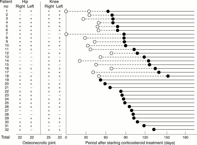
Objectives: To investigate the actual time of onset of osteonecrosis (ON) after high dose corticosteroid treatment in systemic lupus erythematosus (SLE).
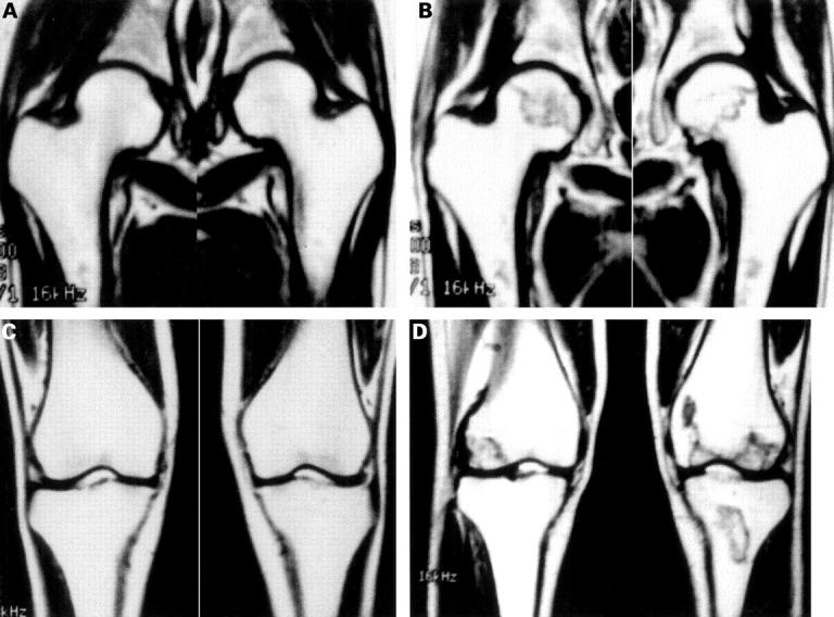
Methods: 72 patients with active SLE, who received high dose corticosteroid for the first time, for the development of ON at hips and knees were monitored by magnetic resonance imaging for at least 12 months.
Results: ON lesions were detected in 32/72 patients (44%) between one and five months (3.1 months on average) after starting high dose corticosteroid treatment. No osteonecrotic lesion was newly detected from the sixth month of treatment until the end of the follow up period.
Conclusion: The findings suggested that the actual time of onset of ON in SLE is within the first month of high dose corticosteroid treatment.
Figures
MeSH terms
Substances
LinkOut - more resources
Full Text Sources
Medical
