

Review
doi: 10.1136/heart.86.suppl_2.ii23.
Transoesophageal echocardiography during interventional cardiac catheterisation in congenital heart disease
Affiliations
- PMID: 11709531
- PMCID: PMC1766548
- DOI: 10.1136/heart.86.suppl_2.ii23
Item in Clipboard
Review
Transoesophageal echocardiography during interventional cardiac catheterisation in congenital heart disease
Heart.
2001 Dec.
No abstract available
Figures
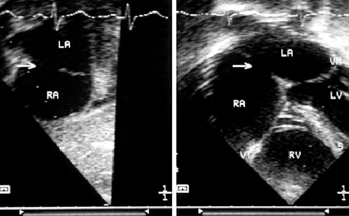
(Left) Short axis section of the atria, showing an oval fossa defect suitable for transcatheter closure. (Right) Four chamber section from the same patient, revealing an adequate rim of septum between the defect and the atrioventricular valves. A, left atrium; RA, right atrium; arrow, atrial septal defect; LV, left ventricle; RV, right ventricle; S, septum.
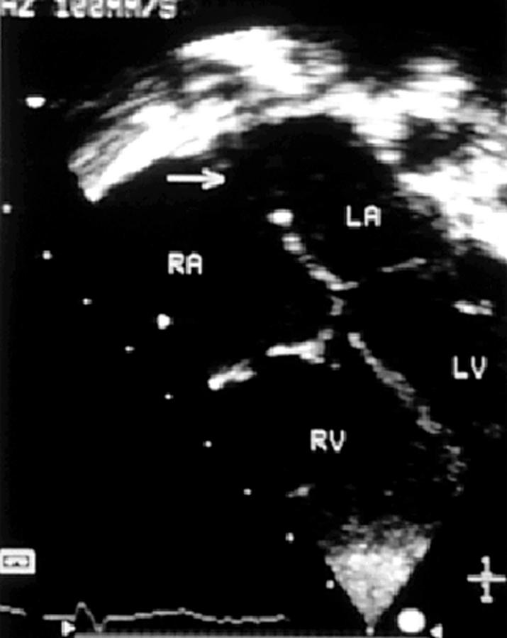
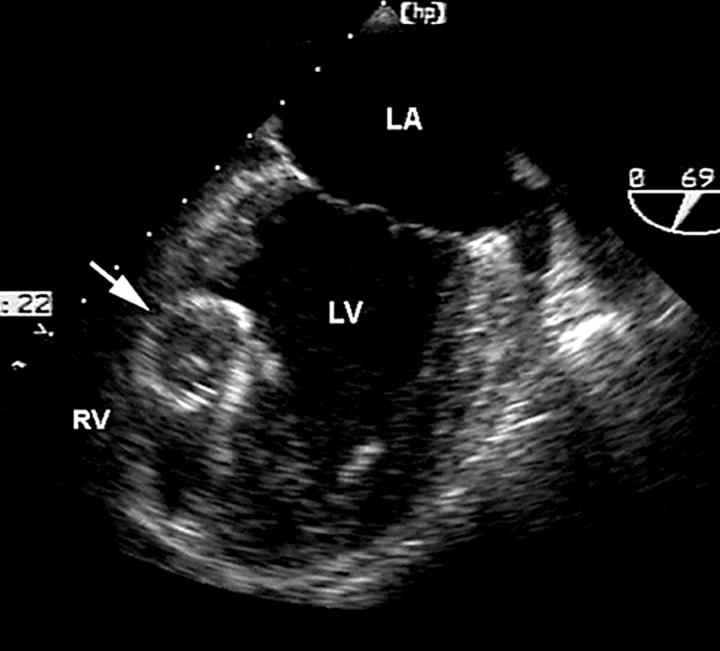
Four chamber section from an oval fossa atrial septal defect, in which there is a deficiency of infolding with an incomplete postero-superior rim. The arrow identifies the defect. LA, left atrium; RA, right atrium; LV, left ventricle; RV, right ventricle.
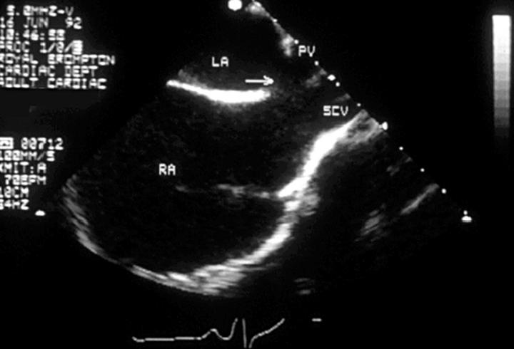
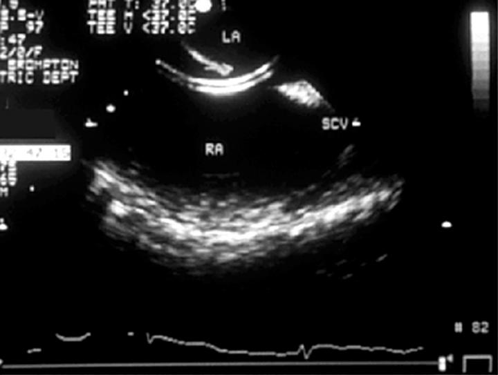
Transoesophageal echocardiogram in the vertical plane from a patient with a sinus venosus atrial septal defect, in which the superior caval vein (SCV) and right upper pulmonary vein (PV) override the atrial septum which divides the left atrium (LA) from right atrium (RA).
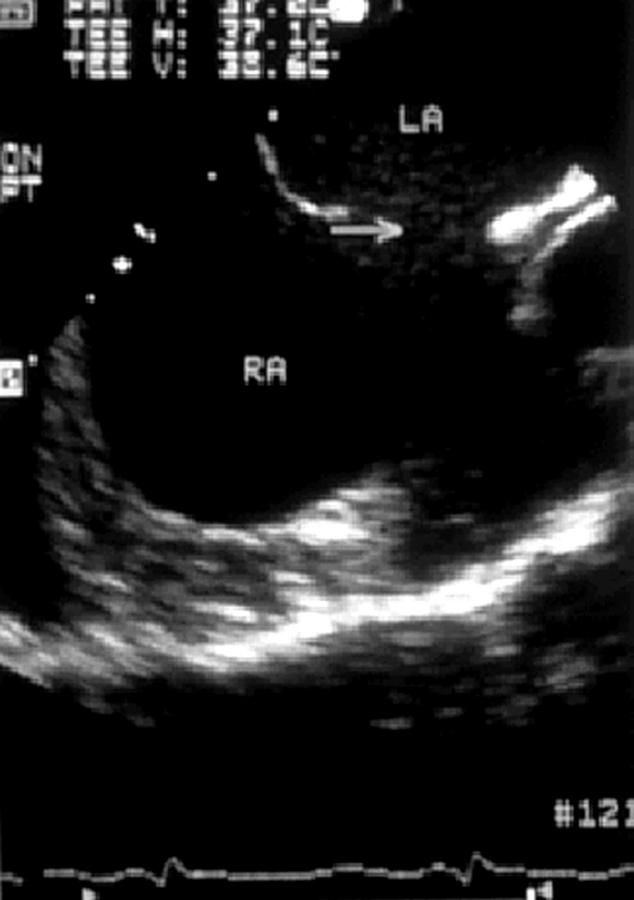
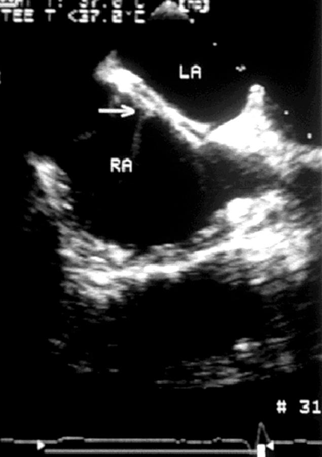
Transoesophageal echocardiogram in which an oval fossa atrial septal defect is indicated by an arrow. LA, left atrium, RA, right atrium.
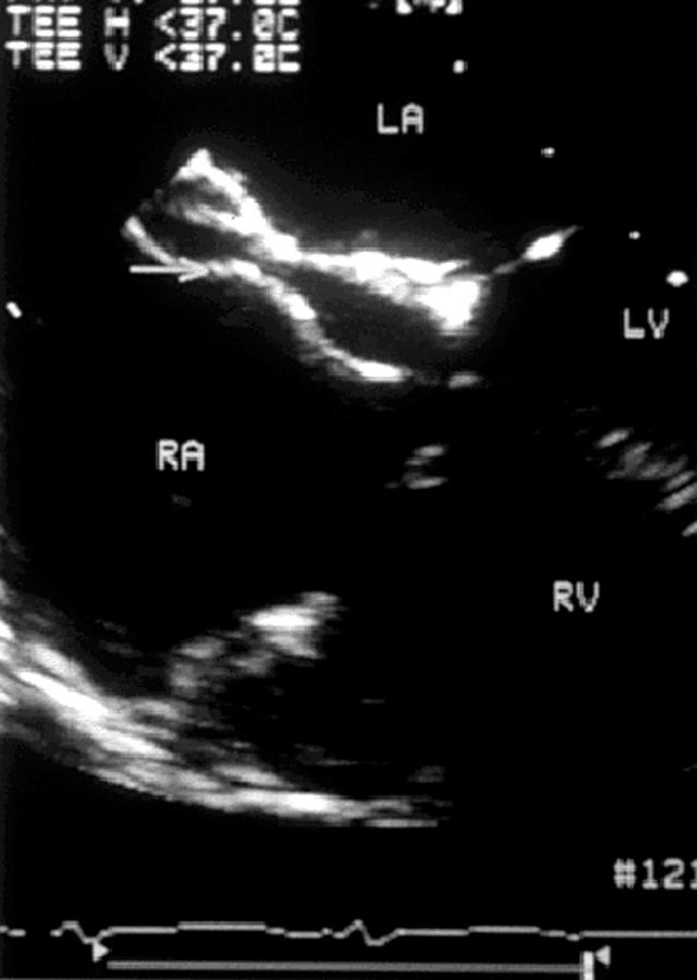
Transoesophageal echocardiogram identifying a catheter passing through a moderately large oval fossa atrial septal defect. LA, left atrium, RA, right atrium; SCV, superior caval vein.
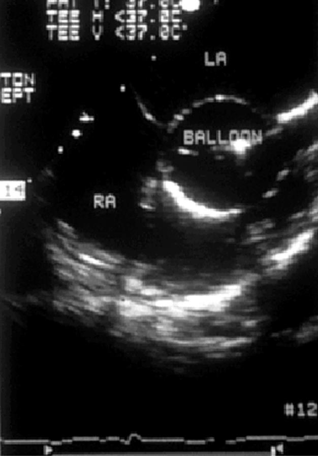
Transoesophageal echocardiogram recorded during balloon sizing of an oval fossa atrial septal defect. LA, left atrium, RA, right atrium.
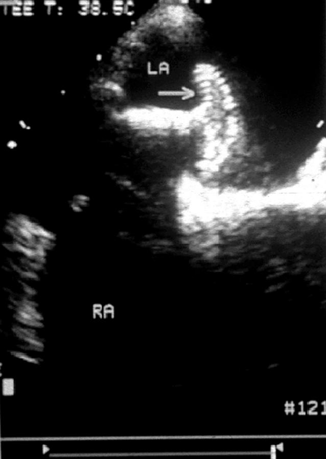
Transoesophageal echocardiogram recorded during deployment of a closure device illustrating the left atrial disc opened in the left atrium (arrow). LA, left atrium, RA, right atrium.
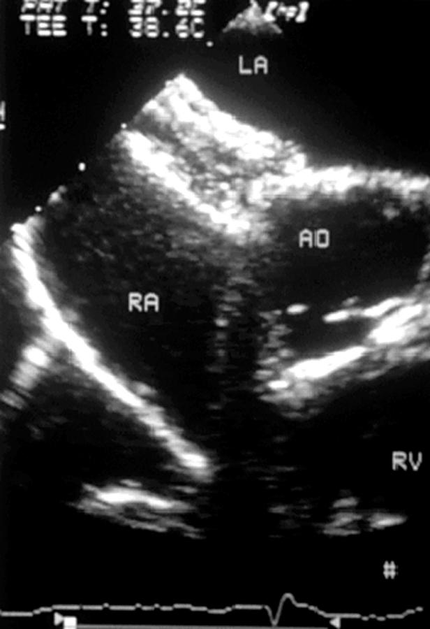
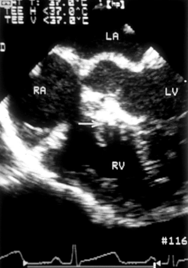
Transoesophageal echocardiogram recorded during device closure of an atrial septal defect with an Amplatzer occluder, showing left atrial and right atrial discs opened in their respective atria. AO, aortic root; LA, left atrium, RA, right atrium; RV, right ventricle.
Transoesophageal echocardiogram recorded during closure of an oval fossa defect with a Cardioseal device (arrow). LA, left atrium; RA, right atrium; LV, left ventricle; RV, right ventricle.
Transoesophageal echocardiogram recorded during closure of an oval fossa atrial septal defect with an Angel Wings device. LA, left atrium; RA, right atrium.
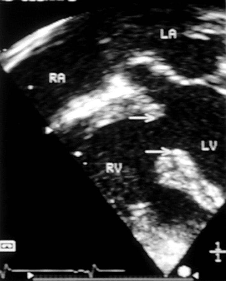
Transoesophageal horizontal section of a large muscular ventricular septal defect, whose borders are identified by the arrows. LA, left atrium; RA, right atrium; LV, left ventricle; RV, right ventricle.
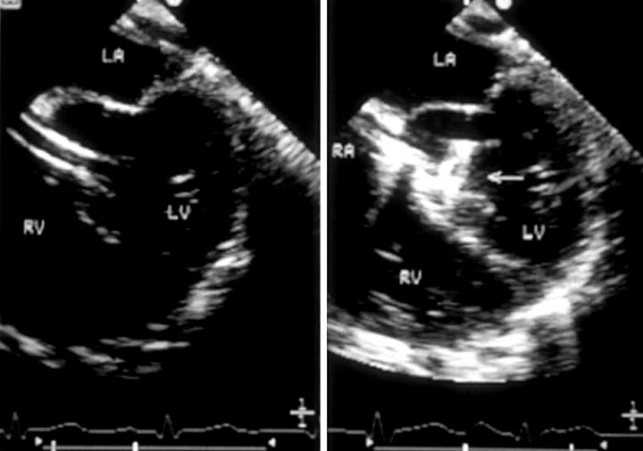
(Left) Transoesophageal echocardiogram recorded during device closure of a perimembranous ventricular septal defect, in which the sheath can be seen crossing the defect from right ventricle (RV) to left ventricle (LV). (Right) Horizontal plane transoesophageal echocardiogram recorded during device closure of a perimembranous ventricular septal defect, in which the left ventricular disc (arrow) can be seen to open within the left ventricle. LA, left atrium; RA, right atrium.
Horizontal transoesophageal echocardiogram following device closure of a perimembranous ventricular septal defect. An arrow identifies the position of the closure device within the ventricular septal defect.
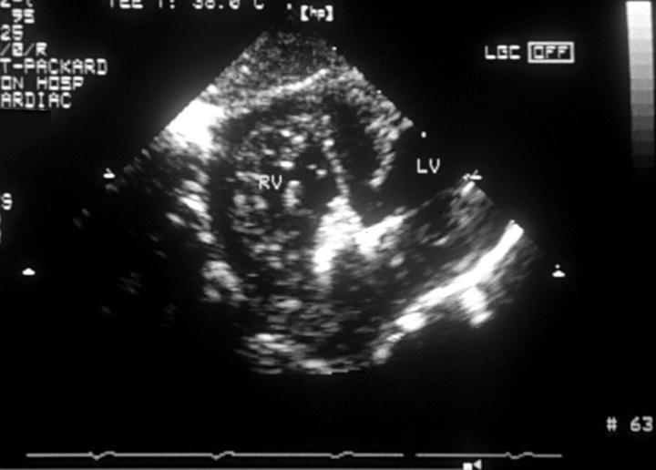
Transgastric short axis echocardiographic section following transcatheter closure of a muscular ventricular septal defect, showing the closure device within the muscular defect. The right ventricular disc is well seen within the right ventricle (RV), although the left ventricular disc within the left ventricle (LV) is imaged less well.
Transoesophageal echocardiogram recorded immediately following device closure of an ischaemic muscular ventricular septal defect in a patient presenting with cardiogenic shock. An arrow identifies the Amplatzer closure device. LA, left atrium; LV, left ventricle; RV, right ventricle.
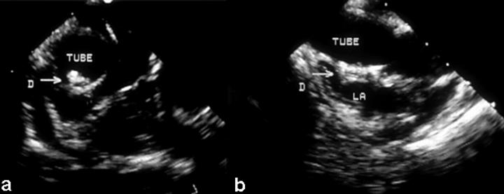
Transoesophageal echocardiograms immediately following transcatheter closure of a fenestrated lateral tunnel (tube) in a patient who had undergone a previous total caval pulmonary connection. The closure device (D, arrow) is seen in (a) horizontal and (b) vertical sections. LA, left atrium.
Similar articles
-
Supravalvar mitral ring with complete atrioventricular septal defect: a case report and three-dimensional echocardiography evaluation.J Am Soc Echocardiogr. 2010 Jul;23(7):792.e1-2. doi: 10.1016/j.echo.2009.11.027. Epub 2010 Feb 1. J Am Soc Echocardiogr. 2010. PMID: 20117914
-
Transcatheter treatment of Lutembacher syndrome: combined balloon mitral valvuloplasty and percutaneous atrial septal defect closure.J Invasive Cardiol. 2004 Nov;16(11):678-9. J Invasive Cardiol. 2004. PMID: 15550747 No abstract available.
-
Transoesophageal echocardiography in adult congenital heart disease.Heart. 2001 Dec;86 Suppl 2(Suppl 2):II30-II40. doi: 10.1136/heart.86.suppl_2.ii30. Heart. 2001. PMID: 11709532 Free PMC article. Review. No abstract available.
-
[Transesophageal and intracardiac echocardiography in therapeutic cardiac catheterizations of structural heart disease].Masui. 2014 Sep;63(9):982-7. Masui. 2014. PMID: 25255660 Japanese.
-
[Catheter interventions in congenital heart defects as alternatives to open surgery].Lakartidningen. 2005 Jul 11-24;102(28-29):2060-3. Lakartidningen. 2005. PMID: 16097174 Review. Swedish. No abstract available.
Cited by
-
The causes of Charcot-Marie-Tooth disease.Cell Mol Life Sci. 2003 Dec;60(12):2547-60. doi: 10.1007/s00018-003-3133-5. Cell Mol Life Sci. 2003. PMID: 14685682 Free PMC article. Review.
-
What is new in pediatric cardiology.Indian J Pediatr. 2003 Jan;70(1):41-9. doi: 10.1007/BF02722744. Indian J Pediatr. 2003. PMID: 12619952 Review.
-
[Catheter interventions for congenital heart disease].Herz. 2008 Dec;33(8):592-600. doi: 10.1007/s00059-008-3133-1. Epub 2009 Jan 8. Herz. 2008. PMID: 19137250 Review. German.
-
Position Statement on Indications for Echocardiography in Fetal and Pediatric Cardiology and Congenital Heart Disease of the Adult - 2020.Arq Bras Cardiol. 2020 Nov;115(5):987-1005. doi: 10.36660/abc.20201122. Arq Bras Cardiol. 2020. PMID: 33295472 Free PMC article. English, Portuguese. No abstract available.
-
Imaging of patients with congenital heart disease.Nat Rev Cardiol. 2011 Nov 1;9(2):101-15. doi: 10.1038/nrcardio.2011.162. Nat Rev Cardiol. 2011. PMID: 22045045 Review.
References
Publication types
MeSH terms
LinkOut - more resources
Full Text Sources
Medical