. 2001:1:24.
doi: 10.1186/1471-2180-1-24. Epub 2001 Oct 8.

Characterization of Norwalk virus GI specific monoclonal antibodies generated against Escherichia coli expressed capsid protein and the reactivity of two broadly reactive monoclonal antibodies generated against GII capsid towards GI recombinant fragments

Affiliations

Characterization of Norwalk virus GI specific monoclonal antibodies generated against Escherichia coli expressed capsid protein and the reactivity of two broadly reactive monoclonal antibodies generated against GII capsid towards GI recombinant fragments

T Yoda et al. BMC Microbiol. 2001.

Abstract

Background: Norwalk virus causes outbreaks of acute non-bacterial gastroenteritis in humans. The virus capsid is composed of a single 60 kDa protein. In a previous study, the capsid protein of recombinant Norwalk virus genogroup II was expressed in an E. coli system and monoclonal antibodies were generated against it. The analysis of the reactivity of those monoclonal antibodies suggested that the N-terminal domain might contain more antigenic epitopes than the C-terminal domain. In the same study, two broadly reactive monoclonal antibodies were observed to react with genogroup I recombinant protein.

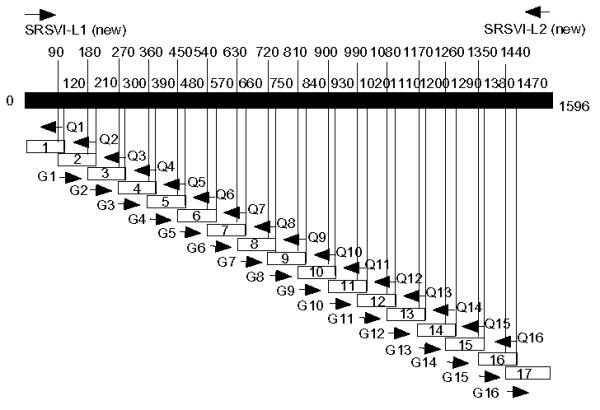
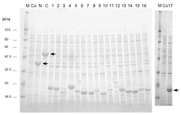
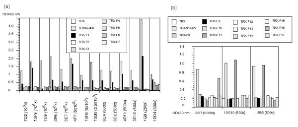
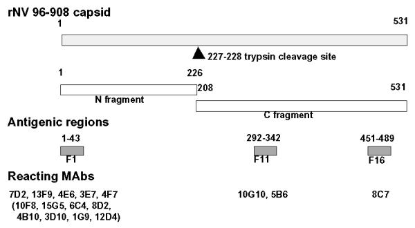
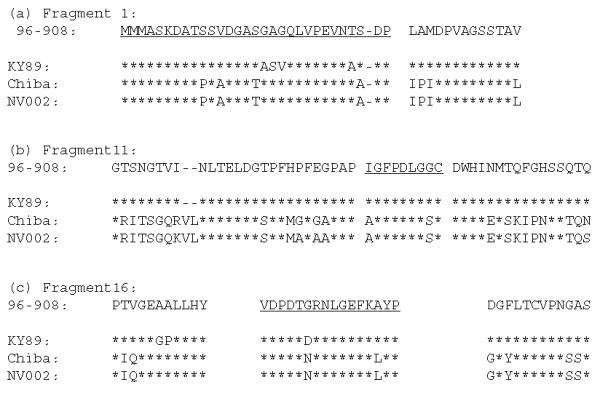
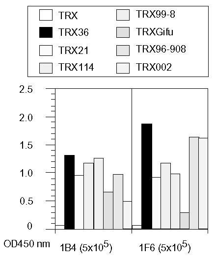
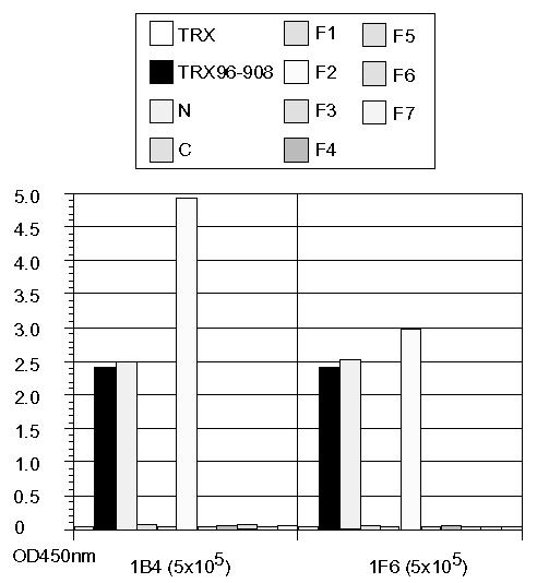
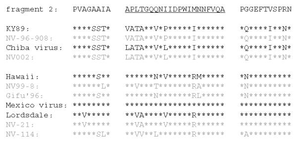
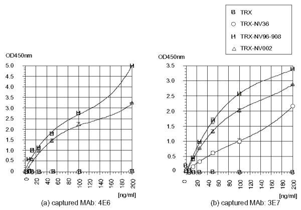
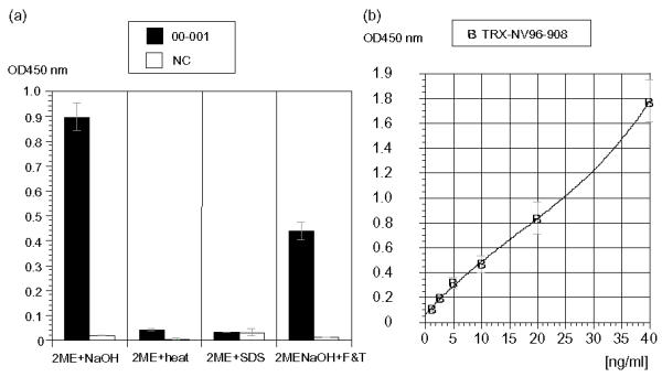
Results: In the present study, we used the recombinant capsid protein of genogroup I and characterized the obtained 17 monoclonal antibodies by using 19 overlapping fragments. Sixteen monoclonal antibodies recognized sequential epitopes on three antigenic regions, and the only exceptional monoclonal antibody recognized a conformational epitope. As for the two broadly reactive monoclonal antibodies generated against genogroup II, we indicated that they recognized fragment 2 of genogroup I. Furthermore, genogroup I antigen from a patient's stool was detected by sandwich enzyme-linked immunosorbent assay using genogroup I specific monoclonal antibody and biotinated broadly reactive monoclonal antibody.

Conclusion: The reactivity analysis of above monoclonal antibodies suggests that the N-terminal domain may contain more antigenic epitopes than the C-terminal domain as suggested in our previous study. The detection of genogroup I antigen from a patient's stool by our system suggested that the monoclonal antibodies generated against E. coli expressed capsid protein can be used to detect genogroup I antigens in clinical material.


Figures

Figure 1
Figure 1
Schematic diagram of rNV96-908 capsid fragments expressed in E. coli. The solid bar represents the full length of the rNV96-908 capsid protein. Numbers besides the bar show nucleotide numbers of NV96-908 capsid. Numbers above the bar indicate the planned position of the fragment edges not the actual positions. Small open boxes with numbers indicate each fragment. The N-terminal fragment consists of fragments 1–7 and the C-terminal fragment consists of fragments 8–17. These fragments were constructed using restriction enzyme sites in primers for ligation convenience (Table 2).
Figure 2
Figure 2
Expression of the rNV96-908 fragments in E. coli. Production of TRX-fused capsid protein fragments in E. coli was analyzed by staining two SDS-PAGE gels. From the left, lane 1 and lane 21 : marker [kDa], lane 2 and lane 2: control recombinant E. coli lysate (before induction). From lane 3 to 23 except 21 and 22 are TRX-NV96-908 N-terminal domain, C-terminal domain, fragment 1, 2, 3, 4, 5, 6, 7, 8, 9, 10, 11, 12, 13, 14, 15, 16, and 17, respectively. The positions of the recombinant NV capsid fragments are indicated by arrows (the small fragments show similar molecular weights).
Figure 3
Figure 3
Antigenic sites on rNV96-908 defined by MAbs using the N-terminal (a) and the C-terminal fragments (b) as antigens. Reactivity of MAbs with rNV96-908 derived fragments was measured by ELISA. Names of the individual MAbs and their dilutions used are shown at the bottom of the figures.
Figure 4
Figure 4
Diagram of rNV antigenic domains. The gray bar at the top represents the full length of the rNV96-908 capsid protein. The arrowhead shows the putative position of the trypsin cleavage site. The N-terminal and C-terminal parts are shown as open bars. The antigenic regions of fragments 1, 11 and 16 are indicated as dark gray color boxes. Reacting MAbs are shown below the corresponding fragments. Numbers on the top of the rNV capsid and the antigenic fragments indicate the first and last residues.
Figure 5
Figure 5
Comparison of amino acid sequences of fragments 1 (a), 11 (b) and 16 (c) in two rNVs of GI strains and the related reference NV strains. The dotted lines show deletions and under lined a.a. sequences are unique parts of the individual fragments. Other parts are overlapping with proximal fragments. Accession numbers for NV96-908, NV002, KY89 and Chiba virus are AB028247, AB063325, L23828 and AB022679, respectively.
Figure 6
Figure 6
Reactivity comparison of the broadly reactive MAbs generated against GII rNV36 towards various rNVs. Reactivity of the MAbs (1B4 and 1F6) was measured by direct ELISA. Among rNVs, strains 002 and 96–908 are GI and the others are GII. Names of the individual MAbs and their dilutions (in the parentheses) are indicated at the bottom of the figure.
Figure 7
Figure 7
Antigenic sites on rNV96-908 were defined by the broadly reactive MAbs using the N- and C-terminal fragments as well as fragments 1 to 7 as antigens. Reactivity of the MAbs towards rNV96-908 derived fragments was measured by ELISA. Names of the MAbs and their dilutions used are shown at the bottom of the figure.
Figure 8
Figure 8
Comparison of amino acid fragment 2 of NV36 with other rNVs and the reference NV strains. The underlined a.a. sequences are unique parts of the individual fragments. Other parts are overlapping with proximal fragments. Accession numbers for NV21, NV114, Gifu'96, Mexico virus and Lordsdale virus are AB028245, AB028246, AB045603, U22498 and X86557, respectively.
Figure 9
Figure 9
A double antibody sandwich ELISA for detecting various rNV antigens. The captured antibody in (a): 4E6 (GI specific MAb) and (b): 3E7 (GI specific MAb), B-1F6 was used as the detecting antibody in both of the reaction.
Figure 10
Figure 10
A double antibody sandwich ELISA for detecting the clinical material (NV from a patient's stool) in (a). GI specific MAb, 3E7 was used as the captured antibody, and B-1F6 was used as the detecting antibody, (a) Comparison of NV GI positive and NV negative clinical materials in 4 types of pre-treatments. (b) A calibration curve to detect rNV96-908 by the same system as (a).

References

    1. Lambden PR, Caul EO, Ashley CR, Clarke IN. Sequence and genome organization of a human small round-structured (Norwalk-like) virus. Science. 1993;259:516–519. - PubMed
    1. Jiang X, Wang M, Wang K, Estes MK. Sequence and genomic organization of Norwalk Virus. Virology. 1993;195:51–61. - PubMed
    1. Glass PJ, White LJ, Ball JM, Leparc-Goffart I, Hardy ME, Estes MK. Norwalk Virus Open Reading Frame 3 Encodes a Minor Structural Protein. J Virol. 2000;74:6581–6591. - PMC - PubMed
    1. Hale AD, Crawford SE, Ciarlet M, Green J, Gallimore C, Brown DWG, Jiang X, Estes MK. Expression and self-assembly of Grimsby Virus: Antigenic distinction from Norwalk and Mexico viruses. Clin Diagn Lab Immunol. 1999;6:142–145. - PMC - PubMed
    1. Leite JPG, Ando T, Noel JS, Jiang B, Humphrey CD, Lew JF, Green KY, Glass RI, Monroe SS. Characterization of Toronto virus capsid protein expressed in baculovirus. Arch Virol. 1996;141:865–875. - PubMed

MeSH terms