. 2001:1:15.
doi: 10.1186/1471-2407-1-15. Epub 2001 Oct 29.

Immunodetection of nmt55/p54nrb isoforms in human breast cancer

Affiliations

Immunodetection of nmt55/p54nrb isoforms in human breast cancer

M Pavao et al. BMC Cancer. 2001.

Abstract

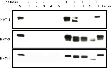
Background: We previously identified and characterized a novel 55 kDa nuclear protein, termed nmt55/p54nrb, whose expression was decreased in a subset of human breast tumors. The objective of this study was to determine if this reduced expression in human breast tumors was attributed to the regulation of mRNA transcription or the presence of altered forms of this protein.

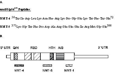
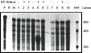
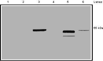
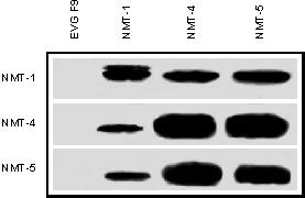
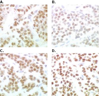
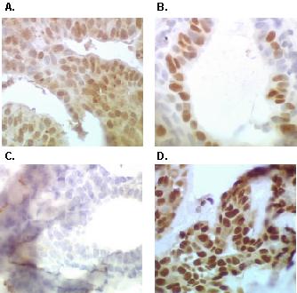
Results: Northern blot analysis and ribonuclease protection assay indicated that nmt55/p54nrb mRNA is expressed at varying levels in estrogen receptor positive (ER+) and estrogen receptor negative (ER-) human breast tumors suggesting that reduced expression of nmt55/p54nrb protein in ER- tumors was not due to transcriptional regulation. To determine if multiple protein isoforms are expressed in breast cancer, we utilized Western blot and immunohistochemical analyses, which revealed the expression of an nmt55/p54nrb protein isoform in a subset of ER+ tumors. This subset of ER+ human breast tumors expressed an altered form of nmt55/p54nrb that was undetectable with an amino-terminal specific antibody suggesting that this isoform contains alterations or modifications within the amino terminal domain.

Conclusions: Our study indicates that nmt55/p54nrb protein is post-transcriptionally regulated in human breast tumors leading to reduced expression in ER- tumors and the expression of an amino terminal altered isoform in a subset of ER+ tumors. The potential involvement of nmt55/p54nrb in RNA binding and pre-mRNA splicing may be important for normal cell growth and function; thus, loss or alteration of protein structure may contribute to tumor growth and progression.


Figures

Figure 1
Figure 1
Amino acid sequence and molecular localization of the synthetic oligopeptides used to generate human nmt55/p54nrb polyclonal antibodies.
Figure 2
Figure 2
Northern blot analysis of nmt55/p54nrb mRNA expression in MCF-7 cells and ER+/ER- Human Breast tumors. Total RNA (20 μg) was prepared from MCF-7 cells (lane M) and twelve different human breast tumors (lanes 1–12). The RNA was subjected to hybridization with a human nmt55/p54nrb 499 bp SacI/BglII radiolabeled probe (top panel) or a human glyceraldehyde-3-phosphate (GAPDH) 545 bp HindIII/Xbal radiolabeled probe to indicate levels of RNA loading (bottom panel). Estrogen receptor (ER) expression levels were determined by ligand binding assay and are indicated along the top of the figure.
Figure 3
Figure 3
Ribonuclease protection assay of nmt55/p54nrb mRNA expression in MCF-7 cells and ER+/ER- Human Breast tumors. Total RNA (20 μg) was prepared from MCF-7 cells (lane M), ten different human breast tumors (lanes 1–10) and calf uterus tissue (lane C). The RNA was subjected to hybridization with a human nmt55/p54nrb 499 bp SacI/BglII radiolabeled probe. The total RNA/radiolabeled probe mixtures were then digested and separated as described in Materials and Methods. Lane P indicates radiolabeled probe alone, which was not subjected to RNase digestion. Lane R represents radiolabeled probe alone that was digested with the RNase mixture. Estrogen receptor (ER) expression levels were determined by ligand binding assay and are indicated along the top of the figure. Estimated molecular weight is indicated in the MW lane.
Figure 4
Figure 4
Assessment of NMT-4 and NMT-5 polyclonal antibody specificity for nmt55/p54nrb. MCF-7 nuclear extract (50 μg) was separated by SDS/PAGE and subjected to Western blot analysis. Lanes 1 and 2 represent immunodetection with NMT-4 pre-immune serum and NMT-5 pre-immune serum, respectively. Lanes 3 and 5 represent immunodetection with polyclonal antibodies NMT-4 and NMT-5, respectively. Lane 4 represents MCF-7 nuclear extract immunoblotted with polyclonal antibody NMT-4 pre-incubated with its corresponding immunogenic peptide. Lane 6 represents MCF-7 nuclear extract immunoblotted with polyclonal antibody NMT-5 pre-incubated with its corresponding immunogenic peptide. Right hand margin represents estimated molecular weight.
Figure 5
Figure 5
Imimunoprecipitation and immunodetection of nmt55/p54nrb protein with specific site-directed antibodies raised against various domains of nmt55/p54nrb. MCF-7 cell nuclear extracts (50 μg) were subjected to immunoprecipitation assays with the antibodies indicated along the top margin. Lane 1 represents immunoprecipitation with monoclonal antibody EVG F9 (raised against human estrogen receptor alpha), lane 2 monoclonal antibody NMT-1, lane 3 polyclonal antibody NMT-4 and lane 4 polyclonal antibody NMT-5. Immunoprecipitated samples were separated by SDS/PAGE and analyzed by Western blot. Left margin indicates the antibodies used for immunodetection. Top panel represents immunodetection with monoclonal antibody NMT-1, middle panel polyclonal antibody NMT-4 and bottom panel polyclonal antibody NMT-5.
Figure 6
Figure 6
Detection of nmt55/p54nrb protein isoforms by Western Blot Analysis using NMT-4, NMT-5 and NMT-1 antibodies. MCF-7 cell nuclear extract (50 μg), lane M, and ten different human breast tumor nuclear extracts (50 μg), lanes 1–10, were separated by SDS/PAGE and subjected to Western blot analysis with polyclonal antibody NMT-4 (Top Panel), polyclonal antibody NMT-5 (Middle Panel) and monoclonal antibody NMT-1 (Bottom Panel). Estrogen receptor (ER) expression levels were determined by ligand binding assay and are indicated along the top of the figure.
Figure 7
Figure 7
Immunohistochemical Analyses of Estrogen Receptor, Progesterone Receptor and nmt55/p54nrb in a Human Breast Tumor. Human breast tumor tissue was fixed, embedded, sectioned and subjected to immunohistochemical analysis with antibodies to human estrogen receptor alpha (hERα), human progesterone receptor (hPR) and nmt55/p54nrb. Panel A represents immunostaining with monoclonal antibody EVG F9 raised against hERα. Panel B represents immunostaining with monoclonal antibody 4.14 raised against hPR. Panels C and D represent immunostaining with anti-nmt55/p54nrb polyclonal antibodies NMT-4 and NMT-5, respectively.
Figure 8
Figure 8
Detection of nmt55/p54nrb protein variants by immunohistochemical analyses with NMT-4 and NMT-5 antibodies in a Human Breast tumor. Human breast tumor tissue was fixed, embedded, sectioned and subjected to immunohistochemical analysis with antibodies to human estrogen receptor alpha (hERα), human progesterone receptor (hPR) and nmt55/p54nrb. Panel A represents immunostaining with monoclonal antibody EVG F9 raised against hERα. Panel B represents immunostaining with monoclonal antibody 4.14 raised against hPR. Panels C and D represent immunostaining with anti-nmt55/p54nrb polyclonal antibodies NMT-4 and NMT-5, respectively.

References

    1. Gould MN. Cellular and molecular aspects of the multistage progression of mammary carcinogenesis in humans and rats. Semin Cancer Biol. 1993;4:161–169. - PubMed
    1. Dickson RB, Lippman ME. Growth factors in breast cancer. Endocr Rev. 1995;16:559–589. - PubMed
    1. Weinberg RA. Tumor suppressor genes. Science. 1991;254:1138–1146. - PubMed
    1. Weinberg RA. The molecular basis of oncogenes and tumor suppressor genes. Ann N Y Acad Sci. 1995;758:331–338. - PubMed
    1. Hanahan D, Weinberg RA. The hallmarks of cancer. Cell. 2000;100:57–70. - PubMed

Publication types

MeSH terms

LinkOut - more resources