. 2001 Nov 15;15(22):2950-66.
doi: 10.1101/gad.925901.

TGF-beta inhibits muscle differentiation through functional repression of myogenic transcription factors by Smad3

Affiliations

TGF-beta inhibits muscle differentiation through functional repression of myogenic transcription factors by Smad3

D Liu et al. Genes Dev. .

Abstract

Transforming growth factor-beta (TGF-beta) is a potent inhibitor of skeletal muscle differentiation, but the molecular mechanism and signaling events that lead to this inhibition are poorly characterized. Here we show that the TGF-beta intracellular effector Smad3, but not Smad2, mediates the inhibition of myogenic differentiation in MyoD-expressing C3H10T1/2 cells and C2C12 myoblasts by repressing the activity of the MyoD family of transcriptional factors. The Smad3-mediated repression was directed at the E-box sequence motif within muscle gene enhancers and the bHLH region of MyoD, the domain required for its association with E-protein partners such as E12 and E47. The repression could be overcome by supplying an excess of E12, and covalent tethering of E47 to MyoD rendered the E-box-dependent transcriptional activity refractory to the effects of Smad3 and TGF-beta. Smad3 physically interacted with the HLH domain of MyoD, and this interaction correlated with the ability of Smad3 to interfere with MyoD/E protein heterodimerization and binding of MyoD complexes to oligomerized E-box sites. Together, these results reveal a model for how TGF-beta, through Smad3-mediated transcriptional repression, inhibits myogenic differentiation.


Figures

Figure 1
Figure 1
Smad3, but not Smad2, inhibits MyoD-induced myogenic differentiation of C3H10T1/2 fibroblasts. 10T1/2 cells were transiently transfected with an expression plasmid for MyoD alone (A,B) or together with an expression plasmid for Smad2 (C,D) or Smad3 (E,F), respectively. At day 1 post transfection, cells were shifted to differentiation medium for an additional 2 d. Cells were fixed and myofiber formation was assessed by immunostaining with an anti-MHC monoclonal antibody (green). MyoD expression in transfected cells was visualized by immunofluorescence using an anti-MyoD antibody, revealing predominantly nuclear staining (red).
Figure 2
Figure 2
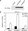
Stable expression of wild-type and dominant-negative Smad3 in C2C12 myoblast cells. (A) Immunoblot analysis (IB) of Smad3 expression in lysates of C2C12 cells stably infected with a viral vector for either an N-terminally (Smad3NF) or a C-terminally Flag-tagged (Smad3CF) Smad3. Lysates from cells infected with the empty vector were used as control. (B) TGF-β-induced transcription from the 3TP-Lux reporter plasmid in transiently transfected C2C12 cells that stably express Smad3NF or Smad3CF, or vector control cells. Luciferase expression values, normalized for transfection efficiency, are shown as fold induction relative to the basal promoter activity.
Figure 3
Figure 3
Smad3 regulates myoblast differentiation and the inhibitory response to TGF-β. C2C12 cells stably infected with control viral vector (A,B) or those harboring coding-sequences of Smad3NF (C,D) or Smad3CF (E,F) were grown to near confluence and then shifted to differentiation medium without or with 1-ng/mL TGF-β, as indicated, for 4 d. Terminal differentiation and myotube formation were assessed by immunofluorescence with an anti-MHC antibody. (G) Expression of myogenin and MHC, as assessed by Western blotting, in control-infected or Smad3NF- or Smad3CF-expressing C2C12 cells. Cells were cultured in growth medium (GM) and shifted to differentiation medium (DM) as in AF. The lower panel shows expression of Smad3 detected by an anti-Smad3 antibody. Flag-tagged Smad3 was expressed at considerably higher levels than endogenous Smad3 represented by the slightly faster migrating band.
Figure 4
Figure 4
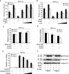
TGF-β signaling through Smad3 inhibits the transcriptional activation of muscle-specific enhancers by myogenic bHLH factors. (A,B) Effects of TGF-β, Smad2, or Smad3 on MyoD- or myogenin-directed transcription from the MCK promoter. 10T1/2 cells were cotransfected with the MCK-Luc reporter plasmid and the indicated expression plasmids. Transfected cells were cultured with or without 1-ng/mL TGF-β before measuring luciferase activities. (C,D) Similar transcriptional assays were performed using the 4R-tk-Luc reporter. Increasing quantities of a Smad3 expression plasmid were cotransfected with MyoD or myogenin to show the dosage dependence. (E) Effects of wild-type and mutant forms of Smad3 on MyoD-directed transcription from the 4R-tk promoter. 10T1/2 cells were cotransfected with 4R-tk-Luc, expression plasmids for MyoD and wild-type Smad3 (WT), Smad3 dominant-negative mutants SA, and ΔC or truncation mutants NL and C. (F) Effect of TGF-β on MyoD transactivation of the 4R-tk-Luc reporter in Smad3−/− mouse embryonic fibroblasts and in Smad3−/− cells engineered to constitutively express wild-type Smad3. All luciferase values, normalized for transfection efficiency, are denoted as a percentage of the activity in cells expressing MyoD or myogenin alone in the absence of TGF-β.
Figure 5
Figure 5
Transcriptional repression by TGF-β/Smad3 signaling is directed at the bHLH domain of MyoD. (A) TGF-β/Smad signaling does not affect the transcriptional activity of E47. 10T1/2 cells were transfected with the reporter plasmid (E2-5)4-TATA-CAT and expression plasmids for E47, Smad2, or Smad3, as indicated. Transfected cells were treated with or without TGF-β as in Fig. 4, and CAT activities were measured. (B) TGF-β/Smad3 signaling does not affect the transcriptional potential of MyoD. 10T1/2 cells were transfected with a plasmid encoding either Gal4 DNA-binding domain (Gal4-DBD) or the fusion of Gal4-DBD with MyoD (Gal4-MyoD) or a similar fusion with deletion of the HLH domain (Gal4-MyoDΔHLH), or Gal4-DBD fused to the VP16 transcriptional activation domain (Gal4-VP16). Cells were cotransfected with reporter construct Gal-Luc, containing five copies of Gal4-binding sites driving luciferase expression, as well as Smad2 or Smad3 expression plasmids. (C) TGF-β/Smad3 signaling regulates the function of the HLH domain of MyoD. 4R-tk-Luc reporter assays were performed in 10T1/2 cells expressing the bHLH domain of MyoD fused to the VP16 TAD (MyoDbHLH-VP16) or a similar fusion protein, in which the basic domain of MyoD was replaced by that of E12 (MyoDHLH-E12b-VP16). In all assays, values for reporter activities are represented as percentage of the activities of each bHLH transcription factor obtained in the absence of TGF-β and coexpressed Smads.
Figure 6
Figure 6
Smad3 interacts with MyoD in vivo and in vitro. (A) Association of Smad3 with MyoD in transfected cells. COS cells were transfected with expression plasmids for Myc-tagged MyoD Flag-tagged full-length Smad2 or Smad3 or Smad3 fragments (Smad3NL and Smad3C). Cell lysates were subjected to Flag immunoprecipitation followed by immunoblotting using anti-Myc tag antibody to detect the coprecipitated MyoD, or anti-Flag antibody to detect the Smad proteins. Expression of Myc-MyoD was assessed by direct immunoblotting of a portion of the cell lysates. (B) Interaction of 35S-labeled in vitro-translated MyoD with GST-Smad fusion proteins. The upper panel shows MyoD protein retained by the indicated GST-Smad beads. The Coomassie Blue-stained gel in the lower panel shows the integrity and equal loading of the GST-fusion proteins. (C) Mapping of the MyoD domain that interacts with Smad3 in vivo. (Top) Schematic diagram shows the location of the MyoD deletion mutants. (Bottom) Myc-tagged full-size MyoD or its deletion mutants were coexpressed with Flag-Smad3 in transfected COS cells. Flag immunoprecipitates were subjected to immunoblotting using anti-Myc antibody as in A. The expression levels of epitope-tagged proteins were analyzed by immunoblotting (middle and lower panel).
Figure 7
Figure 7
TGF-β and Smad3 repress the transcriptional activity of MyoD by limiting its access to the E2A class of bHLH protein partners. (A,B) Increased E12 expression counteracts Smad3-mediated repression of transcription by MyoD. 10T1/2 cells were transfected with the 4R-tk-Luc (A) or MCK-Luc (B) reporter plasmid and expression plasmids for MyoD and Smad3, as well as increasing amounts of an E12 expression plasmid. Luciferase expression levels in the absence or presence of TGF-β were quantitated as in Fig. 4. (C,D) The activity of a covalently tethered MyoD∼E47 dimer is resistant to repression by TGF-β/Smad3 signaling. 10T1/2 cells were transfected with the 4R-tk-Luc (C) or MCK-Luc (D) reporter, together with expression plasmids for MyoD∼E47 and Smad2 or Smad3. Luciferase activities were scored relative to the value of MyoD∼E47 alone in the absence of TGF-β. Cotransfection of equivalent quantities of Smad3 and MyoD expression plasmid resulted in a repression of the MyoD activity by > 80% (see Fig. 4). (E) Mammalian two-hybrid analyses of the association between MyoD and E12. 10T1/2 cells were transfected with the indicated expression plasmids and a Gal4-responsive reporter Gal-Luc in the presence of increasing amounts of Smad3 expression plasmids. The normalized luciferase activities, as a consequence of the interaction between Gal4-E12 and MyoDbHLH-VP16 fusion proteins, are shown. (F) Evaluating the MyoD/E12 interaction by coimmunoprecipitation. HA-tagged MyoD and Myc-tagged E12 were expressed in 10T1/2 cells in the presence or absence of 2ng/mL TGF-β and/or cotransfected Smad3. Cell lysates were subjected to anti-HA immunoprecipitation, and MyoD and coprecipitated E12 in the protein complexes were detected by immunoblotting, using anti-HA (top) or anti-Myc antibodies (middle), respectively. The expression of E12 in the cell lysates was monitored by immunoblotting using anti-Myc antibody (bottom).
Figure 8
Figure 8
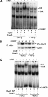
TGF-β/Smad3 signaling diminishes the binding of MyoD protein complexes to E-box DNA sequences. (A) Electrophoretic mobility shift assay (EMSA) using nuclear extracts from 10T1/2 cells transfected with Myc-tagged MyoD and Flag-tagged Smad3, as indicated, in the presence or absence of TGF-β. The oligonucleotide probe contains two direct repeats of an E-box sequence from the MCK enhancer (2xMEF1). In lane 4, anti-Myc antibody, which recognizes Myc-tagged MyoD, was added to the binding reaction. The MyoD-containing DNA-protein complex (Shift) was identified by comparing the gel-shift patterns from extracts of vector-transfected control and MyoD-transfected cells, and by the appearance of a supershifted (SS) tertiary complex in the presence of anti-Myc antibody. The composition of the lower mobility complexes, present in all samples, including control reactions, is not clear; however, these bands were absent when the oligonucleotide contained only one E-box sequence (data not shown). (B) Binding of MyoD to the MEF1 sites was analyzed using biotin-labeled oligonucleotides. The biotinylated wild-type 2xMEF1 (W) or a mutant MEF1 (M) oligonucleotide immobilized on streptavidin beads were incubated with lysates of the transfected 10T1/2 cells as in A, and the DNA-bound MyoD complexes were analyzed by gel electrophoresis followed by immunoblotting using anti-Myc antibody. The expression of MyoD or Smad3 in the cells was detected by immunoblotting of a fraction of the lysates. (C) EMSA was also performed for nuclear extracts of cells transfected with HA-tagged MyoD∼E47 fusion protein. Binding of this protein to the 2xMEF2 site, as identified by the supershifted band following incubation with anti-HA antibody, was not significantly affected by TGF-β treatment and Smad3 coexpression.

Similar articles

Cited by

References

    1. Alliston T, Choy L, Ducy P, Karsenty G, Derynck R. TGF-β-induced repression of CBFA1 by Smad3 decreases cbfa1 and osteocalcin expression and inhibits osteoblast differentiation. EMBO J. 2001;20:2254–2272. - PMC - PubMed
    1. Bailey P, Downes M, Lau P, Harris J, Chen SL, Hamamori Y, Sartorelli V, Muscat GE. The nuclear receptor corepressor N-CoR regulates differentiation: N-CoR directly interacts with MyoD. Mol Endocrinol. 1999;13:1155–1168. - PubMed
    1. Benezra R, Davis RL, Lockshon D, Turner DL, Weintraub H. The protein Id: A negative regulator of helix-loop-helix DNA binding proteins. Cell. 1990;61:49–59. - PubMed
    1. Bengal E, Flores O, Rangarajan PN, Chen A, Weintraub H, Verma IM. Positive control mutations in the MyoD basic region fail to show cooperative DNA binding and transcriptional activation in vitro. Proc Natl Acad Sci. 1994;91:6221–6225. - PMC - PubMed
    1. Black BL, Olson EN. Transcriptional control of muscle development by myocyte enhancer factor-2 (MEF2) proteins. Annu Rev Cell Dev Bi. 1998;14:167–196. - PubMed

Publication types

MeSH terms