Review
. 2001 Dec;86(6):612-8.
doi: 10.1136/heart.86.6.612.

Photodynamic therapy: shedding light on restenosis

Affiliations
Review

Photodynamic therapy: shedding light on restenosis

R Mansfield et al. Heart. 2001 Dec.
No abstract available


Figures

Figure 1
Figure 1
Mechanism of action of photodynamic therapy. Photosensitising agents are activated by light of a specific wavelength. In its excited state the photosensitiser reacts with tissue oxygen to yield various reactive oxidative species, inducing specific cellular and extracellular effects that lead to inhibition of restenosis (see text). The emission of fluorescence as the photosensitiser decays back to its ground state can be used to study the tissue distribution of the sensitiser.
Figure 2
Figure 2
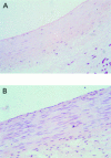
Photomicrograph (×400 magnification) of haematoxylin and eosin stained sections of pig coronary artery. Histological changes observed three days after photodynamic therapy (PDT) (top) include endothelial denudation and medial smooth muscle cell eradication. The PDT regime involved local light delivery (50 J/cm2 at a wavelength of 635 nm from a copper vapour dye laser), using a modified standard angioplasty catheter 4-6 hours after systemic sensitisation with 5-aminolaevulinic acid (ALA). A normal untreated vessel is shown for comparison (bottom).
Figure 3
Figure 3
Modification of a standard transparent Asuka angioplasty balloon for endovascular light delivery. A 100 µm laser fibre with a distal diffuser is placed in the guide wire lumen such that the diffuser is located in the balloon segment, allowing circumferential illumination through the entire balloon surface area.
Figure 4
Figure 4
Photomicrograph (×40 magnification) of elastin van Giessen stained sections of pig circumflex artery 28 days after oversized balloon injury. The balloon injured control artery (top) shows a greater neointimal area and reduced lumen size than the balloon injured artery that received photodynamic therapy (PDT) (bottom). The PDT regimen involved local light delivery (50 J/cm2 at a wavelength of 635 nm from a copper vapour dye laser) using a modified standard angioplasty catheter 4-6 hours after systemic sensitisation with 5-aminolaevulinic acid (ALA).

References

    1. Circulation. 2000 Nov 7;102(19):2322-4 - PubMed
    1. J Vasc Surg. 1994 Feb;19(2):321-9; discussion 329-31 - PubMed
    1. Arterioscler Thromb Vasc Biol. 1999 Sep;19(9):2154-61 - PubMed
    1. Laryngoscope. 1995 Aug;105(8 Pt 1):867-71 - PubMed
    1. J Photochem Photobiol B. 2000 Sep;57(2-3):94-101 - PubMed

Publication types

Substances