

Review
doi: 10.1136/heart.86.6.619.
Constrictive pericarditis in the modern era: a diagnostic dilemma
Affiliations
- PMID: 11711451
- PMCID: PMC1730027
- DOI: 10.1136/heart.86.6.619
Item in Clipboard
Review
Constrictive pericarditis in the modern era: a diagnostic dilemma
Heart.
2001 Dec.
No abstract available
Figures
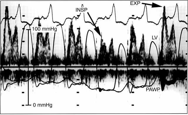
Transmitral flow velocity in a patient with constrictive pericarditis. During peak inspiration (INSP), there is a decrease in the early diastolic driving pressure across the mitral valve, seen as the initial gradient between the pressure in the left ventricle (LV) and the pulmonary artery wedge pressure (PAWP). This results in a decrease in the initial E velocity on the transmitral flow velocity curve. During expiration (EXP), there is an increase in the transmitral gradient between the pressure in the LV and the PAWP, resulting in an increase in the initial E velocity and the transmitral flow velocity curve. Data were obtained simultaneously by Doppler echocardiography and high fidelity manometer tipped catheters.
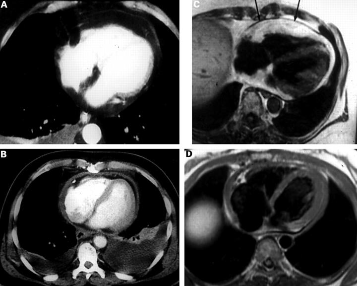
Normal and abnormal pericardium seen on computed tomography (CT) and magnetic resonance imaging (MRI) studies. (A) CT scan of normal pericardium. (B) CT scan of thickened pericardium. (C) MRI scan of normal thick pericardium (arrows). (D) MRI scan of a thickened pericardium. Reproduced from Breen18 with permission of Lippincott Williams & Wilkins.
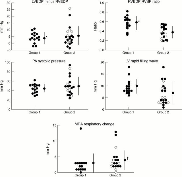
Conventional criteria at cardiac catheterisation. Group 1, patients with constrictive pericarditis; group 2, patients with restrictive cardiomyopathy or other types of cardiomyopathy and a normal pericardium. Although there are significant differences between the two groups, overlap makes it difficult to apply the criteria in an individual case. LVEDP, left ventricular end diastolic pressure; RVEDP, right ventricular end diastolic pressure; RVSP, right ventricular systolic pressure; PA, pulmonary artery; LV, left ventricular; MRA, mean right atrial pressure. Reproduced from Hurrell et al 7 with permission of the American Heart Association.
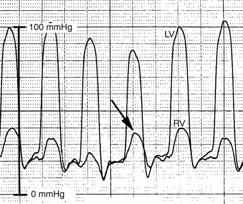
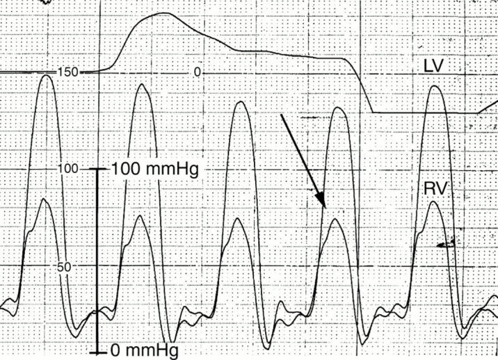
Pressures in the left (LV) and right ventricle (RV) of a patient with constrictive pericarditis. During peak inspiration (arrow), there is a decrease in LV pressure and a concomitant increase in RV pressure, indicating discordance of ventricular pressures.
Pressures in the left (LV) and right ventricle (RV) of a patient with restrictive cardiomyopathy. During peak inspiration (arrow) there is a decrease in LV pressure and a concomitant decrease in RV pressure, indicating concordance of ventricular pressures.
References
Publication types
MeSH terms
LinkOut - more resources
Full Text Sources