. 2001 Nov 15;537(Pt 1):237-50.
doi: 10.1111/j.1469-7793.2001.0237k.x.

Distribution of pacemaker function through the tunica muscularis of the canine gastric antrum

Affiliations

Distribution of pacemaker function through the tunica muscularis of the canine gastric antrum

K Horiguchi et al. J Physiol. .

Abstract

1. Interstitial cells of Cajal (ICC) have been shown to generate pacemaker activity in gastrointestinal (GI) muscles. Experiments were performed to characterize the ICC within the canine gastric antrum and to determine the site(s) of pacemaker activity and whether active propagation pathways exist within the thick-walled tunica muscularis of large mammals. 2. Immunohistochemistry and electron microscopy revealed four populations of ICC within the antral muscularis on the basis of anatomical location. Typical ICC were found in the myenteric region of the small intestine (IC-MY). Intramuscular ICC (IC-IM) were intermingled between muscle fibres of circular and longitudinal muscle layers. ICC were also found within septa (IC-SEP) between muscle bundles and along the submucosal surface of the circular muscle layer (IC-SM). ICC were identified in each location by ultrastructural features. 3. Intracellular electrical recordings demonstrated nifedipine-insensitive slow waves throughout the circular muscle layer. Separation of interior and submucosal circular muscle strips from the dominant (myenteric) pacemaker region dramatically slowed frequency but did not block spontaneous slow waves, suggesting that pacemaker cells populate all regions of the circular muscle. 4. Slow waves could be evoked in interior and submucosal circular muscles at rates above normal antral frequency by electrical pacing or by acetylcholine (0.3 microM). Active slow wave propagation occurred in all regions of the circular muscle, and propagation velocities were similar in each region. 5. In summary, antral muscles of the canine stomach have pacemaker capability throughout the circular muscle. Normally, a dominant pacemaker near the myenteric plexus drives slow waves that actively propagate throughout the circular layer. Pacemaker activity and the active propagation pathway may occur in networks of ICC that are distributed in the region of the myenteric plexus and throughout the circular muscle layer.


Figures

Figure 4
Figure 4. Intracellular electrical recordings from different regions of the tunica muscularis
The upper panel shows the muscle preparation used to examine the electrical activities of different regions of the circular muscle layer (myenteric, interior and submucosal regions). In the example in Fig. 4 the strips were left connected to an intact segment of the entire muscularis. In other studies (see later figures) cuts were made along the line denoted (white dashed line) to separate the 3 regions. Electrical recordings were made from each region at distances up to 20 mm from the intact portion of muscularis to determine the spread of slow waves. Spontaneous slow waves at similar frequencies were recorded at all sites along the myenteric surface (A–E), within the interior circular muscles (F–J) and in the submucosal muscle (K–O). Summary data of slow wave parameters from a series of recordings from preparations of this type are given in Table 1. Scale bar in upper panel = 5 mm.
Figure 1
Figure 1. Kit-like immunoreactivity in ICC at different levels in the canine antrum
A shows a cryostat section through the myenteric plexus region. IC-MY (arrows) were located around myenteric ganglia (mg) on both the longitudinal (lm) and circular muscle (cm) aspects of ganglia. ICC were found within the circular and longitudinal muscle layers (arrowheads; IC-IM). B shows IC-IM within the circular and longitudinal muscle layers (arrowheads). IC-IM ran parallel to the long axis of the smooth muscle cells in both layers. C, ICC also populated septa (s; IC-SEP; arrows) that separated the circular muscle layer into discrete bundles. IC-SEP transversed circular muscle bundles and were often associated with more than a single muscle bundle. D shows ICC located along the submucosal surface of the circular muscle layer (IC-SM; arrows). These cells formed a network and occasionally bridged two or more circular muscle bundles. IC-IM (arrowheads) are denoted. E–H show whole mount preparations of canine gastric antrum. E shows IC-MY (arrows). These cells formed an anastomosing network of cells with short interconnecting processes. F shows IC-SM (arrowheads). IC-SM were similar in structure to IC-SEP and processes that extended from the main axis of the cell body formed a interconnecting network with adjacent IC-SM. G and H are montages of circular and longitudinal muscle slivers, respectively. G reveals circular IC-IM located within smooth muscle bundles (arrowheads). Occasional ICC (IC-SEP; arrows) were also observed running along the outside of muscle bundles and occasionally crossed septa to interconnect adjacent smooth muscle bundles. IC-SEP were also bi-polar with occasional processes extending out to adjacent ICC. H shows longitudinal IC-IM, which were typically spindle-shaped with occasional projections extending perpendicular from bi-polar processes and had a similar morphology to circular IC-IM. These projections formed contacts with adjacent ICC to form a 3-dimensional network within the longitudinal muscle layer. Scale bars are indicated in each panel.
Figure 2
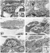
Figure 2. Ultrastructure of IC-MY in the canine antrum
A shows two IC-MY (IC) located near circular muscle cells (CM). IC-MY possessed many mitochondria compared to neighbouring circular smooth muscle cells (see also inset). Inset in A, caveolae and a distinct basal lamina were also present. B shows an IC-MY (IC) forming a close contact with a neighbouring smooth muscle cell (arrow). The inset shows the region at higher magnification revealing that the close contact is a gap junction between the IC-MY and the smooth muscle cell. C shows communication between IC-MY. Both cells have an abundance of mitochondria, extensive endoplasmic reticulum and numerous free ribosomes. A gap junction can be seen between the two IC-MY (arrow and inset). Scale bars are as indicated in each panel and represent 0.1 μm in insets in B and C.
Figure 3
Figure 3. Ultrastructure of IC-IM, IC-SEP and IC-SM
A shows an IC-IM within the circular muscle layer (IC). IC-IM were distinct from neighbouring smooth muscle cells and contained a reduced filament content and lacked dense bodies. IC-IM formed multiple close associations with neighbouring smooth muscle cells (arrowheads). A distinct gap junction between the IC-IM (IC) and a neighbouring circular smooth muscle cell is indicated by the arrow and this region is shown at a higher magnification in the inset. IC-IM were also closely associated and formed gap junctions with each other (arrows). These cells contained many mitochondria, caveolae, rough and smooth ER and free ribosomes (B). IC-IM were also located in the longitudinal muscle layer (C). These cells had similar ultrastructural features to circular muscle IC-IM and possessed caveolae and a distinct basal lamina. D shows an IC-SEP and processes (*) at the interface between two circular muscle bundles. IC-SEP possessed many mitochondria, caveolae and a continuous basal lamina. Varicose nerve fibres (N) are closely associated with the IC-SEP. E and F show IC-SM at the submucosal surface of the circular muscle layer. IC-SM contained clusters of mitochondria and caveolae along the plasma membrane. A gap junction (arrow) between an IC-SM and a neighbouring smooth muscle cell (CM) is shown at higher magnification in the inset in F. IC-SM possessed numerous mitochondria, Golgi apparatus, rough and smooth endoplasmic recticulum and many free ribosomes. Numerous caveolae were present along the plasma membrane and a continuous basal lamina was observed. Scale bars are as indicated in each panel and represent 0.1 μm in insets in A and F.
Figure 5
Figure 5. Electrical activity in interior circular muscle before and after isolation from the intact muscularis
A shows electrical activity in an interior circular muscle strip 15 mm from the intact region of muscularis in a preparation like that shown in Fig. 4 (recording made from point I in Fig. 4). B shows loss of spontaneous slow wave activity (observed in 9 of 11 preparations) after the interior circular muscle was isolated from the intact region of muscularis. In 2 preparations, spontaneous electrical activity was recorded in interior circular muscle strips after isolation from the intact muscularis (C).
Figure 6
Figure 6. Spontaneous and paced slow waves in myenteric, interior and submucosal circular muscle strips (preparations as shown in Fig. 4)
A and B are spontaneous and paced slow waves recorded from myenteric strips. C–F are recordings from interior and submucosal circular muscle strips, respectively. All recordings were made 20 mm from the intact portion of muscularis and 25 mm from the site of EFS.
Figure 7
Figure 7. Spontaneous and paced slow waves in interior circular muscle when this region was attached (A and B) and isolated (C and D) from the intact region of the muscularis
A shows normal slow wave activity and frequency. B, the frequency could be increased by electrical pacing with EFS. Then the interior muscle strip was separated by sharp dissection from the intact region of the muscularis (as shown by the white dashed line in the image in Fig. 4). This caused loss of slow wave activity (C), but pacing of the isolated strip of interior circular muscle restored slow wave activity (D).
Figure 8
Figure 8. Slow wave activity was restored in isolated interior muscle strips by cholinergic stimulation
A shows lack of slow wave activity in an isolated interior muscle strip. B, ACh (0.3 μm) produced slight membrane depolarization and evoked slow wave activity in the same muscle strip. C, washout of ACh restored electrical quiescence. Traces in A–C are excerpts from a continuous recording from the same cell.
Figure 9
Figure 9. Nifedipine did not block spontaneous and evoked slow waves
A and B show spontaneous slow waves recorded from the myenteric region of the circular muscle layer before and after addition of nifedipine (1 μm) for 20 min. C and D show paced slow wave activity before and after nifedipine. E–H show the effects of nifedipine on spontaneous and evoked slow waves recorded from an interior circular muscle strip attached to the intact region of muscularis (as in Fig. 4). Nifedipine partially reduced the amplitude of the plateau phase and reduced the duration of slow waves. Nifedipine had no effect on resting potential (I) and did not block slow waves evoked in isolated interior circular muscle by EFS (J).
Figure 10
Figure 10. Propagation of slow waves in myenteric and interior muscle strips
Slow waves were recorded at various distances (5–25 mm) from the site of stimulation (EFS site). The latencies between the time of stimulation and the time at which slow waves occurred at the various recording sites were plotted as a function of distance from the site of stimulation. Slow waves from several recording sites are superimposed in A (myenteric muscle strip) and B (interior muscle strip). C shows a summary of slow propagation in 5 myenteric muscle strips. The data were fitted by linear regression analysis and the best line had a slope of 0.0602 ± 0.008 (n = 5; r2 = 0.954). The inverse of the slope gave a propagation velocity of 16.6 mm s−1. D shows a summary of slow wave propagation in 5 interior circular muscle strips. Linear regression analysis of the data points gave a slope of 0.052 ± 0.002 (n = 5; r2 = 0.9951). The inverse of the slope gave a propagation velocity of 19.2 mm s−1.

Comment in

Similar articles

Cited by

References

    1. Bauer AJ, Publicover NG, Sanders KM. Origin and spread of slow waves in canine gastric antral circular muscle. American Journal of Physiology. 1985a;249:G800–806. - PubMed
    1. Bauer AJ, Reed JB, Sanders KM. Slow wave heterogeneity within the circular muscle of the canine gastric antrum. Journal of Physiology. 1985b;366:221–232. - PMC - PubMed
    1. Bauer AJ, Sanders KM. Passive and active membrane properties of canine gastric antral circular muscles. American Journal of Physiology. 1986;251:C268–273. - PubMed
    1. Burns AJ, Herbert TM, Ward SM, Sanders KM. Interstitial cells of Cajal in the guinea-pig gastrointestinal tract as revealed by c-Kit immunohistochemistry. Cell and Tissue Research. 1997;290:11–20. - PubMed
    1. Burns AJ, Lomax AE, Torihashi S, Sanders KM, Ward SM. Interstitial cells of Cajal mediate inhibitory neurotransmission in the stomach. Proceedings of the National Academy of Sciences of the USA. 1996;93:12008–12013. - PMC - PubMed

Publication types

Substances

LinkOut - more resources