. 2001 Nov 15;537(Pt 1):267-75.
doi: 10.1111/j.1469-7793.2001.0267k.x.

Rapid and non-genomic reduction of intracellular [Ca(2+)] induced by aldosterone in human bronchial epithelium

Affiliations

Rapid and non-genomic reduction of intracellular [Ca(2+)] induced by aldosterone in human bronchial epithelium

V Urbach et al. J Physiol. .

Abstract

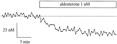
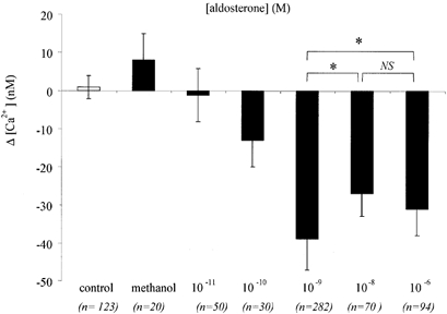
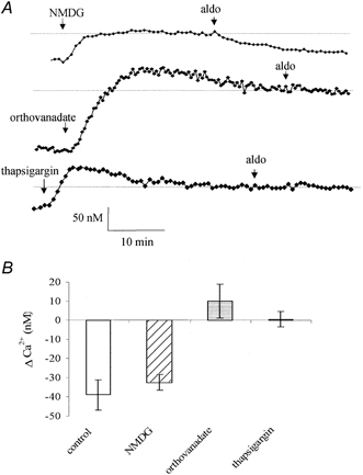
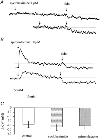
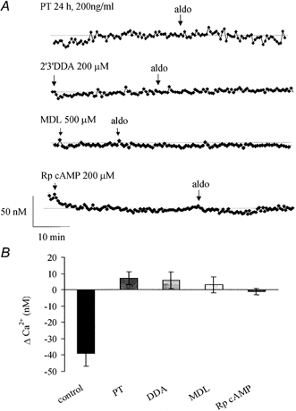
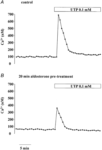
1. Using a Ca(2+) imaging system and fura-2 AM (5 microM) we showed that exposure of polarised monolayers of human bronchial epithelial cells (16HBE14o- cell line) to aldosterone produced a fast intracellular [Ca(2+)] ([Ca(2+)](i)) decrease, in 70 % of cells. Exposure to aldosterone (1 nM) reduced the [Ca(2+)](i) by 39 +/- 9 nM (n = 282, P < 0.0001) within 10 min, from a basal [Ca(2+)](i) of 131 +/- 19 nM (n = 282). 2. The effect of aldosterone on [Ca(2+)](i) was not affected by inhibitors of the classical genomic pathway, cycloheximide (1 microM) or spironolactone (10 microM). The aldosterone-induced [Ca(2+)](i) decrease was inhibited by thapsigargin (1 microM), pertussis toxin (24 h at 200 ng ml(-1)), the adenylate cyclase inhibitors 2',3'-dideoxyadenosine (200 microM) and MDL-12,330A hydrochloride (500 microM), and the protein kinase A inhibitor R(P)-adenosine 3',5'-cyclic monophosphorothioate (200 microM). In addition, treatment of 16HBE14o- monolayers with aldosterone (1 nM) inhibited by approximately 30 % the large and transient [Ca(2+)](i) increase induced by apical exposure to uridine triphosphate (UTP, 0.1 mM), a known secretagogue in airway epithelia. 3. Our results demonstrate for the first time that in human bronchial epithelial cells, aldosterone decreases [Ca(2+)](i) levels via a non-genomic mechanism. The hormone-induced changes to [Ca(2+)](i) involve stimulation of thapsigargin-sensitive Ca(2+)-ATPase, via G-protein-, adenylate cyclase- and protein kinase A-coupled signalling pathways.


Figures

Figure 1
Figure 1. Aldosterone effect on [Ca2+]i in 16HBE14o- monolayers
Typical record of [Ca2+]i variations upon exposure of a 16HBE14o- monolayer to 1 nm aldosterone. The [Ca2+]i was measured in a single cell. The dashed line indicates the average [Ca2+]i measured in the cell before aldosterone treatment.
Figure 2
Figure 2. Dose-response relationship of the aldosterone-induced [Ca2+]i decrease
Variations in [Ca2+]i as a function of aldosterone concentration. Significant changes to [Ca2+]i were obtained upon addition of aldosterone over the range 0.1 nm to 1 μm (overall P < 0.0001, ANOVA). The solvent methanol (0.01 %) alone did not produce any significant [Ca2+]i change. *P < 0.05 from t test.
Figure 3
Figure 3. Glucocorticoid effect on [Ca2+]i
Variations in [Ca2+]i measured 15–20 min after exposure of 16HBE14o- monolayers to the glucocorticoids hydrocortisone and triamcilone acetonide, at two different concentrations (1 nm and 1 mm).
Figure 4
Figure 4. Effects of NMDG, orthovanadate and thapsigargin
A, representative [Ca2+]i change upon replacement of Na+ by NMDG, or treatment of the monolayers to orthovanadate (1 mm) or thapsigargin (1 μm) followed by exposure to aldosterone (aldo, 1 nm). B, average [Ca2+]i variations induced by aldosterone in control conditions, and between 15 and 20 min after Na+ replacement (NMDG), or orthovanadate or thapsigargin treatment.
Figure 5
Figure 5. Effects of cycloheximide and spironolactone
Representative [Ca2+]i change in 16HBE14o- monolayers pre-treated for 15 min with cycloheximide (1 μm, A) or spironolactone (10 μm, B) and exposed to aldosterone (1 nm). C, average [Ca2+]i variations induced by aldosterone in control conditions and after cycloheximide or spironolactone treatment.
Figure 6
Figure 6. Effects of G-protein, adenylate cyclase and PKA inhibition
A, representative [Ca2+]i recording during treatment with pertussis toxin (PT), DDA, MDL or Rp-cAMP followed by exposure to aldosterone (1 nm). B, average [Ca2+]i variations induced by aldosterone in control conditions or after treatment with inhibitor.
Figure 7
Figure 7. Effect of UTP on [Ca2+]i in 16HBE14o- monolayers
Representative [Ca2+]i recording before and after apical exposure to UTP (0.1 mm) in 16HBE14o- monolayers either not treated (A) or pre-treated for 30 min with aldosterone (0.1 nm, B).

References

    1. Al-Dujaili EA, Edwards CR. The development and application of a direct radioimmunoassay for plasma aldosterone using 125I-labeled ligand-comparison of three methods. Journal of Clinical Endocrinology Metabolism. 1978;46:105–113. - PubMed
    1. Ballard PL, Baxter JD, Higgins SJ, Rousseau GG, Tomkins GM. General presence of glucocorticoid receptors in mammalian tissues. Endocrinology. 1974;94:998–1002. - PubMed
    1. Champigny G, Voilley N, Lingueglia E, Friend V, Brabry P, Lazdunski M. Regulation of expression of the lung amiloride sensitive Na+ channel by steroid hormones. EMBO Journal. 1994;13:2177–2181. - PMC - PubMed
    1. Chen SY, Bhargava A, Mastroberardino L, Meijer OC, Wang J, Buse P, Firestone GL, Verrey F, Pearce D. Epithelial sodium channel regulated by aldosterone-induced protein sgk. Proceedings of the National Academy of Sciences of the USA. 1999;96:2514–2519. - PMC - PubMed
    1. Chabra SK, Khanduja A, Jain D. Decreasing sodium-potassium and calcium adenosine triphosphatase activity in asthma: modulation by inhaled and oral corticosteroids. Indian Journal of Chest Disease and Allied Sciences. 1999;41:15–26. - PubMed

Publication types

MeSH terms

LinkOut - more resources