. 2001 Dec;75(24):12058-69.
doi: 10.1128/JVI.75.24.12058-12069.2001.

The major human immunodeficiency virus type 2 (HIV-2) packaging signal is present on all HIV-2 RNA species: cotranslational RNA encapsidation and limitation of Gag protein confer specificity

Affiliations

The major human immunodeficiency virus type 2 (HIV-2) packaging signal is present on all HIV-2 RNA species: cotranslational RNA encapsidation and limitation of Gag protein confer specificity

S D Griffin et al. J Virol. 2001 Dec.

Abstract

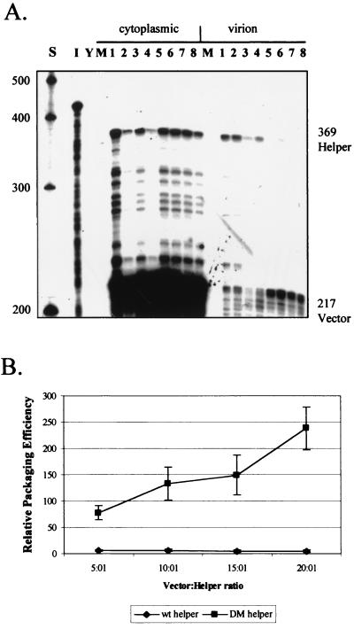
Deletion of a region of the human immunodeficiency virus type 2 (HIV-2) 5' leader RNA reduces genomic RNA encapsidation to about 5% that of wild-type virus with no defect in viral protein production but severely limits virus spread in Jurkat T cells, indicating that this region contains a major cis-acting encapsidation signal, or psi (Psi). Being upstream of the major splice donor, it is present on all viral transcripts. We have shown that HIV-2 selects its genomic RNA for encapsidation cotranslationally, rendering wild-type HIV-2 unable to encapsidate vector RNAs in trans. Virus with Psi deleted, however, encapsidates an HIV-2 vector, demonstrating competition for Gag protein. HIV-2 overcomes the lack of packaging signal location specificity by two novel mechanisms, cotranslational packaging and competition for limiting Gag polyprotein.


Figures

FIG. 1
FIG. 1
Deletion mutations of the HIV-2 leader region. Deletions were introduced into the infectious proviral clone pSVR by site-directed mutagenesis, and their effects on RNA encapsidation were assessed by RPAs. (A) Schematic representation of the locations of DM and Ψ1 deletions in the HIV-2 leader, as well as those characterized by previous studies. LTR, long terminal repeat; SD, splice donor; RRE, Rev-responsive element; PBS, primer binding site. (B) RPA of RNA from COS-1 cells transiently transfected with new mutants using the KSΨ2KE riboprobe. Wt, wild type. Lanes: 1, pSVR; 2, pSVRΨ1; 3, pSVRDM; 4, pSVRDM/Ψ1; I, input riboprobe diluted 1/100; Y, yeast RNA plus RNase (control); M, RNA from mock-transfected cells. The leftmost lane contains RNA size markers (Century Plus; Ambion).
FIG. 2
FIG. 2
Protein production by new HIV-2 deletion mutants. COS-1 cells transfected with proviral clones were metabolically labeled with [35S]methionine, and viral proteins were immunoprecipitated with pooled HIV-2-positive patient sera. Proteins from cellular and virion fractions were visualized by sodium dodecyl sulfate-polyacrylamide gel electrophoresis and autoradiography. Lanes: M, protein from mock-transfected cells; 1, pSVR; 2, pSVRΨ1; 3, pSVRDM; 4, pSVRDM/Ψ1. gp, glycoprotein; pr, protein; MA, matrix; Env, envelope; Gag, group antigen; SU, surface; TM, transmembrane.
FIG. 3
FIG. 3
Replication of HIV-2 deletion mutants in Jurkat T cells. Replicate wells of 50,000 Jurkat T cells were infected with normalized amounts of concentrated supernatants from provirus-transfected COS-1 cells. Every 3 to 4 days, a 10-μl sample of medium was removed prior to feeding of the cells, and viral replication was assessed by measuring the RT activity of the sample.
FIG. 4
FIG. 4
Reduction of mutant encapsidation efficiencies in competition with wild-type HIV-2. COS-1 cells were transfected with leader region mutant HIV-2 proviruses either alone or with an equal amount of wild-type HIV-2 provirus. RNA was collected, and encapsidation efficiencies were assessed by RPAs. (A) Representative RPA using the KSΨ2KE probe with cytoplasmic and virion RNAs from transfected COS-1 cells. Lanes: S, markers; I, input probe diluted 1/100; Y, yeast RNA plus RNase; M, RNA from mock-transfected cells; 1, pSVR; 2, pSVRΔ1; 3, pSVR plus pSVRΔ1; 4, pSVRΔ2; 5, pSVR plus pSVRΔ2; 6, pSVRΔ4; 7, pSVR plus pSVRΔ4; 8, pSVRΨ1; 9, pSVR plus pSVRΨ1; 10, pSVRDM; 11, pSVR plus pSVRDM. wt, wild type (pSVR); mutant proviruses are abbreviated by their deletion names, e.g., Δ1 represents pSVRΔ1. (B) Bar chart showing quantification of encapsidation efficiencies of mutants, with and without competition, relative to wild-type virus. Results are averages of at least three separate experiments (numbers vary for each mutant); error bars represent the standard error of the mean between experiments. Numbering is as described for panel A.
FIG. 5
FIG. 5
Gag availability is limiting for HIV-2 RNA encapsidation. The encapsidation of wild-type HIV-2 was assessed by RPAs in cotransfections with a DM mutant virus that could produce Gag protein compared to one that could not. (A) Representative RPA using the KSΨ2KE riboprobe and showing an increase in wild-type encapsidation in competition with a DM virus that makes its own Gag protein. Lanes: S, RNA size markers (Century Plus); I, input probe diluted 1/100; Y, yeast RNA plus RNase; M, RNA from mock-transfected cells; 1, pSVR; 2, pSVRDM; 3, pSVR plus pSVRDM; 4, pSVR plus pSVRDMΔH. wt, wild type (pSVR); DMΔH, pSVRDMΔH; DM, pSVRDM. (B) Bar chart showing quantification of experiments. The encapsidation efficiency of pSVR in competition with a non-Gag-producing virus, pSVRDMΔH, is taken as 100%. Results are averages of four separate experiments; error bars represent the standard error of the mean between experiments. Bars: 1, pSVR-pSVRDM; 2, pSVR-pSVRDMΔH.
FIG. 6
FIG. 6
Packaging region HIV-2 mutants are able to encapsidate HIV-2 vectors efficiently in trans. (A) Representative RPA with the KSΨ2KE riboprobe showing encapsidation of wild-type HIV-2 pSVR and HIV-2 vector RNA pSVRΔH. Lanes: 1, pSVR; 2, pSVRΔH. Cyt, cytoplasmic RNA; Vir, virion RNA. The structure of pSVRΔH is shown at the right. wt, wild type. See the legend to Fig. 1 for other definitions. (B) Representative RPAs showing trans-acting encapsidation of pSVRΔH by packaging region mutants. Lanes: 1, pSVRΔ1 plus pSVRΔH; 2, pSVRΔ2 plus pSVRΔH; 3, pSVRΔ4 plus pSVRΔH; 4, pSVRΨ1 plus pSVRΔH; 5, pSVRDM plus pSVRΔH. Cyt, cytoplasmic RNA; Vir, virion RNA.
FIG. 7
FIG. 7
First-generation HIV-2 vector system. (A) Structures of two HIV-2 helper constructs as well as two HIV-2 vectors containing a puromycin resistance gene cassette under the control of the simian virus 40 promoter. See the legend to Fig. 1 for definitions. (B) Results of transduction experiments with HeLa CD4+ LTR-βgal cells and different combinations of the helper and the vector described above, pseudotyped with the VSV G glycoprotein envelope. Results are the means of three experiments and are expressed as CFU/10,000RTU; error bars represent the standard error of the mean between experiments. Bars: 1, pSVRΔNB-pSVRΔNBPuroΔH; 2, pSVRΔNB-pSVRΔNBPuroΔE; 3, pSVRDM-pSVRΔNBPuroΔH; 4, pSVRDM-pSVRΔNBPuroΔE. (C) Quantification of vector RNA encapsidation by an RPA using the SKH2CA riboprobe and different helper-vector combinations. Results are the means of three separate experiments; error bars represent the standard error of the mean between experiments. Bars are as described for panel B.
FIG. 8
FIG. 8
trans-Acting encapsidation by pSVRDM of HIV-2 vectors containing various amounts of the gag ORF. (A) Representative RPA using the KS2ES riboprobe and cytoplasmic and virion RNAs from COS-1 cells transfected with equal amounts of pSVRDM and vector. Lanes: S, RNA size markers (Century Plus); I, input probe diluted 1/100; Y, yeast RNA plus RNase; M, RNA from mock-transfected cells; 1, pSVRDM; 2, pSVRDM plus pSVRΔX; 3, pSVRDM plus pSVRΔAX; 4, pSVRDM plus pSVRΔHX; 5, pSVRDM plus pSVRΔpol; 6, pSVRDM plus pSVRΔHΔpol; 7, pSVRDM plus pSVRΔpolncm. (B) Quantification of vector encapsidation efficiencies, relative to that of the DM helper, in the experiments detailed above. Results are averages of at least two separate experiments; error bars represent the standard error of the mean between experiments. Numbering is as described for panel A.
FIG. 9
FIG. 9
Titration of helper encapsidation by competition for limiting amounts of Gag polyprotein. COS-1 cells were transfected with 1 μg of helper virus, either pSVR or pSVRDM, and increasing amounts of HIV-2 vector pSVRΔHΔpol along with stuffer DNA. RNA from cytoplasmic and virion fractions was then analyzed by RPAs. (A) Representative RPA with the KS2ES riboprobe and RNA from titration experiments. Lanes: S, RNA size markers (Century Plus); I, input probe diluted 1/100; Y, yeast RNA plus RNase; M, RNA from mock-transfected cells; 1, 1 μg of pSVR plus 5 μg of pSVRΔHΔpol; 2, 1 μg of pSVR plus 10 μg of pSVRΔHΔpol; 3, 1 μg of pSVR plus 15 μg of pSVRΔHΔpol; 4, 1 μg of pSVR plus 20 μg of pSVRΔHΔpol; 5, 1 μg of pSVRDM plus 5 μg of pSVRΔHΔpol; 6, 1 μg of pSVRDM plus 10 μg of pSVRΔHΔpol; 7, 1 μg of pSVRDM plus 15 μg of pSVRΔHΔpol; 8, 1 μg of pSVRDM plus 20 μg of pSVRΔHΔpol. (B) Quantification of vector encapsidation efficiencies in the experiments detailed above. wt, wild type. Results are the averages of three separate experiments; error bars represent the standard error of the mean between experiments.
FIG. 10
FIG. 10
Alternative secondary structure for the region of the DM deletion in the HIV-2 leader generated with the Mfold program. The free energy of the structure was calculated to be −7.6 kcal/mol.

Similar articles

Cited by

References

    1. Aldovini A, Young R A. Mutations of RNA and protein sequences involved in human immunodeficiency virus type 1 packaging result in production of noninfectious virus. J Virol. 1990;64:1920–1926. - PMC - PubMed
    1. Arya S K, Zamani M, Kundra P. Human immunodeficiency virus type 2 lentivirus vectors for gene transfer: expression and potential for helper virus-free packaging. Hum Gene Ther. 1998;9:1371–1380. - PubMed
    1. Berkhout B, Schoneveld I. Secondary structure of the HIV-2 leader RNA comprising the tRNA-primer binding site. Nucleic Acids Res. 1993;21:1171–1178. - PMC - PubMed
    1. Berkhout B, van Wamel J L. The leader of the HIV-1 RNA genome forms a compactly folded tertiary structure. RNA. 2000;6:282–295. - PMC - PubMed
    1. Berkowitz R D, Hammarskjold M L, Helga-Maria C, Rekosh D, Goff S P. 5′ regions of HIV-1 RNAs are not sufficient for encapsidation: implications for the HIV-1 packaging signal. Virology. 1995;212:718–723. - PubMed

Publication types

LinkOut - more resources