. 2001 Dec;75(24):12241-51.
doi: 10.1128/JVI.75.24.12241-12251.2001.

Transcription and RNA replication of tacaribe virus genome and antigenome analogs require N and L proteins: Z protein is an inhibitor of these processes

Affiliations

Transcription and RNA replication of tacaribe virus genome and antigenome analogs require N and L proteins: Z protein is an inhibitor of these processes

N López et al. J Virol. 2001 Dec.

Abstract

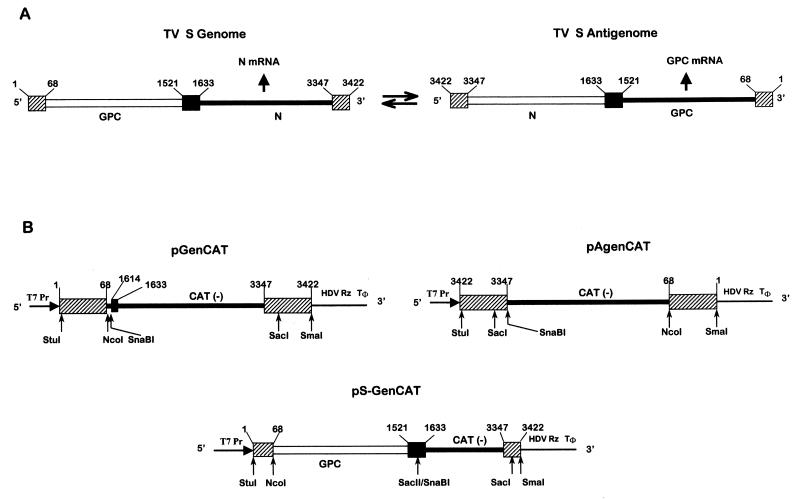
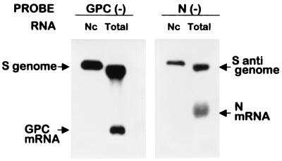
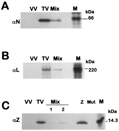
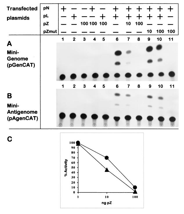
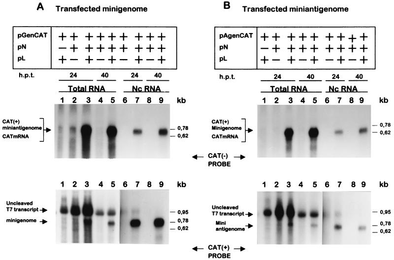
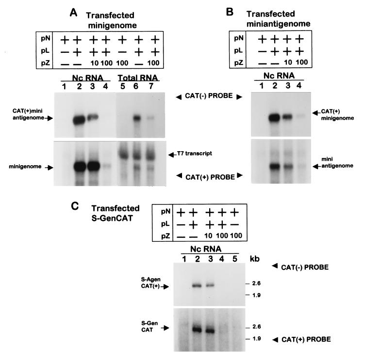
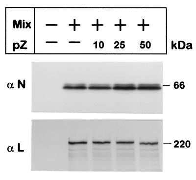
Tacaribe virus (TV), the prototype of the New World group of arenaviruses, comprises a single phylogenetic lineage together with four South American pathogenic producers of hemorrhagic disease. The TV genome consists of two single-stranded RNA segments called S and L. A reconstituted transcription-replication system based on plasmid-supplied TV-like RNAs and TV proteins was established. Plasmid expression was driven by T7 RNA polymerase supplied by a recombinant vaccinia virus. Plasmids were constructed to produce TV S segment analogs containing the negative-sense copy of chloramphenicol acetyltransferase (CAT) flanked at the 5' and 3' termini by sequences corresponding to those of the 5' and 3' noncoding regions of the S genome (minigenome) or the S antigenome (miniantigenome). In cells expressing N and L proteins, input minigenome or miniantigenome produced, respectively, encapsidated miniantigenome or minigenome which in turn produced progeny minigenome or progeny miniantigenome. Both minigenome and miniantigenome in the presence of N and L mediated transcription, which was analyzed as CAT expression. Coexpression of the small RING finger Z (p11) protein was highly inhibitory to both transcription and replication mediated by the minigenome or the miniantigenome. The effect depended on synthesis of Z protein rather than on plasmid or the RNA and was not ascribed to decreased amounts of plasmid-supplied template or proteins (N or L). N and L proteins were sufficient to support full-cycle RNA replication of a plasmid-supplied S genome analog in which CAT replaced the N gene. Replication of this RNA was also inhibited by Z expression.


Figures

FIG. 1
FIG. 1
Schematic diagram (not to scale) showing the TV S RNA organization and expression (arrows show the direction of RNA synthesis) (A) and plasmids used to generate TV RNA analogs (B). The position of T7 polymerase promoter (T7Pr), the HDV Rz, and the T7 RNA polymerase terminator (Tφ) are indicated. Constructions were performed as indicated in Materials and Methods. pGenCAT transcript (842 nt) contains (5′ to 3′) the entire TV S genome 5′ NCR sequence (68 nt), a short linker (16 nt) including a SnaBI site, a short sequence (nt 1614 to 1633) of the S genome intergenic region (IGR) which is not involved in the hairpin structure (18), CAT ORF in an antisense orientation (660 nt), and the complete S genome 3′ NCR sequence (76 nt). pAgenCAT transcript (810 nt) contains (5′ to 3′) the entire TV S antigenome 5′ NCR sequence, a SnaBI site, CAT ORF in an antisense orientation, and the complete S antigenome 3′ NCR. pS-GenCAT (2,343 nt), contains (5′ to 3′) nt 1 to 1633 of the TV S genome followed by the negative-sense copy of CAT and the entire S genome 3′ NCR sequence. Sizes of transcripts refer to the processed RNA, which includes two nonviral Gs at the 5′ end (23). Nucleotides were numbered considering as 1 the 5′ end of the S genome. Open lines represent positive-sense coding regions; negative-sense coding regions are indicated in blackened thicker lines. S RNA IGR is represented as a black box, and the 3′ and 5′ NCR termini are indicated as shaded boxes. Key restriction endonuclease sites used in the assembly of the DNA constructs are shown.
FIG. 2
FIG. 2
Northern blot analysis of immunoselected and total RNA from TV-infected cells. Cells were infected with TV as described in Materials and Methods. Immunoselection of nucleocapsids (Nc) and extraction of Nc-associated and total RNA from infected cell extracts were performed as indicated in Materials and Methods. RNAs were analyzed by Northern blotting using 32P-labeled negative-sense GPC or N riboprobes which detected, respectively, S genome and GPC mRNA or S antigenome and N mRNA. Comigration of 28S rRNA with genome and antigenome RNA in the total RNA preparation leads to a higher mobility of these RNAs than that observed for the genome and antigenome RNA extracted from immunoselected nucleocapsids.
FIG. 3
FIG. 3
Analysis of TV proteins expressed in virus-infected and plasmid-transfected cells. CV1 cells were infected with TV (lanes TV), vTF7-3 (lanes VV), or vTF7-3 and transfected with a mixture of pGenCAT, pL, and pN as indicated in Materials and Methods (lanes Mix) without (A and B) or with the addition of 10 ng (C, lane 1) or 100 ng (C, lane 2) of pZ. Lanes Z and Mut in C, correspond to cells infected with vTF7-3 and transfected, respectively, with 1 μg of pZ or 1 μg of pZmut. Transfected cells were incubated for 18 h and TV-infected cells were incubated for 40 h and then labeled for 1.5 h with [35S]cysteine (150 μCi/ml) and cell extracts were prepared. At the time of labeling, both TV-infected and transfected cells were actively engaged in transcription and replication (see Materials and Methods). Cytoplasmic extracts were immunoprecipitated with monospecific serum against N (panel A), L (panel B), or Z (panel C) proteins. Immunoprecipitated proteins (corresponding to 2 × 10⁵ cells per lane) were separated on an SDS-PAGE gel containing 10% (A), 7% (B), or 17% (C) polyacrylamide, followed by direct exposure to X-ray film for 2, 6, and 4 days (panels A, B, and C, respectively), using intensifying screen (Biomax, Kodak). Molecular masses of 14C-labeled markers (lanes M) are indicated in kilodaltons.
FIG. 4
FIG. 4
Detection of CAT activity mediated by the minigenome or the miniantigenome. Duplicate dishes were transfected with pGenCAT (A) or pAgenCAT (B) with (+) or without (−) the addition of pN and pL as indicated. When pZ or pZmut was included, its amount is indicated in nanograms. At 24 h posttransfection, cell extracts were prepared, CAT activity was assayed, and the reaction products were analyzed as described in Materials and Methods. Films were exposed for 18 h (A) or 48 h (B). (C) CAT activity expressed from the minigenome (▴) or the miniantigenome (●) was quantified in three independent experiments in which cells were transfected as in lanes 6 to 8. The mean values obtained are shown as the percentage of the corresponding value without added pZ.
FIG. 5
FIG. 5
Replication of plasmid-supplied minigenome or miniantigenome. Subconfluent cultures of CV1 cells were infected with vTF7-3 and transfected with pGenCAT (A) or pAgenCAT (B) and pN and/or pL, as indicated. At 24 h (lanes 1 to 3, 6, and 7) and 40 h (lanes 4, 5, 8, and 9) posttransfection (h.p.t.), total RNA (lanes 1 to 5) or nucleocapsid-associated RNA (Nc RNA, lanes 6 to 9) was purified and analyzed by Northern blotting. Duplicate blots were hybridized either with an antisense CAT riboprobe [CAT(−)] (upper panels) or a sense CAT riboprobe [CAT(+)] (lower panels). Films were exposed for 30 h with the exception of lanes 1 to 5 in lower panels that were exposed for 3 h. Arrows on the left side of the panels indicate the position of the RNAs synthesized by the TV polymerase or the T7 polymerase. Sizes (in kilobases) of marker RNAs (Promega) are indicated on the right side of the panels.
FIG. 6
FIG. 6
Effect of pZ cotransfection on RNA replication. Subconfluent cultures of CV1 cells previously infected with vTF7-3 were transfected with minigenome (A), miniantigenome (B), or pS-GenCAT (C) together with pN with or without pL. When pZ was included, its amount is indicated in nanograms. Total RNA (A, lanes 5 to 7) was obtained at 24 h posttransfection, whereas nucleocapsid-associated RNA (Nc RNA; A and B, lanes 1 to 4; and C, lanes 1 to 5) was immunoselected at 40 h posttransfection. Purified RNAs were then analyzed by Northern blotting. Duplicate blots were hybridized either with an antisense CAT riboprobe [CAT(−)] (upper panels) or a sense CAT riboprobe [CAT(+)] (lower panels). Arrows show the positions of the RNAs synthesized by the TV polymerase or the T7 polymerase (T7 transcript). “kb” indicates the sizes of RNA markers resolved in parallel. In panels A and B, films were exposed for 20 h, with the exception of lanes 5 to 7 (A), which were exposed for 2 h. In panel C, films were exposed for 4 days.
FIG. 7
FIG. 7
Effect of Z protein expression on the synthesis of N or L proteins. CV1 cells (4 × 10⁵) were infected with vTF7-3 and those indicated (+) were transfected with: 2 μg of pGenCAT, 2 μg of pN, and 50 ng of pL (Mix) without or with the addition of 10, 25, or 50 ng of pZ as indicated. At 24 h posttransfection, cells were labeled for 1.5 h with a mixture of [35S]cysteine (80 μCi/ml) and [35S]methionine (80 μCi/ml), cytoplasmic extracts were prepared, and proteins were immunoprecipitated with monospecific serum directed to N (αN) or L (αL) proteins. Immunoprecipitated proteins (corresponding to 2 × 10⁵ cells per lane) were subjected to SDS-PAGE gels containing 12% (αN) or 7% (αL) polyacrylamide. Gels were exposed to X-ray film using an intensifying screen (Biomax, Kodak). Molecular masses according to 14C-labeled markers are indicated on the right.

References

    1. Atreya P L, Peeples M E, Collins P L. The NS1 protein of human respiratory syncytial virus is a potent inhibitor of minigenome transcription and RNA replication. J Virol. 1998;72:1452–1461. - PMC - PubMed
    1. Bishop D H L, Auperin D D. Arenavirus gene structure and organization. Curr Top Microbiol Immunol. 1987;133:5–17. - PubMed
    1. Borden K L B, Campbell Dwyer E J, Salvato M S. An arenavirus RING (zinc-binding) protein binds the oncoprotein promyelocyte leukemia protein (PML) and relocates PML nuclear bodies to the cytoplasm. J Virol. 1998;72:758–766. - PMC - PubMed
    1. Borden K L B, Campbell Dwyer E J, Carlile G W, Djavani M, Salvato M S. Two RING finger proteins, the oncoprotein PML and the arenavirus Z protein, colocalize with the nuclear fraction of the ribosomal P proteins. J Virol. 1998;72:3819–3826. - PMC - PubMed
    1. Bowen M D, Peters C J, Nichol S T. The phylogeny of New World (Tacaribe complex) arenaviruses. Virology. 1996;219:285–290. - PubMed

Publication types

LinkOut - more resources