

Identification of BV/ODV-C42, an Autographa californica nucleopolyhedrovirus orf101-encoded structural protein detected in infected-cell complexes with ODV-EC27 and p78/83
- PMID: 11711623
- PMCID: PMC116129
- DOI: 10.1128/JVI.75.24.12331-12338.2001
Identification of BV/ODV-C42, an Autographa californica nucleopolyhedrovirus orf101-encoded structural protein detected in infected-cell complexes with ODV-EC27 and p78/83
Abstract
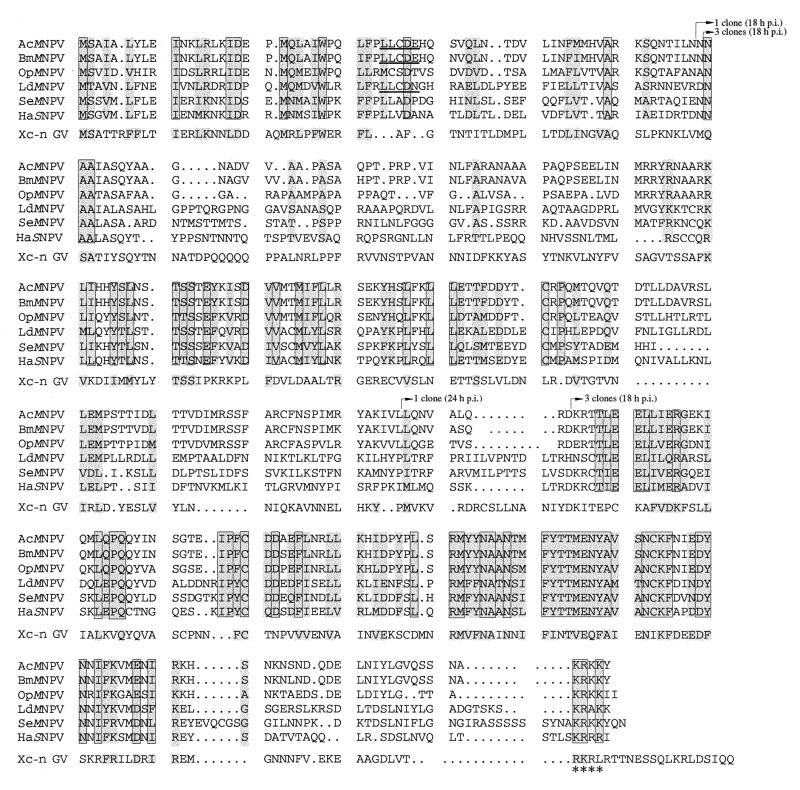
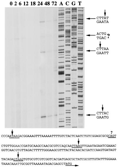
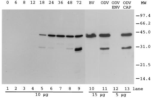
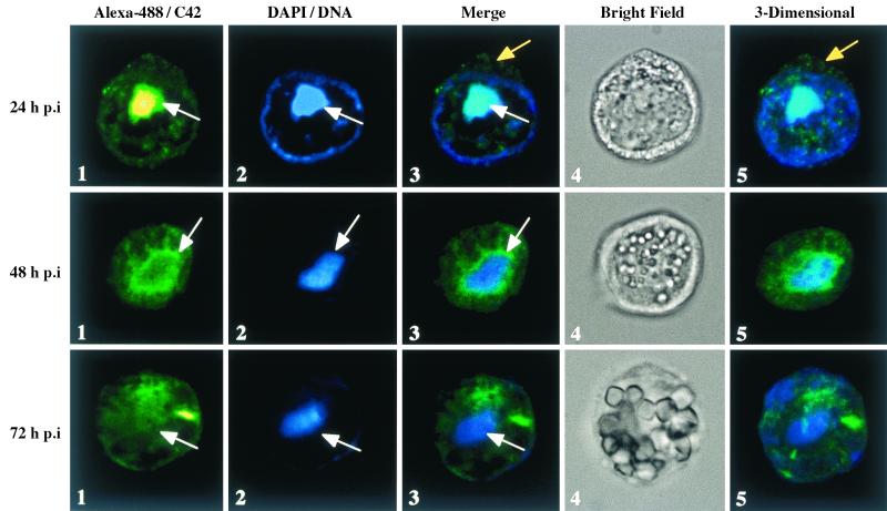
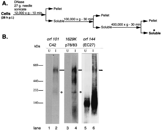
orf101 is a late gene of Autographa californica nucleopolyhedrovirus (AcMNPV). It encodes a protein of 42 kDa which is a component of the nucleocapsid of budded virus (BV) and occlusion-derived virus (ODV). To reflect this viral localization, the product of orf101 was named BV/ODV-C42 (C42). C42 is predominantly detected within the infected-cell nucleus: at 24 h postinfection (p.i.), it is coincident with the virogenic stroma, but by 72 h p.i., the stroma is minimally labeled while C42 is more uniformly located throughout the nucleus. Yeast two-hybrid screens indicate that C42 is capable of directly interacting with the viral proteins p78/83 (1629K) and ODV-EC27 (orf144). These interactions were confirmed using blue native gels and Western blot analyses. At 28 h p.i., C42 and p78/83 are detected in two complexes: one at approximately 180 kDa and a high-molecular-mass complex (500 to 600 kDa) which also contains EC27.
Figures

Similar articles
-
Ac102 Participates in Nuclear Actin Polymerization by Modulating BV/ODV-C42 Ubiquitination during Autographa californica Multiple Nucleopolyhedrovirus Infection.J Virol. 2018 May 29;92(12):e00005-18. doi: 10.1128/JVI.00005-18. Print 2018 Jun 15. J Virol. 2018. PMID: 29618641 Free PMC article.
-
The Autographa californica Multiple Nucleopolyhedrovirus ac54 Gene Is Crucial for Localization of the Major Capsid Protein VP39 at the Site of Nucleocapsid Assembly.J Virol. 2016 Mar 28;90(8):4115-4126. doi: 10.1128/JVI.02885-15. Print 2016 Apr. J Virol. 2016. PMID: 26865720 Free PMC article.
-
Transcription, translation, and cellular localization of three Autographa californica nuclear polyhedrosis virus structural proteins: ODV-E18, ODV-E35, and ODV-EC27.Virology. 1996 Aug 1;222(1):100-14. doi: 10.1006/viro.1996.0401. Virology. 1996. PMID: 8806491
-
Autographa californica nuclear polyhedrosis virus: subcellular localization and protein trafficking of BV/ODV-E26 to intranuclear membranes and viral envelopes.Virology. 1998 Jan 5;240(1):64-75. doi: 10.1006/viro.1997.8903. Virology. 1998. PMID: 9448690
-
Molecular biology of the baculovirus occlusion-derived virus envelope.Curr Drug Targets. 2007 Oct;8(10):1084-95. doi: 10.2174/138945007782151315. Curr Drug Targets. 2007. PMID: 17979668 Review.
Cited by
-
Proteomics of the Autographa californica nucleopolyhedrovirus budded virions.J Virol. 2010 Jul;84(14):7233-42. doi: 10.1128/JVI.00040-10. Epub 2010 May 5. J Virol. 2010. PMID: 20444894 Free PMC article.
-
Nuclear Translocation Sequence and Region in Autographa californica Multiple Nucleopolyhedrovirus ME53 That Are Important for Optimal Baculovirus Production.J Virol. 2016 Mar 28;90(8):3953-3965. doi: 10.1128/JVI.03115-15. Print 2016 Apr. J Virol. 2016. PMID: 26842471 Free PMC article.
-
Protein composition analysis of polyhedra matrix of Bombyx mori nucleopolyhedrovirus (BmNPV) showed powerful capacity of polyhedra to encapsulate foreign proteins.Sci Rep. 2017 Aug 18;7(1):8768. doi: 10.1038/s41598-017-08987-8. Sci Rep. 2017. PMID: 28821766 Free PMC article.
-
Nucleocapsid Assembly of Baculoviruses.Viruses. 2019 Jul 1;11(7):595. doi: 10.3390/v11070595. Viruses. 2019. PMID: 31266177 Free PMC article. Review.
-
Identification of a novel regulatory sequence of actin nucleation promoting factor encoded by Autographa californica multiple nucleopolyhedrovirus.J Biol Chem. 2015 Apr 10;290(15):9533-41. doi: 10.1074/jbc.M114.635441. Epub 2015 Feb 17. J Biol Chem. 2015. PMID: 25691574 Free PMC article.
References
-
- Bozzola J J, Russell L D. Electron microscopy. Boston, Mass: Jones and Bartlett; 1992.
-
- Bradford M M. A rapid and sensitive method for the quantitation of microgram quantities of protein utilizing the principle of protein-dye binding. Anal Biochem. 1976;72:248–254. - PubMed
-
- Braunagel S C, Parr R, Belyavskyi M, Summers M D. Autographa californica nucleopolyhedrovirus infection results in Sf9 cell cycle arrest at G2/M phase. Virology. 1998;244:195–211. - PubMed
-
- Braunagel S C, Summers M D. Autographa californica nuclear polyhedrosis virus, PDV, and ECV viral envelopes and nucleocapsids: structural proteins, antigens, lipid and fatty acid profiles. Virology. 1994;202:315–328. - PubMed
Publication types
MeSH terms
Substances
Grants and funding
LinkOut - more resources
Full Text Sources
Molecular Biology Databases
