

doi: 10.1128/JVI.75.24.12431-12438.2001.
Glycoprotein K specified by herpes simplex virus type 1 is expressed on virions as a Golgi complex-dependent glycosylated species and functions in virion entry
Affiliations
- PMID: 11711633
- PMCID: PMC116139
- DOI: 10.1128/JVI.75.24.12431-12438.2001
Item in Clipboard
Glycoprotein K specified by herpes simplex virus type 1 is expressed on virions as a Golgi complex-dependent glycosylated species and functions in virion entry
J Virol.
2001 Dec.
Abstract
To facilitate detection of glycoprotein K (gK) specified by herpes simplex virus, a 12-amino-acid epitope tag was inserted within gK domain III. Recombinant virus gKprotC-DIII, expressing the tagged gK, was isolated. This virus formed wild-type plaques and replicated as efficiently as the wild-type KOS virus in Vero cells. Anti-protein C MAb detected high-mannose and Golgi complex-dependent glycosylated gK within cells as well as on purified virions. The gK-null virus DeltagK (gK(-/-)) entered Vero cells substantially more slowly than the wild-type KOS (gK(+/+)), while DeltagK virus grown in complementing VK302 cells (gK(-/+)) entered with entry kinetics similar to those of the KOS virus.
Figures
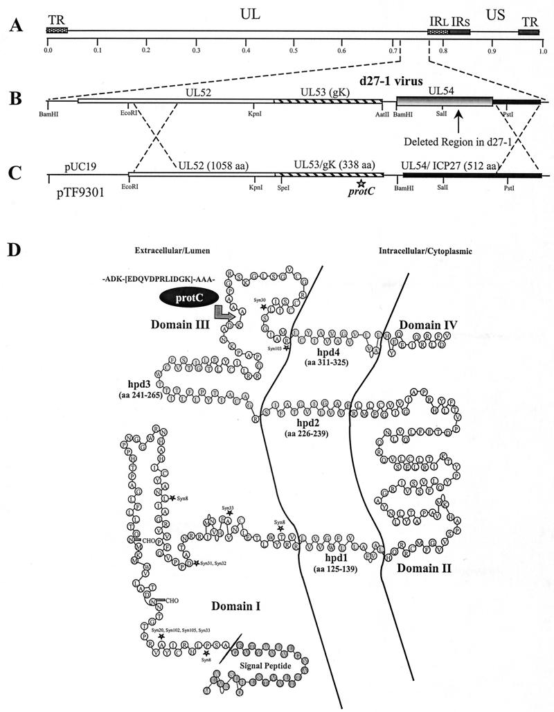
Construction of recombinant virus gKprotC-DIII, specifying gK containing a protein C epitope tag. (A) The top line represents the prototypic arrangement of the HSV-1 genome with the unique long (UL) and unique short (US) regions flanked by the terminal repeat (TR) and internal repeat (IR) regions. (B) Shown below is the region of the mutant virus HSV-1 d27-1 genome (between map units 0.7 and 0.8) containing the UL52, UL53, and the partially deleted UL54 open reading frames (shaded and boxed regions of the UL54 gene pointed to by an arrow) with relevant restriction endonuclease sites. (C) Plasmid construct, pTF9301, containing the recombinant gK-protC gene and flanking UL52 and UL54 sequences used to generate recombinant virus gKprotC-DIII. (D) Schematic model of the predicted secondary structure of gK (10). The predicted putative hydrophobic domains (hpd) of gK that transverse the membrane are shown embedded within the membrane. The arrow points to the site of the protC epitope tag insertion. The primary structure of the epitope tag is shown. Known syncytial mutations are denoted by asterisks.
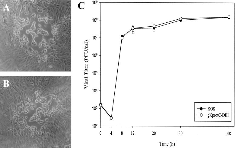
Comparison of plaque morphology and replication characteristics of gKprotC-DIII and KOS viruses. Comparison of virus plaque morphologies formed on Vero cells at 48 h postinfection. (A) KOS. (B) gKprotC-DIII. (C) Time-dependent kinetics of infectious virus production after infection of Vero cells at an MOI of 5 and incubation at 37°C. The graph depicts one of three separate experiments with similar results. Each separate experiment was repeated in triplicate to obtain standard deviations.
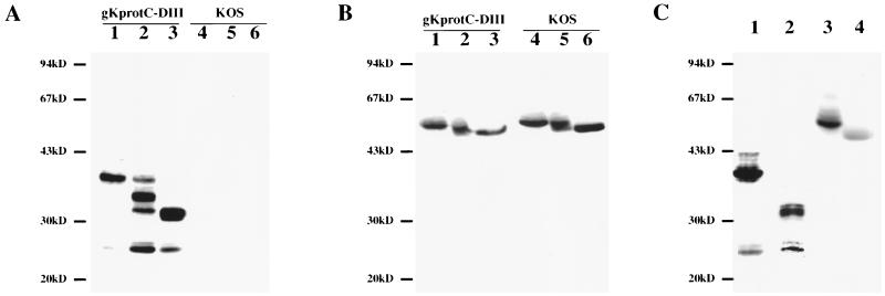
Characterization of synthesis and processing of protC-tagged gK specified by gKprotC-DIII. Immunoblots of gKprotC-DIII- (lanes 1 to 3) and KOS (lanes 4 to 6)-infected cell extracts reacted with either anti-protC MAb HPC-4 (A) or anti-gD MAb 1103 (B). Cellular extracts were treated with Endo-H (lanes 2 and 5), PNGase-F (lanes 3 and 6), or mock treated (lanes 1 and 4). (C) Cellular extracts obtained from Vero cells infected with gKprotC-DIII in the presence (lanes 2 and 4) or absence (lanes 1 and 3) of TM were probed with either anti-protC MAb (lanes 1 and 2) or anti-gD MAb (lanes 3 and 4).
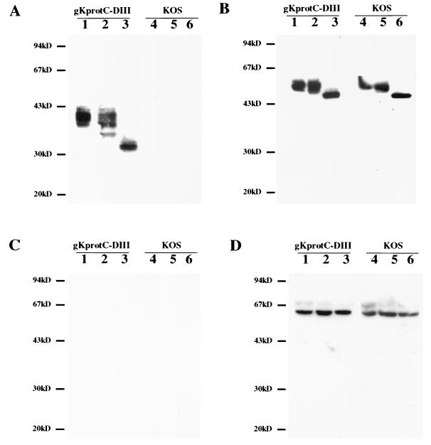
Detection and characterization of protC-tagged gK expressed on purified virions. Immunoblots of gKprotC-DIII (lanes 1 to 3) or KOS (lanes 4 to 6) purified virion preparations reacted with anti-protC MAb HPC-4 (A), anti-gD MAb 1103 (B), and anti-ICP27 MAb 1113 (C). Purified virion preparations were treated with Endo-H (lanes 2 and 5), PNGase-F (lanes 3 and 6), or mock treated (lanes 1 and 4). (D) Cellular extracts from Vero cells infected with either gKprotC-DIII (lanes 1 to 3) or KOS (lanes 4 to 6) were reacted with anti-ICP27 MAb. Cellular extracts were treated with Endo-H (lanes 2 and 5), PNGase-F (lanes 3 and 6), or mock treated (lanes 1 and 4).
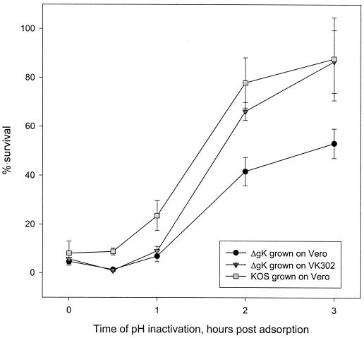
Penetration kinetics of KOS and ΔgK viruses into VK302 (Vero) cells. The penetration kinetics of KOS virus grown on Vero cells and ΔgK virus grown on either Vero or VK302 cells were obtained by determining the percentage of PFU surviving low-pH treatment relative to PBS-treated controls at different times postadsorption. Mean values and standard deviations of three independent experiments are shown.
Similar articles
-
Plasma membrane topology of syncytial domains of herpes simplex virus type 1 glycoprotein K (gK): the UL20 protein enables cell surface localization of gK but not gK-mediated cell-to-cell fusion.J Virol. 2003 Jan;77(1):499-510. doi: 10.1128/jvi.77.1.499-510.2003. J Virol. 2003. PMID: 12477855 Free PMC article.
-
Herpes simplex virus type 1 glycoprotein K is not essential for infectious virus production in actively replicating cells but is required for efficient envelopment and translocation of infectious virions from the cytoplasm to the extracellular space.J Virol. 1997 Jul;71(7):5012-24. doi: 10.1128/JVI.71.7.5012-5024.1997. J Virol. 1997. PMID: 9188566 Free PMC article.
-
Genetic analysis of the role of herpes simplex virus type 1 glycoprotein K in infectious virus production and egress.J Virol. 1999 Oct;73(10):8457-68. doi: 10.1128/JVI.73.10.8457-8468.1999. J Virol. 1999. PMID: 10482598 Free PMC article.
-
Glycoprotein K of herpes simplex virus: a transmembrane protein encoded by the UL53 gene which regulates membrane fusion.Virus Genes. 1999;18(1):81-90. doi: 10.1023/a:1008025520655. Virus Genes. 1999. PMID: 10334040 Review.
-
The role of herpes simplex virus glycoproteins in the virus replication cycle.Acta Virol. 1998 Apr;42(2):103-18. Acta Virol. 1998. PMID: 9770079 Review.
Cited by
-
The herpes simplex virus type 1 UL20 protein and the amino terminus of glycoprotein K (gK) physically interact with gB.J Virol. 2010 Sep;84(17):8596-606. doi: 10.1128/JVI.00298-10. Epub 2010 Jun 23. J Virol. 2010. PMID: 20573833 Free PMC article.
-
The herpes simplex virus type 1 glycoprotein D (gD) cytoplasmic terminus and full-length gE are not essential and do not function in a redundant manner for cytoplasmic virion envelopment and egress.J Virol. 2009 Jun;83(12):6115-24. doi: 10.1128/JVI.00128-09. Epub 2009 Apr 8. J Virol. 2009. PMID: 19357164 Free PMC article.
-
Comprehensive characterization of extracellular herpes simplex virus type 1 virions.J Virol. 2008 Sep;82(17):8605-18. doi: 10.1128/JVI.00904-08. Epub 2008 Jul 2. J Virol. 2008. PMID: 18596102 Free PMC article.
-
The Amino Terminus of Herpes Simplex Virus 1 Glycoprotein K (gK) Is Required for gB Binding to Akt, Release of Intracellular Calcium, and Fusion of the Viral Envelope with Plasma Membranes.J Virol. 2018 Feb 26;92(6):e01842-17. doi: 10.1128/JVI.01842-17. Print 2018 Mar 15. J Virol. 2018. PMID: 29321326 Free PMC article.
-
UL20 protein functions precede and are required for the UL11 functions of herpes simplex virus type 1 cytoplasmic virion envelopment.J Virol. 2007 Apr;81(7):3097-108. doi: 10.1128/JVI.02201-06. Epub 2007 Jan 10. J Virol. 2007. PMID: 17215291 Free PMC article.
References
-
- Bond V C, Person S. Fine structure physical map locations of alterations that affect cell fusion in herpes simplex virus type 1. Virology. 1984;132:368–376. - PubMed
-
- Bond V C, Person S, Warner S C. The isolation and characterization of mutants of herpes simplex virus type 1 that induce cell fusion. J Gen Virol. 1982;61:245–254. - PubMed
-
- Bzik D J, Fox B A, DeLuca N A, Person S. Nucleotide sequence of a region of the herpes simplex virus type 1 gB glycoprotein gene: mutations affecting rate of virus entry and cell fusion. Virology. 1984;137:185–190. - PubMed
Publication types
MeSH terms
Substances
Grants and funding
LinkOut - more resources
Full Text Sources
