. 2001 Dec;21(24):8385-97.
doi: 10.1128/MCB.21.24.8385-8397.2001.

Neuropeptide-induced androgen independence in prostate cancer cells: roles of nonreceptor tyrosine kinases Etk/Bmx, Src, and focal adhesion kinase

Affiliations

Neuropeptide-induced androgen independence in prostate cancer cells: roles of nonreceptor tyrosine kinases Etk/Bmx, Src, and focal adhesion kinase

L F Lee et al. Mol Cell Biol. 2001 Dec.

Abstract

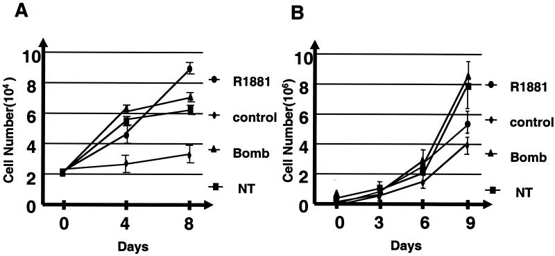
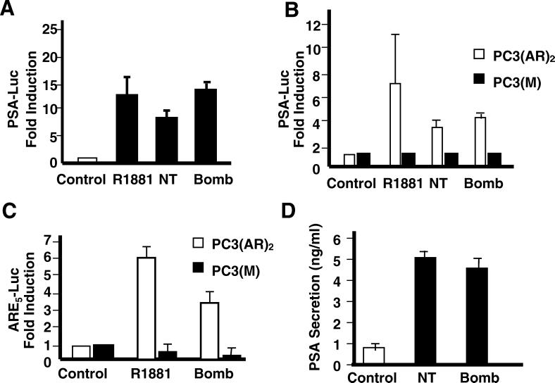
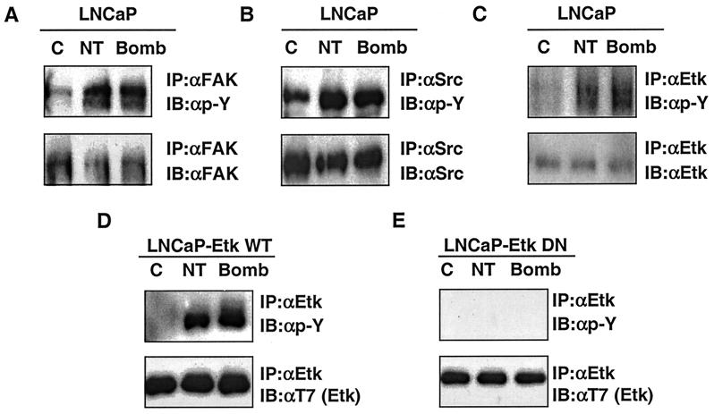
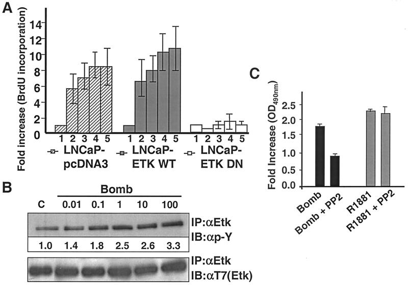
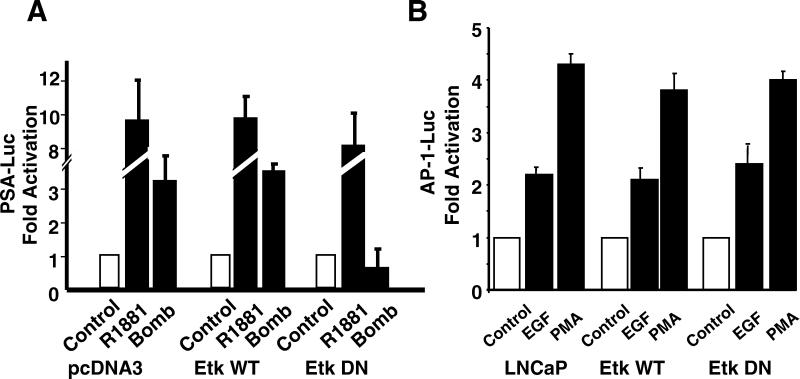
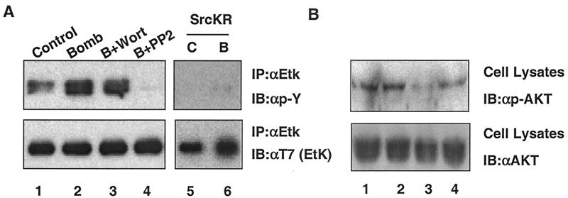
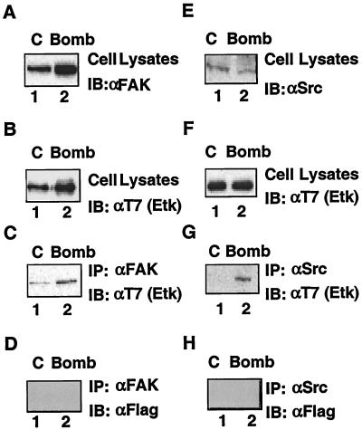
The bombesin/gastrin-releasing peptide (GRP) family of neuropeptides has been implicated in various in vitro and in vivo models of human malignancies including prostate cancers. It was previously shown that bombesin and/or neurotensin (NT) acts as a survival and migratory factor(s) for androgen-independent prostate cancers. However, a role in the transition from an androgen-dependent to -refractory state has not been addressed. In this study, we investigate the biological effects and signal pathways of bombesin and NT on LNCaP, a prostate cancer cell line which requires androgen for growth. We show that both neurotrophic factors can induce LNCaP growth in the absence of androgen. Concurrent transactivation of reporter genes driven by the prostate-specific antigen promoter or a promoter carrying an androgen-responsive element (ARE) indicate that growth stimulation is accompanied by androgen receptor (AR) activation. Furthermore, neurotrophic factor-induced gene activation was also present in PC3 cells transfected with the AR but not in the parental line which lacks the AR. Given that bombesin does not directly bind to the AR and is known to engage a G-protein-coupled receptor, we investigated downstream signaling events that could possibly interact with the AR pathway. We found that three nonreceptor tyrosine kinases, focal adhesion kinase (FAK), Src, and Etk/BMX play important parts in this process. Etk/Bmx activation requires FAK and Src and is critical for neurotrophic factor-induced growth, as LNCaP cells transfected with a dominant-negative Etk/BMX fail to respond to bombesin. Etk's activation requires FAK, Src, but not phosphatidylinositol 3-kinase. Likewise, bombesin-induced AR activation is inhibited by the dominant-negative mutant of either Src or FAK. Thus, in addition to defining a new G-protein pathway, this report makes the following points regarding prostate cancer. (i) Neurotrophic factors can activate the AR, thus circumventing the normal growth inhibition caused by androgen ablation. (ii) Tyrosine kinases are involved in neurotrophic factor-mediated AR activation and, as such, may serve as targets of future therapeutics, to be used in conjunction with current antihormone and antineuropeptide therapies.


Figures

FIG. 1
FIG. 1
Effects of bombesin (Bomb) and NT on growth of androgen-dependent prostate cancer cells. Parental LNCaP (A) and CWR22R (B) cells were plated in medium supplemented with 10% charcoal-stripped FBS with 1 nM R1881 or with 50 nM bombesin or NT, and the numbers of cells were counted at the indicated times. These results represent the averages of two independent experiments. Error bars indicate standard errors.
FIG. 2
FIG. 2
Effects of bombesin (Bomb) and NT on androgen-dependent PSA transcription. Stimulation of reporter gene activity in LNCaP and PC3 cells stably expressing AR [PC3(AR)2] or mock-transfected cells [PC3(M)] transfected with the reporter plasmids. (A) LNCaP cells were transiently transfected with the PSA (−630/+12 Luc) plasmid and then treated with either R1881 (1 nM), bombesin (100 nM), or NT (100 nM) for 24 h in medium with 10% charcoal-stripped FBS. (B) PC3(AR)2 and PC3(M) cells were transfected with the PSA (−630/+12 Luc) plasmid. After transfection, cells were treated with either R1881(1 nM), bombesin (100 nM), or NT (100 nM) for 24 h in medium containing 5% charcoal-stripped FBS and 50 μg of hygromycin per ml. (C) PC3(AR)2 and PC3(M) cells were transfected with ARE5-Luc and treated with either R1881 (1 nM), bombesin (100 nM), or NT (100 nM) for 24 h in 5% charcoal-stripped FBS. The results are taken from three independent experiments. The ratio of ARE luciferase to pRL-tk luciferase represents relative luciferase activity. The fold increase indicates the ratio of the normalized luciferase activities between the cells cultured without androgen and with androgen or bombesin. (D) Regulation of PSA secretion in LNCaP cells by bombesin or NT. The cells were incubated in the presence of bombesin or NT for 72 h.
FIG. 3
FIG. 3
Inhibitory effect of flutamide on bombesin-induced cell proliferation. LNCaP cells were preincubated in the absence or presence of flutamide (Flu) for 30 min before the addition of R1881 (1 nM) or bombesin (Bomb) (100 nM) for 72 h under charcoal-stripped serum conditions. Then 20 μl of MTS was added to each well for 2 h at 37°C. After incubation, the absorbance or optical density at a wavelength of 490 nm (OD490nm) was read as described in Materials and Methods.
FIG. 4
FIG. 4
Bombesin (Bomb) and NT stimulate the tyrosine phosphorylation of FAK, Src, and Etk compared to control. Antiphosphotyrosine Western blots of immunoprecipitates of FAK (A), Src (B), and Etk kinase (top blots of panels C to E). The protein expression level was confirmed by immunoblotting with antibodies to individual signaling molecules (bottom blots). (D and E) LNCaP cells were transfected with T7-Etk or T7-EtkDN (E42K and K444Q), a dominant-negative mutant, and selected by using 600 μg of G418 per ml. C, control; IP, immunoprecipitation; IB, immunoblotting; αFAK, anti-FAK antibody.
FIG. 5
FIG. 5
Dominant-negative Etk (A) and Src (C) inhibits bombesin-mediated cell growth using the BrdU labeling proliferation assay. (A) LNCaP-pcDNA3, LNCaP-EtkWT, and LNCaP-EtkDN cells were incubated in the absence (bar 1) or presence of different concentrations of bombesin as follows: 0.1 nM (bar 2), 1 nM (bar 3), 100 nM (bar 4), and 1 μM (bar 5) in 10% charcoal-stripped FBS for 72 h. At the end of incubation, cells were fixed and stained with BrdU as specified by the manufacturer's protocol and fold increase was measured by ELISA. Each experiment was carried out in triplicate, and the error bars represent standard deviations. For each bar, the fold increase was normalized to the value for the control group. (B) LNCaP cells were not treated (lane C) or treated with different concentrations (nanomolar) of bombesin (Bomb) as indicated for 30 min. Tyrosine phosphorylation of Etk was analyzed by immunoprecipitation using anti-Etk antibody (IP:αEtk) followed by Western blotting (immunoblotting) with anti-pY antibody (IB:αp-Y). Anti-phospho-tyrosine (top blot) (αEtk) (active Etk) and anti-T7 (bottom blot) (αT7) (total Etk) antibodies were used in Western blots (immunoblots [IB]) of Etk immunoprecipitates. Numbers under the bands indicate the fold activation of Etk, as quantitated by video image densitometry. (C) LNCaP cells were preincubated in the absence or presence of PP2 (10 μM) for 30 min before the addition of R1881 (1 nM), or bombesin (Bomb) (100 nM) for 72 h under charcoal-stripped serum conditions. Then 20 μl of MTS was added to each well for 2 h at 37°C. After incubation, the absorbance or optical density at a wavelength of 490 nm (OD490nm) was read.
FIG. 6
FIG. 6
Dominant-negative mutant Etk (EtkDN) blocks bombesin-induced AR pathway but not AP-1 luciferase activity. (A) LNCaP-pcDNA3, LNCaP-EtkWT, or LNCaP-EtkDN cells were cotransfected with pRL-tk vector and PSA-Luc reporter. Cells were then treated with R1881 (1 nM) or bombesin (Bomb) (100 nM) for 24 h in 5% charcoal-stripped FBS. (B) LNCaP, LNCaP-EtkWT, or LNCaP-EtkDN cells were cotransfected with pRL-tk vector and AP-1-Luc reporter (pUXLUC2X(−126/−120). Cells were then treated with EGF (10 ng/ml) or PMA (1 nM) for 24 h. The fold increase represents the ratio of the normalized luciferase activities between the cells cultured without and with EGF or PMA. The results are taken from three independent experiments.
FIG. 7
FIG. 7
Dominant-negative mutant of Src (SrcKR) and FAK (FRNK) blocks bombesin-induced AR pathway. LNCaP cells were cotransfected with the PSA-Luc reporter and a pRL-tk reporter plus SrcKR or FRNK or an empty vector and cultured in 5% charcoal-stripped FBS. The ratio of ARE luciferase to pRL-tk luciferase represents relative luciferase activity. The fold increase indicates the ratio of the normalized luciferase activities between the cells cultured without bombesin and with bombesin (Bomb). The results are taken from three independent experiments.
FIG. 8
FIG. 8
Dominant-negative mutants of FAK, HA-FAKY397F, and HA-FRNK, but not HA-FAKD395A, blocked Etk activation in response to bombesin. (A) Cells were cotransfected with wild-type Etk or T7-Etk with one of the following plasmids: vector, wild-type FAK, HA-FAK, or dominant-negative mutant HA-FAKD395A, HA-FAKY397F, or HA-FRNK. After transfection, LNCaP cells were then treated with 100 nM bombesin (lanes B) or untreated control (lanes C) for 30 min as indicated and subsequently lysed. Tyrosine phosphorylation of Etk was analyzed by immunoprecipitation using anti-Etk antibody (IP:αEtk) followed by Western blotting (immunoblotting) with anti-pY antibody (IB:αp-Y) 4G10 (top blots). The membrane was analyzed further by Western blotting using T7 antibody [IB:αT7(Etk)] (bottom blots). Half of the cell lysates described for Etk were immunoprecipitated with HA antibody [IP:αHA(FAK)] followed by blotting with FAK polyclonal antibody (IB:αFAK) (middle blots). (B) LNCaP cells were transfected with FAKY397F or empty vector, pcDNA3, and the lysates were immunoprecipitated with monoclonal Src antibody (IP:αSrc) and immunoblotted with anti-pY antibody (IB:αp-Y) (top blot) and anti-Src polyclonal antibody (IB:αSrc) (bottom blot).
FIG. 9
FIG. 9
Src, but not PI3K, is critical in bombesin-induced Etk activation. (A and B) PP2 blocks the activation of Etk by bombesin (Bomb). LNCaP-EtkWT cells were serum starved for 24 h. The cells were then pretreated with 100 nM wortmannin (Wort), 10 μM PP2, or dimethyl sulfoxide (control or C [lanes 1 and 5]) for 30 min and then treated with bombesin for 30 min as indicated (Bomb or B) (lanes 2 to 4). A dominant-negative Src (c-SrcKR) blocks the activation of Etk by bombesin (lanes 5 and 6). LNCaP cells were cotransfected with Src and Etk dominant-negative mutant (EtkDN and c-SrcKR). At 24 h posttransfection, cells were serum starved for 24 h and treated with bombesin (lane B) for 30 min as indicated. The cell extracts were immunoprecipitated with anti-Etk (IP:αEtk) and then immunoblotted with anti-pY antibody (IB:αp-Y) (top blots) and anti-T7 antibody [IB:αT7 (EtK)] (bottom blots). (B) The cell extracts were immunoblotted with anti-phospho-AKT antibody (IB:αp-AKT) (top blot) and anti-AKT antibody (IB:αAKT) (bottom blot).
FIG. 10
FIG. 10
Etk interacts with FAK and Src in cells. LNCaP-EtkWT cells were either not treated or treated with 100 nM bombesin for 30 min. After 48 h posttransfection, cells were not treated (control [C]) (lanes 1) or treated with 100 nM bombesin (Bomb) (lanes 2). The expression of FAK, Etk, or Src was analyzed by Western blotting (immunoblotting [IB]) using either anti-FAK (αFAK) (A), anti-T7 (Etk) [αT7 (Etk)] (B and F), or anti-Src (αSrc) (E) antibodies, respectively. Half of the cell lysates used above for panels A and D were incubated with anti-FAK antibody and anti-Src antibody, and the immunoprecipitates were then Western blotted (immunoblotted) with anti-T7 (Etk) antibody [IB: αT7 (Etk)] (C and G), respectively, to detect the association of Etk with FAK and Src. The blots from panels C and G were stripped and then Western blotted with anti-Flag antibody (IB: αFLAG) (D and H).
FIG. 11
FIG. 11
Summary of the signaling pathway that connects neuropeptides to the AR.

Similar articles

Cited by

References

    1. Abi-Aad A S, Opsomer R J. Prostate cancer–treatment of disseminated disease. Acta Urol Belg. 1996;64:67–76. - PubMed
    1. Abrahamsson P A. Neuroendocrine cells in tumour growth of the prostate. Endocr Relat Cancer. 1999;6:503–519. - PubMed
    1. Abrahamsson P A. Neuroendocrine differentiation in prostatic carcinoma. Prostate. 1999;39:135–148. - PubMed
    1. Abrahamsson P A, Falkmer S, Falt K, Grimelius L. The course of neuroendocrine differentiation in prostatic carcinomas. An immunohistochemical study testing chromogranin A as an “endocrine marker.”. Pathol Res Pract. 1989;185:373–380. - PubMed
    1. Abrahamsson P A, Wadstrom L B, Alumets J, Falkmer S, Grimelius L. Peptide-hormone- and serotonin-immunoreactive tumour cells in carcinoma of the prostate. Pathol Res Pract. 1987;182:298–307. - PubMed

Publication types

MeSH terms