. 2001 Dec;21(24):8428-36.
doi: 10.1128/MCB.21.24.8428-8436.2001.

The putative oncoprotein Bcl-3 induces cyclin D1 to stimulate G(1) transition

Affiliations

The putative oncoprotein Bcl-3 induces cyclin D1 to stimulate G(1) transition

S D Westerheide et al. Mol Cell Biol. 2001 Dec.

Abstract

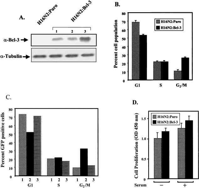
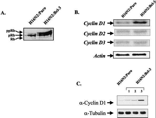
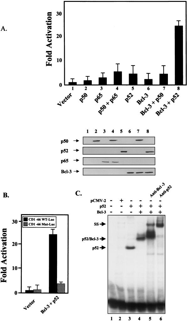
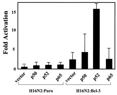
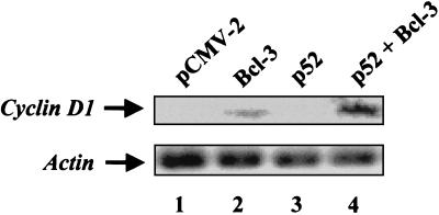
Bcl-3 is a distinctive member of the IkappaB family of NF-kappaB inhibitors because it can function to coactivate transcription. A potential involvement of Bcl-3 in oncogenesis is highlighted by the fact that it was cloned due to its location at a breakpoint junction in some cases of human B-cell chronic lymphocytic leukemia and that it is highly expressed in human breast tumor tissue. To analyze the effects of Bcl-3 dysregulation in breast epithelial cells, we created stable immortalized human breast epithelial cell lines either expressing Bcl-3 or carrying the corresponding vector control plasmid. Analysis of the Bcl-3-expressing cells suggests that these cells have a shortened G(1) phase of the cell cycle as well as a significant increase in hyperphosphorylation of the retinoblastoma protein. Additionally, the cyclin D1 gene was found to be highly expressed in these cells. Upon further analysis, Bcl-3, acting as a coactivator with NF-kappaB p52 homodimers, was demonstrated to directly activate the cyclin D1 promoter through an NF-kappaB binding site. Therefore, our results demonstrate that dysregulated expression of Bcl-3 potentiates the G(1) transition of the cell cycle by stimulating the transcription of the cyclin D1 gene in human breast epithelial cells.


Figures

FIG. 1
FIG. 1
Stable expression of Bcl-3 in breast epithelial cells promotes the G1 transition. (A) Creation o f stable cell lines expressing Bcl-3. H16N2 immortalized human breast epithelial cells were transfected with pFlag-CMV2 or pFlag-Bcl-3, and stable cell lines were generated (H16N2:Puro and H16N2:Bcl-3). Western analysis with anti-Bcl-3 shows expression of Bcl-3 protein in H16N2:Bcl-3 clones 1, 2, and 3. (B) FACS analysis shows that Bcl-3 expression leads to an accelerated G1 phase. Proliferating cells were harvested and analyzed by FACS. The percentage of cells in each stage of the cell cycle is indicated. Data represent the mean ± standard deviation of three independent experiments. (C) Cyclin D1 is required for the induction of G1 phase in H16N2:Bcl-3 cells. Cells were cotransfected with pPCMVEGFP-spectrin and either vector control plasmid (pBPSTR-1) or a cyclin D1 antisense plasmid (pBPSTR-1 CD1 AS). GFP-positive cells were then analyzed by FACS. In sample 1, H16N2:Puro cells were transfected with a vector control plasmid. In sample 2, H16N2:Bcl-3 cells were transfected with a vector control plasmid. In sample 3, H16N2:Bcl-3 cells were transfected with a cyclin D1 antisense plasmid. (D) Expression of Bcl-3 does not lead to increased cellular proliferation in H16N2 cells. H16N2:Puro or H16N2:Bcl-3 cells were grown in the presence or absence of serum in triplicate wells of a 96-well plate. Twenty-four hours after plating, the cells were incubated for 4 h with BrdU and analyzed by ELISA for BrdU incorporation. OD, optical density. Results represent the mean ± standard deviation of three independent experiments.
FIG. 2
FIG. 2
Bcl-3 expression leads to hyperphosphorylation of Rb and induction of endogenous cyclin D1 levels. (A) Fifty micrograms of total cellular protein from the H16N2:Puro and H16N2:Bcl-3 cell lines was run on SDS-PAGE. After transfer to nitrocellulose, the membrane was probed with an anti-Rb antibody. Of the three Rb bands detected, the upper-molecular-weight band represents hyperphosphorylated Rb (ppRB). (B) The H16N2:Bcl-3 cell line shows increased levels of cyclin D1 RNA by Northern analysis. Cyclin D2 and cyclin D3 mRNA levels are unchanged. Equal RNA loading was verified by using a probe for actin. (C) Cell extracts were isolated from the H16N2:Puro and H16N2:Bcl-3 cell lines and run on SDS-PAGE. After transfer to nitrocellulose, the membranes were probed with an anti-cyclin D1 antibody. Equal protein loading was verified with antitubulin antibody.
FIG. 3
FIG. 3
Bcl-3 and p52 synergistically activate the cyclin D1 promoter. (A) NIH 3T3 cells were transiently cotransfected with the full-length cyclin D1 promoter luciferase construct (CD1 −963 WT-Luc) and with expression vectors encoding p50, p65, p52, and Bcl-3 proteins alone or in combination. Forty-eight hours posttransfection, cells were harvested and luciferase assays were performed. The data presented represent the mean ± standard deviation of three independent experiments performed in triplicate. The fold inductions, as compared to the vector control, were plotted. (Lower panel) Western analysis showing equivalent expression of transiently transfected constructs. (B) Bcl-3 and p52 activate transcription through the proximal NF-κB site in transient transfection assays. NIH-3T3 cells were transiently cotransfected with a cyclin D1 promoter luciferase construct containing the wild-type proximal NF-κB site (CD1 −66 WT-Luc) or a mutated site (CD1 −66 Mut-Luc) together with expression vectors encoding p52 and Bcl-3 proteins and assayed as described above. (C) Bcl-3 and p52 proteins bind to the proximal NF-κB site of the cyclin D1 promoter. COS-7 cells were transfected with various combinations of Bcl-3, p52, or vector control expression constructs as indicted. Nuclear extracts were analyzed by gel shift. The identities of the bound proteins were verified by supershift (SS) with antibodies against Bcl-3 and p52, as indicated.
FIG. 4
FIG. 4
Activation of the cyclin D1 promoter by p52 in Bcl-3 stable breast epithelial cells. H16N2:Puro and H16N2:Bcl-3 cell lines were transiently transfected with the cyclin D1 promoter luciferase construct (CD1 −963 WT-Luc), a lacZ expression vector, and with expression vectors encoding p50, p52, and p65 proteins alone or in combination. Forty-eight hours posttransfection, cells were harvested and luciferase assays were performed. Transfection efficiency for the two cell lines was found to be equivalent, as determined by counting β-galactosidase-positive cells (data not shown). The data presented represent the mean ± standard deviation of luciferase expressions of three independent experiments performed in triplicate. The fold inductions, as compared to that of the vector control, were plotted.
FIG. 5
FIG. 5
Bcl-3 and p52 proteins upregulate endogenous cyclin D1 mRNA in 293T cells. 293T cells were transfected with various combinations of Bcl-3, p52, or vector control expression constructs as indicated. RNA was isolated, and Northern blot analysis was used to probe for levels of cyclin D1 mRNA. An actin probe was used to verify equal RNA loading.

References

    1. Albanese C, Johnson G, Watanabe N, Eklund N, Vu D, Arnold A, Pestell R G. Transforming p21ras mutants and c-Ets-2 activate the cyclin D1 promoter through distinguishable regions. J Biol Chem. 1995;270:23589–23597. - PubMed
    1. Baldwin A S., Jr The NF-κB and IκB proteins: new discoveries and insights. Annu Rev Immunol. 1996;14:649–683. - PubMed
    1. Beijersbergen R L, Bernards R. Cell cycle regulation by the retinoblastoma family of growth inhibitory proteins. Biochim Biophys Acta. 1996;1287:103–120. - PubMed
    1. Bours V, Franzoso G, Azarenko V, Park S, Kanno T, Brown K, Siebenlist U. The oncoprotein Bcl-3 directly transactivates through kappa B motifs via association with DNA-binding p50B homodimers. Cell. 1993;72:729–739. - PubMed
    1. Brasier A R, Lu M, Hai T, Lu Y, Boldogh I. NF-κB inducible BCL-3 expression is an autoregulatory loop controlling nuclear p50/NF-κB1 residence. J Biol Chem. 2001;276:32080–32093. - PubMed

Publication types

MeSH terms