. 2001 Dec;21(24):8437-51.
doi: 10.1128/MCB.21.24.8437-8451.2001.

Transcriptional consequences of topoisomerase inhibition

Affiliations

Transcriptional consequences of topoisomerase inhibition

I Collins et al. Mol Cell Biol. 2001 Dec.

Abstract

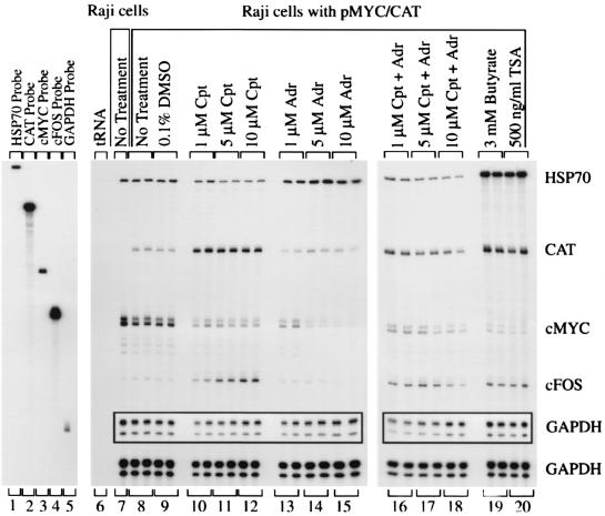
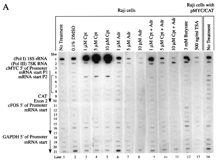
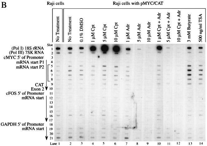
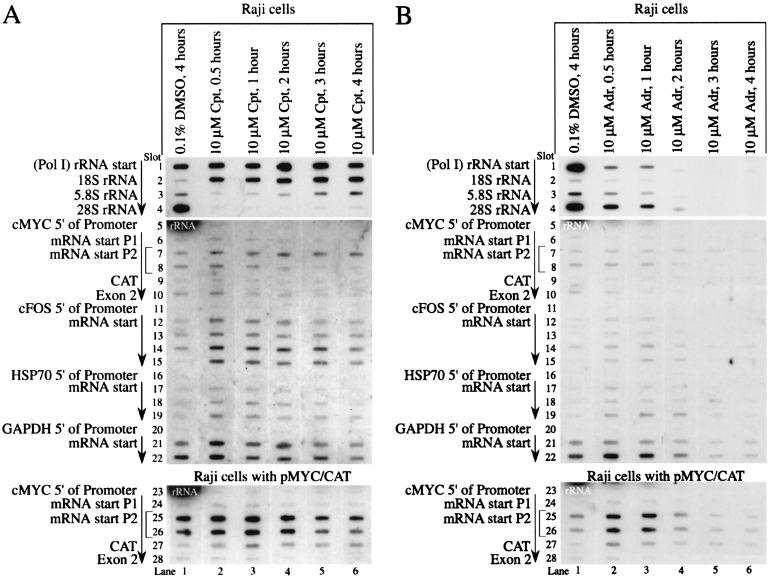
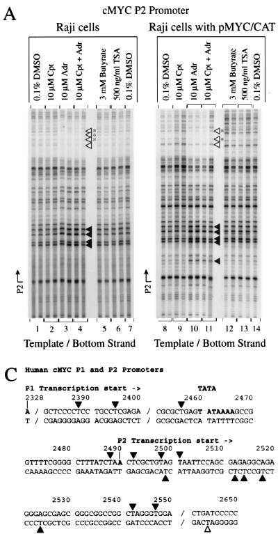
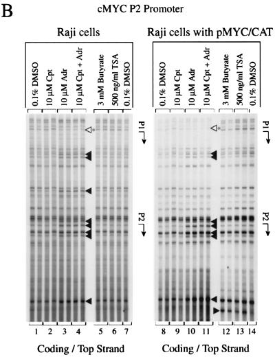
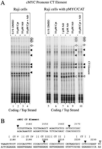
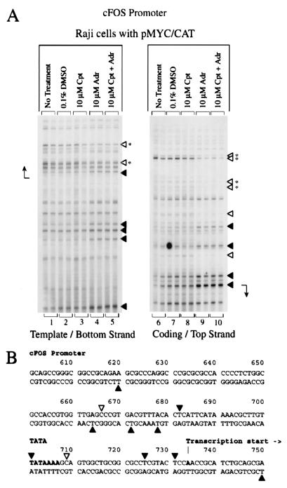
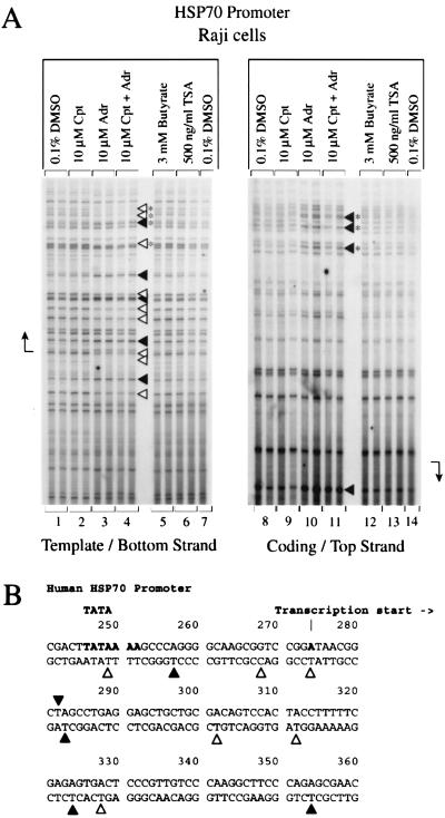
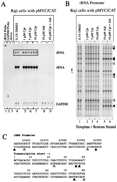
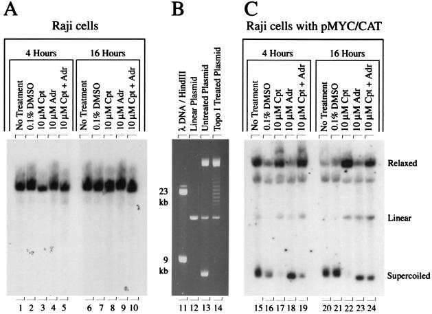
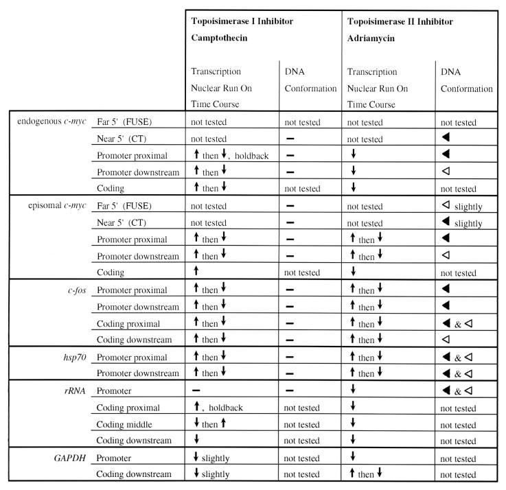
In principle, the generation, transmission, and dissipation of supercoiling forces are determined by the arrangement of the physical barriers defining topological boundaries and the disposition of enzymes creating (polymerases and helicases, etc.) or releasing (topoisomerases) torsional strain in DNA. These features are likely to be characteristic for individual genes. By using topoisomerase inhibitors to alter the balance between supercoiling forces in vivo, we monitored changes in the basal transcriptional activity and DNA conformation for several genes. Every gene examined displayed an individualized profile in response to inhibition of topoisomerase I or II. The expression changes elicited by camptothecin (topoisomerase I inhibitor) or adriamycin (topoisomerase II inhibitor) were not equivalent. Camptothecin generally caused transcription complexes to stall in the midst of transcription units, while provoking little response at promoters. Adriamycin, in contrast, caused dramatic changes at or near promoters and prevented transcription. The response to topoisomerase inhibition was also context dependent, differing between chromosomal or episomal c-myc promoters. In addition to being well-characterized DNA-damaging agents, topoisomerase inhibitors may evoke a biological response determined in part from transcriptional effects. The results have ramifications for the use of these drugs as antineoplastic agents.


Figures

FIG. 1
FIG. 1
The steady-state level of each mRNA displays a distinctive profile in response to inhibition of topoisomerase I or II, by monitoring RNase protection. The c-myc exon 2 versus CAT probes used in this experiment distinguish between the c-myc RNAs encoded by the endogenous c-myc gene and the hybrid MYC/CAT mRNA encoded by the episome pMYC/CAT. Cpt, camptothecin; Adr, adriamycin; TSA, trichostatin A. The cells were incubated with inhibitors for 4 h. The duplicate lanes are each from different independent experiments. A 3-day exposure made with Kodak XAR film is shown, along with a 13-h exposure for the full-length probes and the GAPDH (glyceraldehyde-3-phosphate dehydrogenase) inset.
FIG. 2
FIG. 2
Heterogeneous response of promoter activity to topoisomerase inhibition. (A) Nuclear run-ons of Raji cells. (B) Nuclear run-ons of Raji cells with pMYC/CAT. c-myc exon 2 (slot 10) and CAT (slot 9) distinguish between the c-myc RNAs encoded by the endogenous c-myc gene and the hybrid MYC/CAT mRNA encoded by the episome pMYC/CAT. The arrow indicates the direction of transcription. Cpt, camptothecin; Adr, adriamycin; TSA, trichostatin A. The cells were incubated with inhibitors for 4 h. A 10-min run-on reaction was performed.
FIG. 2
FIG. 2
Heterogeneous response of promoter activity to topoisomerase inhibition. (A) Nuclear run-ons of Raji cells. (B) Nuclear run-ons of Raji cells with pMYC/CAT. c-myc exon 2 (slot 10) and CAT (slot 9) distinguish between the c-myc RNAs encoded by the endogenous c-myc gene and the hybrid MYC/CAT mRNA encoded by the episome pMYC/CAT. The arrow indicates the direction of transcription. Cpt, camptothecin; Adr, adriamycin; TSA, trichostatin A. The cells were incubated with inhibitors for 4 h. A 10-min run-on reaction was performed.
FIG. 3
FIG. 3
Rapid response of promoter activity to topoisomerase inhibition as shown by nuclear run-on. (A) Time course of camptothecin inhibition. In lane 1, slots 5 and 23 show the strong 28S rRNA signal spreading from the slot above. (B) Time course of adriamycin inhibition. c-myc exon 2 (slot 10) and CAT (slot 9) distinguish between the c-myc RNAs encoded by the endogenous c-myc gene and the hybrid MYC/CAT mRNA encoded by the episome pMYC/CAT. The arrow indicates the direction of transcription. Cpt, camptothecin; Adr, adriamycin. Lane 1 shows cells treated with DMSO for 4 h. Lanes 2 to 6 were incubated with either camptothecin or adriamycin for 0.5, 1, 2, 3, or 4 h before nuclei were harvested. A 10-min run-on reaction was performed.
FIG. 4
FIG. 4
Response of c-myc promoter structure to topoisomerase inhibition. In vivo potassium permanganate footprint. (A) Bottom DNA strand. (B) Top DNA strand. Cpt, camptothecin; Adr, adriamycin; TSA, trichostatin A. The cells were incubated with inhibitors for 4 h. The arrow indicates the transcription start site. Bases made partially hyposensitive by camptothecin are present but are not annotated (panel B, lane 2). (C) Nucleotides with altered intensities in the footprint are identified. Symbols: ◂, hypersensitivity; ◃, hyposensitivity. The asterisk indicates reactivity at bases that were not exactly mapped due to DNA compression in this region of the gel. The duplicate lanes are each from different independent experiments.
FIG. 4
FIG. 4
Response of c-myc promoter structure to topoisomerase inhibition. In vivo potassium permanganate footprint. (A) Bottom DNA strand. (B) Top DNA strand. Cpt, camptothecin; Adr, adriamycin; TSA, trichostatin A. The cells were incubated with inhibitors for 4 h. The arrow indicates the transcription start site. Bases made partially hyposensitive by camptothecin are present but are not annotated (panel B, lane 2). (C) Nucleotides with altered intensities in the footprint are identified. Symbols: ◂, hypersensitivity; ◃, hyposensitivity. The asterisk indicates reactivity at bases that were not exactly mapped due to DNA compression in this region of the gel. The duplicate lanes are each from different independent experiments.
FIG. 5
FIG. 5
Topoisomerase II inhibition provokes conformational changes at the CT-element of the endogenus c-myc gene. (A) In vivo potassium permanganate footprint. Cpt, camptothecin; Adr, adriamycin. The cells were incubated with inhibitors for 4 h. (B) Nucleotides with altered intensities in the footprint are identified. Symbols are as defined for Fig. 4. The duplicate lanes are each from different independent experiments.
FIG. 6
FIG. 6
Topoisomerase II inhibition alters the structure of the c-fos promoter. (A) In vivo potassium permanganate footprint. Cpt, camptothecin; Adr, adriamycin. The cells were incubated with inhibitors for 4 h. (B) Nucleotides with altered intensities in the footprint are identified. Symbols are as defined for Fig. 4. The duplicate lanes are each from different independent experiments.
FIG. 7
FIG. 7
Topoisomerase II inhibition alters the structure of the hsp70 promoter. (A) In vivo potassium permanganate footprint. An alternating pattern of increased and decreased intensity near the hsp70 transcription start site is seen. Cpt, camptothecin; Adr, adriamycin; TSA, trichostatin A. The cells were incubated with inhibitors for 4 h. (B) Nucleotides with altered intensities in the footprint are identified. Symbols are as in Fig. 4. The duplicate lanes are each from different independent experiments.
FIG. 8
FIG. 8
Adriamycin, but not camptothecin, depresses rRNA promoter activity and alters promoter structure. (A) RNase Protection with a probe for the rRNA transcription start site. A 13-h Kodak XAR film exposure and a 2-h exposure (inset) are shown. (B) In vivo potassium permanganate footprint. Cpt, camptothecin; Adr, adriamycin. The cells were incubated with inhibitors for 4 h. Symbols are as in Fig. 4. (C) Nucleotides with altered intensities in the footprint are identified. The duplicate lanes are each from different independent experiments.
FIG. 9
FIG. 9
DNA damage is unlikely to account fully for the transcriptional response to topoisomerase inhibition. Southern blots showed that brief camptothecin treatment yielded relaxed, but not linear plasmid. Adriamycin yielded hypersupercoiled episomal DNA. (A) Raji cells probed for c-myc exon 2. (B) Pure preparation of pMYC/CAT and the effects of topoisomerase I treatment. (C) Raji pMYC/CAT cell line probed with CAT sequences. Cpt, camptothecin; Adr, adriamycin. The cells were incubated with inhibitors for 4 and 16 h.
FIG. 10
FIG. 10
Summary of response to camptothecin or adriamycin treatment. Symbols: ↑, increased transcription; ↓, decreased transcription. A ↑ then ↓ combination indicates that in the run-on time course experiment, transcription was induced at the early time point but then repressed by the late time point. The dash (—) reflects no change in transcription. The black triangle (◂) indicates hypersensitivity, while the open triangle (◃) indicates hyposensitivity in the in vivo potassium permanganate footprint. The combination “◂ and ◃” indicates a mixed hypersensitivity and hyposensitivity in the in vivo potassium permanganate footprint.

References

    1. Albert T, Mautner J, Funk J O, Hortnagel K, Pullner A, Eick D. Nucleosomal structures of c-myc promoters with transcriptionally engaged RNA polymerase II. Mol Cell Biol. 1997;17:4363–4371. - PMC - PubMed
    1. Aller P, Rius C, Mata F, Zorrilla A, Cabanas C, Bellon T, Bernabeu C. Camptothecin induces differentiation and stimulates the expression of differentiation-related genes in U-937 human promonocytic leukemia cells. Cancer Res. 1992;52:1245–1251. - PubMed
    1. Amara F M, Entwistle J, Kuschak T I, Turley E A, Wright J A. Transforming growth factor-β1 stimulates multiple protein interactions at a unique cis-element in the 3′-untranslated region of the hyaluronan receptor RHAMM mRNA. J Biol Chem. 1996;271:15279–15284. - PubMed
    1. Bates A D, Maxwell A. DNA Topology. Oxford, England: IRL Press; 1993.
    1. Bhat U G, Raychaudhuri P, Beck W T. Functional interaction between human topoisomerase IIα and retinoblastoma protein. Proc Natl Acad Sci USA. 1999;96:7859–7864. - PMC - PubMed

Publication types

MeSH terms

LinkOut - more resources