. 2001 Dec;21(24):8657-70.
doi: 10.1128/MCB.21.24.8657-8670.2001.

Ribosomal pausing at a frameshifter RNA pseudoknot is sensitive to reading phase but shows little correlation with frameshift efficiency

Affiliations

Ribosomal pausing at a frameshifter RNA pseudoknot is sensitive to reading phase but shows little correlation with frameshift efficiency

H Kontos et al. Mol Cell Biol. 2001 Dec.

Abstract

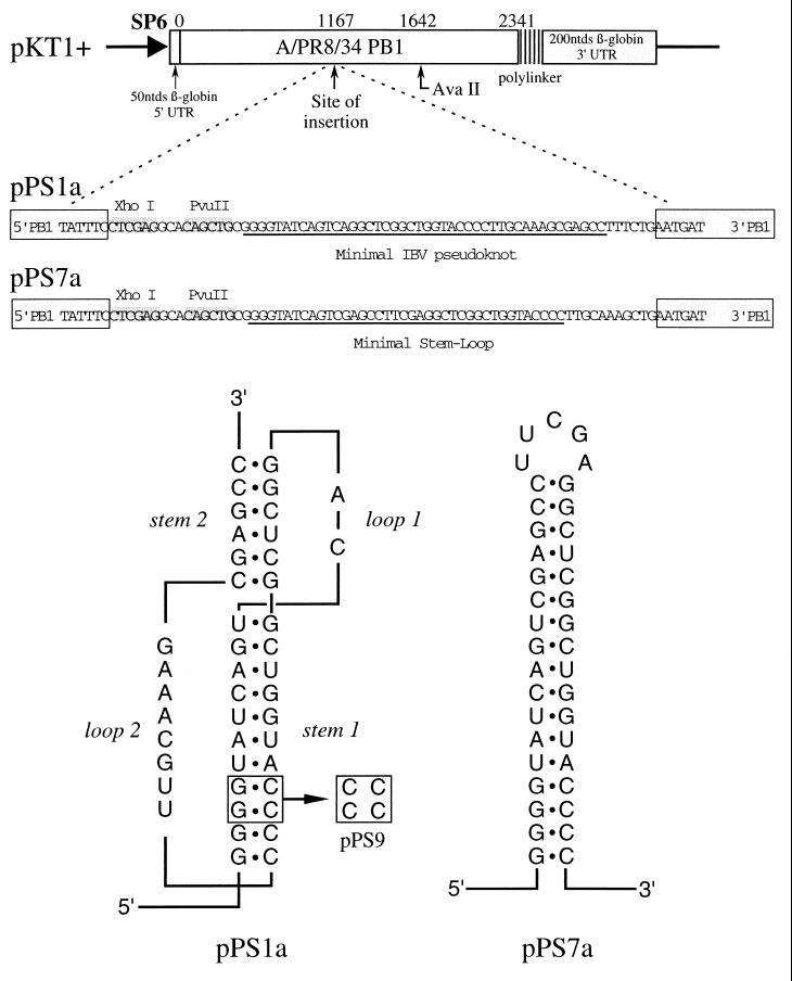
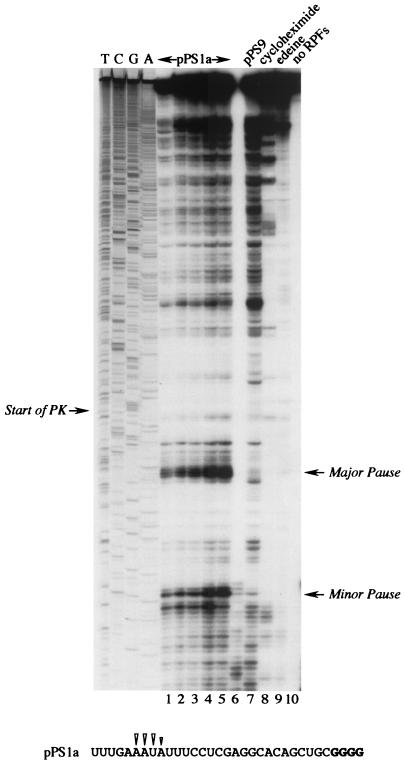
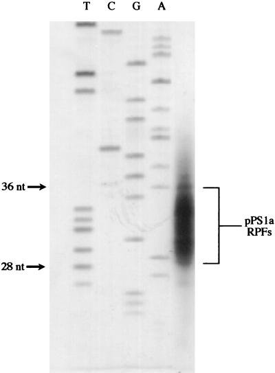
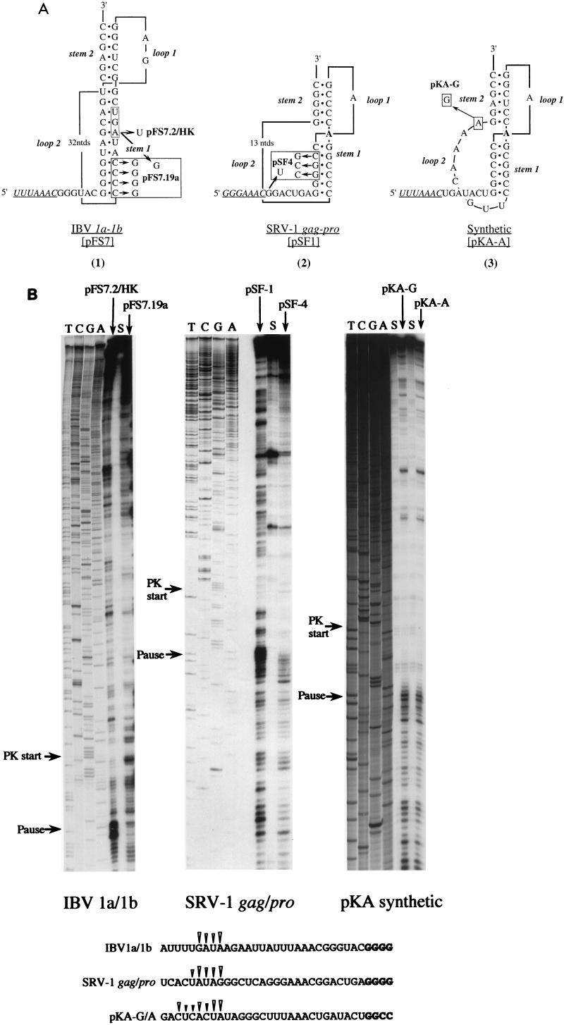
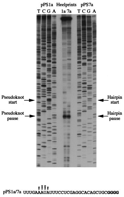
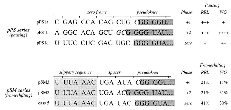
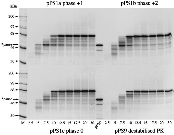
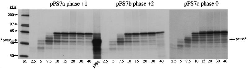
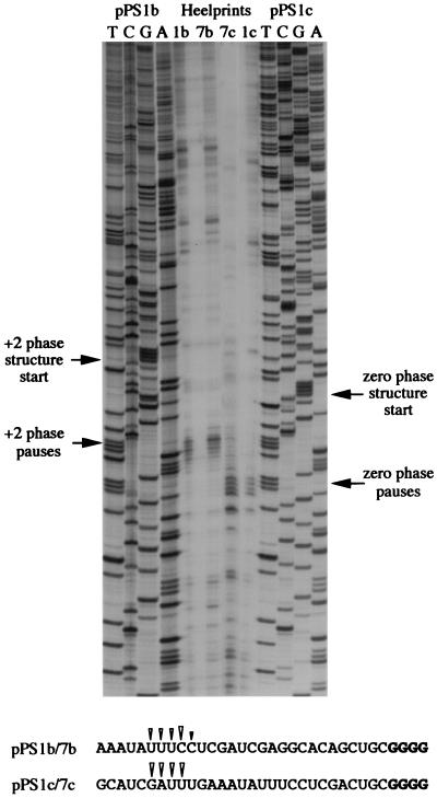
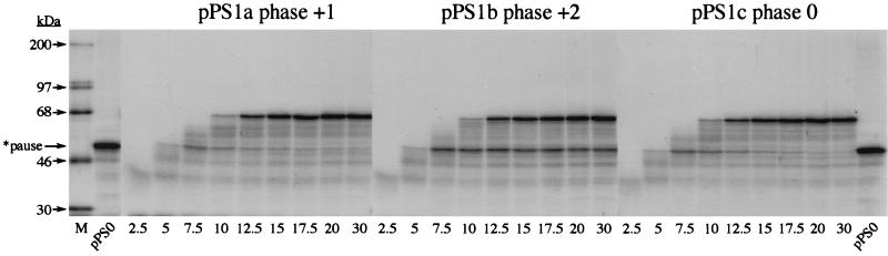
Here we investigated ribosomal pausing at sites of programmed -1 ribosomal frameshifting, using translational elongation and ribosome heelprint assays. The site of pausing at the frameshift signal of infectious bronchitis virus (IBV) was determined and was consistent with an RNA pseudoknot-induced pause that placed the ribosomal P- and A-sites over the slippery sequence. Similarly, pausing at the simian retrovirus 1 gag/pol signal, which contains a different kind of frameshifter pseudoknot, also placed the ribosome over the slippery sequence, supporting a role for pausing in frameshifting. However, a simple correlation between pausing and frameshifting was lacking. Firstly, a stem-loop structure closely related to the IBV pseudoknot, although unable to stimulate efficient frameshifting, paused ribosomes to a similar extent and at the same place on the mRNA as a parental pseudoknot. Secondly, an identical pausing pattern was induced by two pseudoknots differing only by a single loop 2 nucleotide yet with different functionalities in frameshifting. The final observation arose from an assessment of the impact of reading phase on pausing. Given that ribosomes advance in triplet fashion, we tested whether the reading frame in which ribosomes encounter an RNA structure (the reading phase) would influence pausing. We found that the reading phase did influence pausing but unexpectedly, the mRNA with the pseudoknot in the phase which gave the least pausing was found to promote frameshifting more efficiently than the other variants. Overall, these experiments support the view that pausing alone is insufficient to mediate frameshifting and additional events are required. The phase dependence of pausing may be indicative of an activity in the ribosome that requires an optimal contact with mRNA secondary structures for efficient unwinding.


Figures

FIG. 1
FIG. 1
Pausing constructs based on the IBV frameshift signal. Plasmids pPS1a and pPS7a (formerly pPS1 and pPS7 [36]) contain, respectively, the minimal IBV pseudoknot and a related stem-loop structure (3, 4) cloned into the influenza PB1 gene in an SP6 promoter-based transcription vector. Plasmid pPS9 is a derivative of pPS1a in which stem 1 is destabilized by a complementary mutation.
FIG. 2
FIG. 2
Ribosomal pausing at the minimal IBV pseudoknot. mRNA from AvaII-digested pPS1a was subjected to heelprint analysis as detailed in Materials and Methods. Heelprints of the minimal IBV pseudoknot (pPS1a; lanes 1 to 6 and 8 to 10) and a mutant derivative (pPS9, lane 7) are shown alongside a sequencing ladder (TCGA). Each reaction mixture contained 20 ng of the relevant single-stranded DNA template. In lanes 1 to 5, the concentrations of primer and RPFs were varied: lane 1, 0.1 ng of primer, 3 μl of RPFs; lane 2, 0.2 ng of primer, 3 μl of RPFs; lane 3, 0.4 ng of primer, 3 μl of RPFs; lane 4, 0.4 ng of primer, 4 μl of RPF; lane 5, 0.4 ng of primer, 4.5 μl of RPFs. All other lanes (except lane 10) contained 0.4 ng of primer and 3 μl of RPFs. Lanes 8 and 9 were control reactions in which cycloheximide (lane 8) or edeine (lane 9) was added (to 1 mM and 5 μM concentrations, respectively) prior to addition of mRNA to the translation reaction mixture. In lane 10, RPFs were omitted from the primer extension reaction. The start of the pseudoknot (the first G in a block of four reading up the gel) and the position of two clear pause sites are indicated with arrows. Lane 6 was identical to lane 5, except T4 DNA polymerase replaced T7 DNA polymerase (unsuccessfully). The primary sequence of the mRNA upstream of the pseudoknot is shown at the bottom, and the positions of the pseudoknot-dependent heelprints are indicated with arrowheads (the first four G residues of the pseudoknot are shown in bold type).
FIG. 3
FIG. 3
Sizing of RPFs. 32P-trace-labeled AvaII-digested pPS1a mRNA was subjected to heelprinting, and the RPFs were analyzed on an 8% denaturing polyacrylamide gel. A sequencing ladder (TCGA) was run alongside to provide approximate size standards.
FIG. 4
FIG. 4
Ribosomal pausing at natural and synthetic frameshift signals. (A) Predicted secondary structures of natural and synthetic frameshift signals tested in heelprint assays (1). Plasmid pFS7 contains the wild-type IBV slippery sequence (UUUAAAC, underlined) and pseudoknot (2). The 1a termination codon (UGA), which forms part of stem 1, is boxed. Plasmids pFS7.2/HK and pFS7.19a are mutant derivatives in which the 1a termination codon has been changed to UGU or UGG, respectively. Plasmid pFS7.19a contains an additional mutation, a complementary change that destabilizes stem 1 of the pseudoknot (2). Plasmid pSF1 contains the SRV-1 gag/pro frameshift region (42). A derivative, pSF4, has a destabilizing mutation in stem 1 and, additionally, a termination codon (UGA) immediately downstream of the slippery sequence (GGGAAAC, underlined). The unpaired A residue (in bold) between the stems is drawn on the basis that the pseudoknot is similar to that of MMTV gag/pro (see text). Recent NMR studies have challenged this belief (14, 29) and suggest that the A is in fact paired with the most 3′ base of loop 2 (a U). The synthetic frameshift site pKA-A (25) has an IBV-like slippery sequence (UUUAAAC, underlined) and an MMTV-like stimulatory pseudoknot. Plasmid pKA-G differs solely in the identity of the last residue of loop 2. (B) mRNAs from SmaI-digested pFS7.2/HK and pFS7.19a or BamHI-digested pSF-1, pSF-4, pKA-A, and pKA-G were subjected to heelprint analysis as detailed in Materials and Methods. Heelprints of each RNA are shown alongside a sequencing ladder (TCGA) prepared from the relevant plasmid. Each reaction mixture contained 20 ng of the relevant single-stranded DNA template, 0.4 ng of primer, and 3 μl of RPFs. Lanes marked S indicate heelprints in which RPFs were replaced by an equivalent amount (vol/vol) of material harvested from the supernatant produced in the airfuge centrifugation step (see Materials and Methods). The start of each pseudoknot and the position of pseudoknot-dependent ribosomal pauses are indicated by arrows. The primary sequences of the mRNA upstream of the various pseudoknots are shown at the bottom and the position of the pseudoknot-dependent heelprints are indicated with arrowheads (the first four pseudoknot residues are shown in bold type in each case).
FIG. 5
FIG. 5
Comparison of pseudoknot- and hairpin-induced ribosomal pauses. mRNAs from AvaII-digested pPS1a and pPS7a were subjected to heelprint analyses as detailed in Materials and Methods. Heelprints of each RNA are shown alongside a sequencing ladder (TCGA) prepared from each plasmid. Each reaction mixture contained 20 ng of the relevant single-stranded DNA template, 0.4 ng of primer, and 3 μl of RPFs. The start of the pseudoknot (pPS1a) and hairpin (pPS7a) and the position of corresponding ribosomal pauses are indicated with arrows. The primary sequence of the mRNA upstream of the pseudoknot or hairpin is identical and is shown at the bottom. The position of the structure-dependent heelprints are indicated with arrowheads (the first four residues of the pseudoknot and hairpin are shown in bold type).
FIG. 6
FIG. 6
Pseudoknot encounter phase in pausing and frameshifting constructs. The nucleotide sequence of the mRNA in the vicinity of the IBV minimal pseudoknot in pausing (pPS series) and frameshifting (pSM series [4]) constructs is shown. In all mRNAs, only the 5′ portion of the pseudoknot is displayed (in grey). The phase is defined by the number of nucleotides between the last in-frame codon and the start of the pseudoknot. In pPS1a, for example, the single nucleotide (C, italicized) present between the reading frame codon CUG and the start of the pseudoknot defines the phase as +1. In the pSM series, the number of nucleotides that separate the IBV slippery sequence (boxed) and the pseudoknot (spacer region) varies. The wild-type spacer is 6 nt (cass 5); in pSM3 an additional A residue (bold) is present, and in pSM2 a U has been deleted (4). Also shown (on right) is a summary of the pausing level and frameshifting efficiencies specified by the constructs. The frameshift efficiencies in RRL were from Brierley and colleagues (4); those in WG were determined here (translations not shown).
FIG. 7
FIG. 7
Reading-phase dependence of ribosomal pausing. Time courses of translation of AvaII-derived pPS1a, -b, and -c and pPS9 mRNAs in reticulocyte lysates. Translation was allowed to proceed at 26°C in the presence of [35S]methionine for 5 min prior to addition of edeine to a final concentration of 5 μM. Samples were withdrawn at the indicated times (in minutes) post-edeine addition, and translation products were separated on SDS–10% polyacrylamide gels. Labeled polypeptides were detected by autoradiography. [14C]-labeled molecular mass standards (M) were from Amersham Pharmacia Biotech. The pPS0 tracks mark the expected position of a pseudoknot-induced ribosomal pause product and were prepared by translating XhoI-derived pPS0 mRNA at 26°C for 1 h. The pause product is indicated by an arrow. Although the size of this protein as predicted from the nucleic acid sequence of the PB1 reporter gene is 43 kDa, it migrates somewhat slower in SDS-polyacrylamide gels because of the highly basic nature of the PB1 protein (2).
FIG. 8
FIG. 8
Pausing at hairpin phase variants. Time courses of translation of AvaII-derived pPS7a, -b, and -c mRNAs in reticulocyte lysates are shown. Translation products were prepared, labeled, and analyzed as described in the legend to Fig. 7. M, molecular mass standards.
FIG. 9
FIG. 9
Heelprinting of hairpin phase variants. mRNAs from AvaII-digested pPS1b, pPS7b, pPS1c, and pPS7c were subjected to heelprint analysis as detailed in Materials and Methods. Heelprints of each RNA are shown alongside a sequencing ladder (TCGA) prepared from two of the plasmids. Each reaction mixture contained 20 ng of the relevant single-stranded DNA template, 0.4 ng of primer, and 3 μl of RPFs. The position of the start of the +2 and zero-phase pseudoknots and hairpins and the site of the corresponding ribosomal pauses are indicated by arrows. The primary sequences of the mRNA upstream of the various structures are shown at the bottom, and the position of the pseudoknot- or hairpin-dependent heelprints are indicated with arrowheads (the first four pseudoknot and hairpin residues are shown in bold type in each case).
FIG. 10
FIG. 10
Reading-phase-dependent ribosomal pausing in the WG system. Shown are the time courses of translation of AvaII-derived pPS1a, -b, and -c mRNAs in the WG system. Translation products were prepared, labeled, and analyzed as described in the legend to Fig. 7, except that the translations were carried out at 15°C. M, molecular mass standards.

References

    1. Alam S L, Wills N M, Ingram J A, Atkins J F, Gesteland R F. Structural studies of the RNA pseudoknot required for readthrough of the gag-termination codon of murine leukemia virus. J Mol Biol. 1999;288:837–852. - PubMed
    1. Brierley I, Digard P, Inglis S C. Characterisation of an efficient coronavirus ribosomal frameshifting signal: requirement for an RNA pseudoknot. Cell. 1989;57:537–547. - PMC - PubMed
    1. Brierley I, Rolley N J, Jenner A J, Inglis S C. Mutational analysis of the RNA pseudoknot component of a coronavirus ribosomal frameshifting signal. J Mol Biol. 1991;220:889–902. - PMC - PubMed
    1. Brierley I, Jenner A J, Inglis S C. Mutational analysis of the “slippery sequence” component of a coronavirus ribosomal frameshifting signal. J Mol Biol. 1992;227:463–479. - PMC - PubMed
    1. Brierley I. Ribosomal frameshifting on viral RNAs. J Gen Virol. 1995;76:1885–1892. - PubMed

Publication types

LinkOut - more resources